Posts filed under Pharmacology

Breastfeeding Pharmacy: Analgesics

Written by: Courtney Premer-Barragan, MD, PhD (NUEM ‘25)
Edited by: Adam Payne, MD (NUEM ‘24)
Expert Commentary by: Kelsea Caruso, PharmD


Expert Commentary

“Pump and dump” is definitely the easy way out for the emergency medicine provider, but this practice can have detrimental effects on the baby and on the mother. There is a false pretense that many medications are harmful to the breastfeeding infant, but this is not the case. The other consideration to have when thinking about medication use in breastfeeding is the medication effects on the mother’s lactation and the medication impact on breast milk production.

Ibuprofen by far has the most supporting evidence for use in breastfeeding women and this is a reasonable first line agent for treating many types of pain. Ketorolac is used frequently immediately after delivery and limited amount of drug is excreted in colostrum, but more may be excreted as milk supply increases thus increasing the risk of bleeding in the infant. Aspirin is excreted into breastmilk, and long-term use of high doses may cause bleeding along with metabolic abnormalities in the infant. That said, long-term use of low dose aspirin is likely safe.

If opioids are required for pain control, fentanyl is a reasonable choice for immediate pain control. Combination hydrocodone and acetaminophen is also an option when oral pain medications need to be utilized. The jury is (sort of) out on if oxycodone is safe during breastfeeding, and the baby should be monitored closely if oxycodone is selected for pain management.

Local anesthetics are very poorly absorbed by the infant, but still remain diligent about checking the specific maximum recommended dose for adults. My favorite database to find information on medications in lactation is LactMed, a database funded by the NIH. It is always safest to check this database before prescribing a medication to a lactating patient.

Kelsea Caruso, PharmD

Clinical Pharmacist

Department of Emergency Medicine

Northwestern Memorial Hospital


How To Cite This Post:

[Peer-Reviewed, Web Publication] Premer-Barragan, C. Payne, A. (2023, Jul 31). Breastfeeding Pharmacy Analgesics. [NUEM Blog. Expert Commentary by Caruso, K]. Retrieved from http://www.nuemblog.com/blog/breastfeeding-pharm-analgesics



Other Posts You May Enjoy

Posted on July 31, 2023 and filed under Pharmacology.

Breastfeeding Pharmacy: Antibiotics

Written by: Courtney Premer-Barragan, MD, PhD (NUEM ‘25)
Edited by: Ben Kiesel, MD (NUEM ‘23)
Expert Commentary by: Kelsea Caruso, PharmD


Expert Commentary

Emergency Medicine practitioners may be quick to recommend patients to “pump and dump” when on antibiotics, but this can have downstream detrimental effects on the baby and on the mother. With the numerous indications for antibiotics, including some dealing with breastfeeding itself, it is imperative that EM providers recognize that most antibiotics are considered safe.

Most beta-lactam antibiotics are considered safe in breastfeeding women and can be used to treat many infections. Depending on the type of infection, anaerobic coverage may be warranted. Case reports have documented the potential for metronidazole to cause Candida infections and diarrhea in the infant. Clindamycin has the highest potential to cause GI issues in the breastfed infant. If these medications are indicated, it is best to have a risk-benefit discussion with the patient about the best option.

Tetracyclines have historically been feared in breastfeeding mothers due to the potential for bone deposition and staining of the dental enamel. As more literature has reviewed their safety, tetracyclines are considered safe for short term use, but limit courses to fewer than 21 days. Tetracycline absorption is also inhibited by calcium, which is contained in breast milk, so the amount actually absorbed by the infant should be low.

My favorite database to find information on medications in lactation is LactMed, a database funded by the NIH. Always the safest bet is to check this database before prescribing a medication to a lactating patient.

Kelsea Caruso, PharmD

Clinical Pharmacist

Department of Emergency Medicine

Northwestern Memorial Hospital


How To Cite This Post:

[Peer-Reviewed, Web Publication] Premer-Barragan, C. Payne, A. (2023, Jul 25). Breastfeeding Pharmacy Antibiotics. [NUEM Blog. Expert Commentary by Caruso, K]. Retrieved from http://www.nuemblog.com/blog/breastfeeding-pharm-antibiotics


Other Posts You May Enjoy

Posted on July 25, 2023 and filed under Pharmacology.

Toxic Alcohols

Written by: Rafael Lima, MD (NUEM ‘23) Edited by: Laurie Aluce, MD (NUEM ‘21)
Expert Commentary by: Zachary Schmitz (NUEM ‘21)


Methanol Toxicity

Methanol itself is not toxic to the body. Methanol’s metabolite, formic acid, causes toxicity at serum levels greater than 20mg/dl [1].

Clinical Findings of Methanol Poisoning

  • CNS sedation

  • Seizures

  • Rapid, Deep Breathing

  • Hypotension

  • Ocular findings: 

    • Blindness 

    • Afferent pupillary defect

    • Optic disk hyperemia

    • Mydriasis

Ethylene Glycol Toxicity

Similarly, the toxic metabolites of ethylene glycol cause end-organ damage at levels greater than 20mg/dl. The most notable toxic metabolites are glycolic acid and oxalic acid.” [1] .

Clinical Findings of Ethylene Glycol Poisoning

  • CNS sedation

  • Seizures

  • Cranial nerve palsies

  • Rapid, deep breathing

  • Hypotension

  • Hypocalcemia (can result in tetany) 

  • Renal findings: 

    • Oliguria

    • Acute renal failure

    • Flank pain

    • Hematuria

    • Oxalate crystals in the urine under fluorescence

Isopropyl Alcohol Toxicity

Found in hand sanitizers and disinfectants, isopropyl alcohol is a less common source of alcohol poisoning.  The parent molecule does exhibit toxic effects here, unlike methanol and ethylene glycol. If untreated, the lethal dose is between 4-8 g/kg [2].

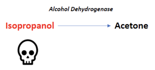
Alcohol dehydrogenase metabolizes isopropyl alcohol into acetone. Because acetone is a ketone, and ketones are not oxidized into carboxylic acids, isopropyl alcohol poisoning does not result in anion gap metabolic acidosis. 

Clinical Findings of Isopropyl Alcohol Poisoning

  • CNS sedation

  • Disconjugate gaze

  • Fruity breath odor

  • Hypotension

  • Hematemesis

  • Pulmonary edema

Plasma Osmolal Gap

One of the most reliable laboratory markers of toxic alcohol poisoning is a large osmolal gap. The osmolal gap is defined as the difference between the measured serum osmolality and the calculated, or expected, plasma osmalality:

OSMOLAR GAP = Measured plasma osmolality – calculated/expected plasma osmolality 

The common equation for calculating the expected plasma osmolality is listed below [3]. Of note, there are other formulas with slight variations. Using an online calculator can be helpful. 

Expected Serum Osmolality=2[Na]+BUN/2.8+Glucose/18

A gap < 10 is considered normal. Any elevation above 10 should raise the clinician’s suspicion of toxic alcohol ingestion.

Note: this tool is not helpful in late presentations as the metabolized forms of the different alcohols do not contribute to the osmolal gap. The calculated gap will be falsely low in late-stage poisoning.

Treatment of Toxic Alcohol Ingestions

Consult your medical toxicologist or poison control center if toxic alcohol ingestion is suspected.

The national poison control center hotline telephone number is 1(800)-222-1222.

Fomepizole

Fomepizole should be used only for methanol and ethylene glycol ingestions. It is not indicated for isopropyl alcohol intoxications [4]. It is an inhibitor of alcohol dehydrogenase (ADH). Evidence shows that it is a superior antidote to ethanol [5]. 

  • Loading dose 15 mg/kg IV

  • Then 10 mg/kg every 12 hours

Continue until blood pH is normal and serum alcohol concentration is less than 20 mg/dL in the presence of retinal or renal injury.

Ethanol

Ethanol works as a competitive inhibitor of ADH, having a higher affinity for the enzyme compared to the other alcohols. Ethanol was used historically before the effects of fomepizole were studied. Fomepizole is now the preferred treatment because the administration of ethanol is more difficult, ethanol causes sedation, and titration of the therapy is challenging in co-ingestions [6]. If ethanol must be used, the preferred route is IV and the studied therapeutic target level is 100 mg/dL [7]. 

Supplemental Therapy

Methanol poisoning patients should also receive folic acid (50mg IV every 6 hours) [7].

Ethylene glycol poisoning patients should also receive thiamine  (100mg IV) and pyridoxine (50mg IV) [8].

Hemodialysis

Consult your nephrologist early if you are considering hemodialysis. Renal replacement therapy should be considered in the following situations [9]:

  • Anion gap metabolic acidosis with known toxic alcohol ingestion

  • End-organ damage

    • Renal failure

    • Vision changes

  • Unexplained anion gap metabolic acidosis with elevated osmolal gap in suspected toxic alcohol ingestion


References

1. Liesivuori, J. and H. Savolainen, Methanol and formic acid toxicity: biochemical mechanisms. Pharmacol Toxicol, 1991. 69(3): p. 157-63.

2. Slaughter, R.J., et al., Isopropanol poisoning. Clin Toxicol (Phila), 2014. 52(5): p. 470-8.

3. Bhagat, C.I., et al., Calculated vs measured plasma osmolalities revisited. Clin Chem, 1984. 30(10): p. 1703-5.

4. Su, M., R.S. Hoffman, and L.S. Nelson, Error in an emergency medicine textbook: isopropyl alcohol toxicity. Acad Emerg Med, 2002. 9(2): p. 175.

5. McMartin, K., D. Jacobsen, and K.E. Hovda, Antidotes for poisoning by alcohols that form toxic metabolites. Br J Clin Pharmacol, 2016. 81(3): p. 505-15.

6. Zakharov, S., et al., Fomepizole versus ethanol in the treatment of acute methanol poisoning: Comparison of clinical effectiveness in a mass poisoning outbreak. Clin Toxicol (Phila), 2015. 53(8): p. 797-806.

7. Barceloux, D.G., et al., American Academy of Clinical Toxicology practice guidelines on the treatment of methanol poisoning. J Toxicol Clin Toxicol, 2002. 40(4): p. 415-46.

8. Ghosh, A. and R. Boyd, Leucovorin (calcium folinate) in "antifreeze" poisoning. Emerg Med J, 2003. 20(5): p. 466.

9. Moreau, C.L., et al., Glycolate kinetics and hemodialysis clearance in ethylene glycol poisoning. META Study Group. J Toxicol Clin Toxicol, 1998. 36(7): p. 659-66.


Expert Commentary

Thank you for this great review of a difficult subject! The combination of a lack of quick, confirmatory testing with delayed onset of symptoms makes toxic alcohol poisoning an incredibly difficult diagnosis to make. Additionally, even small ingestion can lead to major complications. For example, if a typical four-year-old (19kg) child drank windshield washer fluid that contained 50% methanol (a fairly standard formulation), it would take only 5.7 mL to potentially produce a methanol serum concentration of 25 mg/dL. Given the average 4-year-old’s mouthful is 8.9 mL, you can run into trouble quickly.[1]

We frequently see misuse or misunderstanding of osmol and anion gaps in diagnosing toxic alcohol ingestion when history is unclear. First, although a normal osmol gap is generally less than 10, baseline osmol gaps range from -10 to +14.[2] Therefore, a gap of 16 may represent a true gap of +2 in one person and +26 in another. Second, ethanol must be included in the osmol gap equation. An ethanol concentration of 200 mg/dL would increase your osmol gap by 43.5. Third, given metabolism over time, all values included in an anion gap calculation need to be drawn off of the same blood sample.

These considerations make finding the diagnosis even more complicated, but there are a few things that can help you out. First, an osmol gap > 50 is highly concerning for toxic alcohol. Second, an ethanol concentration > 100 mg/dL is sufficient to block ADH, meaning that few toxic metabolites from methanol or ethylene glycol could be made.[3] This means that an anion gap present with an ethanol > 100 mg/dL is not from toxic alcohol (unless the patient drank the ethanol after the toxic alcohol, which is very rare). Third, sequential values over time can be helpful. Metabolism of toxic alcohols should lead to a decreased osmol gap and increased anion gap over time. Proper use of the osmol and anion gap can help identify patients at high risk for morbidity and mortality while decreasing unnecessary administration of fomepizole, which typically costs thousands of dollars.

References

  1. Ratnapalan S, Potylitsina Y, Tan LH, Roifman M, Koren G. Measuring a toddler's mouthful: toxicologic considerations. Journal of Pediatrics. 2003 Jun;142(6):729-30. doi: 10.1067/mpd.2003.216

  2. Hoffman RS, Smilkstein MJ, Howland MA, Goldfrank LR. Osmol gaps revisited: normal values and limitations. J Toxicol Clin Toxicol. 1993;31(1):81-93.  doi: 10.3109/15563659309000375.

  3. Jacobsen D, McMartin KE. Methanol and ethylene glycol poisonings: mechanism of toxicity, clinical course, diagnosis and treatment. Med Toxicol. 1986;1:309-334.

Zachary Schmitz, MD

Zachary Schmitz, MD

Toxicology Fellow

Ronald O. Perelman Department of Emergency Medicine

NYU Langone Health


How To Cite This Post:

[Peer-Reviewed, Web Publication] Lima, R. Aluce, L. (2022, Jan 24). Toxic Alcohols. [NUEM Blog. Expert Commentary by Schmitz, Z]. Retrieved from http://www.nuemblog.com/blog/toxic-alcohols


Other Posts You May Enjoy

Posted on March 28, 2022 and filed under Pharmacology, Toxicology.

Review of the ATHOS 3 trial

Written by: Saabir Kaskar, MD (NUEM ‘23) Edited by: Amanda Randolph, MD (NUEM ‘20)
Expert Commentary by: Matt McCauley, MD (NUEM’ 21)


Review of the ATHOS 3 Trial: Angiotensin II for the Treatment of Vasodilatory Shock

Angiotensin, first isolated in the late 1930s, in recent years has become the new innovative vasopressor used in intensive care units, a change driven largely by the results of the ATHOS-3 trial. The ATHOS-3 trial in 2017 explored the efficacy of angiotensin II as a vasopressor for severe vasodilatory shock.  Severe shock is defined as persistent hypotension requiring vasopressors to maintain a mean arterial pressure of 65mmHg and serum lactate <2 despite adequate volume resuscitation.  Two classes of vasopressors have been used in the past for hypotension. They are catecholamines and vasopressin-like peptides. The human body, however, employs a third class which is angiotensin.  Angiotensin II is an octapeptide hormone and a potent vasopressor that is an integral component of the renin-angiotensin-aldosterone system. It works by activating the ANGII type 1 receptor which subsequently activates a G coupled protein pathway and phospholipase C, thereby inducing vasoconstriction. 

The ATHOS-3 trial compared the efficacy and safety of angiotensin II versus placebo in catecholamine-resistant hypotension, which is defined as an inadequate response to standard doses of vasopressors. The study was designed as a phase III multicenter randomized placebo control trial taking place across 75 intensive care units in the United States from 2015 to 2017. The three main inclusion criteria were catecholamine-resistant hypotension (defined as >0.2ug/kg/min of norepinephrine or equivalent for 6-48 hours to maintain a MAP 55-70 mmHg), adequate volume resuscitation (25mL/kg of crystalloid), and features of vasodilatory shock (mixed venous O2 >70% and CVP >8mmHg or cardiac index >2.3 L/min/m2).

Patients in vasodilatory shock that met the criteria of catecholamine-resistant hypotension were randomized to treatment with angiotensin II or placebo. Angiotensin II was initiated at an infusion rate of 20ng/kg/min and adjusted during the first three hours to increase MAP to at least 75mmHg. The primary outcome of the study was the response in MAP three hours after the start of angiotensin II infusion. A response was deemed as a MAP increase of 10mmHg from baseline or a MAP over 75mmHg without an increase in baseline vasopressor infusions. During the first three hours, the angiotensin II group had a significantly greater increase in MAP than placebo (12.5mmHg vs 2.9 mmHg). Angiotensin II also allowed for rapid increases in MAP which permitted decreases in doses of baseline catecholamine vasopressor. Additionally, improvement in the cardiovascular SOFA score was significantly greater in the angiotensin II group than in the placebo group. However, the overall SOFA score did not differ between groups. Rates of adverse events such as tachyarrhythmias, distal ischemia, ventricular tachycardia, and atrial fibrillation were similar in the angiotensin II and placebo groups. Overall serious adverse events that included infectious, cardiac, respiratory, gastrointestinal, or neurologic events were reported in 60.7% of patients who received angiotensin II and 67.1% of patients who received placebo. 

The strengths and limitations of the ATHOS 3 trial are critical to how its author’s conclusions should be interpreted. The strengths of the study include that it was a randomized double-blind control trial examining a new class of vasopressor for refractory vasodilatory shock. Refractory shock is a common condition with high mortality, and so the investigation of an additional treatment modality can be of great clinical impact. However, one limitation of the study was that it was underpowered to demonstrate a mortality difference. It showed improvement in blood pressure which is a clinically important parameter but not a patient-oriented outcome. Interestingly, when vasopressin was studied in 2008, it similarly did not show a mortality benefit when added to norepinephrine infusion in septic shock2. It did, however, show a decrease in norepinephrine dosing which parallels the findings of the ATHOS 3 trial.

An additional point of contention with the ATHOS 3 trial is that the manuscript does not report an increase in thrombotic risk. It has been shown that angiotensin II increases thrombin formation and impairs thrombolysis3. The FDA even reports angiotensin II has a risk for thrombosis as there was a higher incidence (13% vs 5%) of arterial and venous thrombotic events in the angiotensin II vs placebo group in the ATHOS 3 trial itself. For this reason, the FDA recommends concurrent VTE prophylaxis with the use of angiotensin II. Further data regarding the thrombotic risk of angiotensin II would be helpful to determine which patient populations the vasopressor should be avoided in. 

Overall, the author’s conclusion in the ATHOS 3 trial is that angiotensin II increased blood pressure in patients with a vasodilatory shock that did not respond to high doses of conventional vasopressors. It has been shown to raise mean arterial pressure over 75 mm Hg or by an increase of 10 mm Hg within three hours. The ATHOS 3 trial, however, did not demonstrate a mortality benefit when using angiotensin II. Further studies are needed to elucidate whether Angiotensin II truly improves patient outcomes in vasodilatory shock. 


Expert Commentary

Thank you for this great summary of the ATHOS 3 trial. While this trial paved the way for the clinical use of angiotensin II as a vasopressor, you’ve raised some salient points as to why we should approach this emerging intervention with skepticism. The biggest shortcoming in my mind is the primary outcome of the study; it’s not particularly impressive that a vasopressor resulted in higher blood pressures compared to a placebo. Mortality benefit is an extremely elusive goal in critical care research1 but that doesn’t discount the fact that ATHOS 3 wasn’t designed to demonstrate an improvement in any patient-oriented outcome. ICU length of stay, hospital length of stay, ventilator-dependent days, or rate of renal replacement therapy: these are all things that matter to our patients and to our health systems and they are more fruitful targets when we investigate interventions. 

There’s been some study of angiotensin II in the years since it has landed in our hospital formularies and there has not been robust data supporting its use. Some of the most recent data come from a multi-center retrospective study that includes patients from Northwestern. This review of 270 patients receiving angiotensin II demonstrated that 67% of patients were able to maintain a MAP of 65 with stable or reduced vasopressor doses. Univariate analysis showed that these patients that responded did have a statistically significant mortality benefit over the patients deemed nonresponders (41% vs 25%)2. If we are going to find a benefit of this drug, further study predicting which patients will be responders is necessary but this study did note that patients already receiving vasopressin and those with lower lactates (6.5 vs 9.5) were more likely to respond. Outside of septic shock, there is interest in the use of angiotensin II in refractory vasoplegia associated with post-cardiac surgery3 and anti-hypertensive overdose4. These are, of course, only hypothesis-generating. 

But what does that mean to us clinically in the ED and ICU? This data shows us that angiotensin II can make the blood pressure better but I would never let it distract you from the things we know matter in sepsis resuscitation. Source control timely antibiotics, rational fluid resuscitation, and ruling out other causes of vasopressor refractory shock to include anaphylaxis, hemorrhage, adrenal insufficiency, LVOT obstruction, and any other cause of cardiogenic shock need to be ruled out and addressed. In my personal practice, I make sure to optimize these and start vasopressin shortly after the initiation of norepinephrine. In a patient already on vaso that has stopped responding to escalating doses of norepinephrine, I reach for my ultrasound probe and reassure myself that there isn’t significant sepsis-related myocardial dysfunction because those patients may benefit from a trial of an inotrope like epinephrine. In those with a good cardiac squeeze, I think it’s appropriate to discuss with your intensivist and clinical pharmacist the utility of adding angiotensin II as part of a kitchen-sink approach. Until we have more data about the benefits of this extremely expensive intervention, I wouldn’t lose sleep if you’re unable to secure it for your patient.

References

  1. Chawla LS et al. Intravenous Angiotensin II for the Treatment of High-Output Shock (ATHOS Trial): A Pilot Study. Crit Care 2014; 18(5): 534. PMID: 25286986

  2. Russell JA et al. Vasopressin Versus Norepinephrine Infusion in Patients with Septic Shock. NEJM 2008; 358(9): 877 – 87. PMID: 18305265

  3. Celi A et al. Angiotensin II, Tissue Factor and the Thrombotic Paradox of Hypertension. Expert Review of Cardiovascular Therapy 2010; 8(12): 1723-9 PMID: 21108554

  4. Santacruz CA, Pereira AJ, Celis E, Vincent JL. Which Multicenter Randomized Controlled Trials in Critical Care Medicine Have Shown Reduced Mortality? A Systematic Review. Crit Care Med. 2019;47(12):1680-1691. doi:10.1097/CCM.0000000000004000

  5. Wieruszewski PM, Wittwer ED, Kashani KB, et al. Angiotensin II Infusion for Shock: A Multicenter Study of Postmarketing Use. Chest. 2021;159(2):596-605. doi:10.1016/j.chest.2020.08.2074

  6. Papazisi O, Palmen M, Danser AHJ. The Use of Angiotensin II for the Treatment of Post-cardiopulmonary Bypass Vasoplegia. Cardiovasc Drugs Ther. Published online October 21, 2020. doi:10.1007/s10557-020-07098-3

  7. Carpenter JE, Murray BP, Saghafi R, et al. Successful Treatment of Antihypertensive Overdose Using Intravenous Angiotensin II. J Emerg Med. 2019;57(3):339-344. doi:10.1016/j.jemermed.2019.05.027

Matt McCauley, MD


How To Cite This Post:

[Peer-Reviewed, Web Publication] Kaskar, S. Randolph, A. (2022, Feb 14). Review of ATHOS 3 trial. [NUEM Blog. Expert Commentary by McCauley, M]. Retrieved from http://www.nuemblog.com/blog/review-athos3-trial.


Other Posts You May Enjoy

Vasopressor Nonresponse

Written by: Elizabeth Stulpin, MD (NUEM ‘23) Edited by: Aaron Wibberly, MD (NUEM ‘22)
Expert Commentary by: Joshua Zimmerman, MD (NUEM ‘17)


Non-Response to Vasopressors

Shock is defined as a state of cellular and tissue hypoxia resulting in end organ dysfunction. This state may arise due to impaired oxygen delivery to tissues, impaired oxygen utilization by the tissues themselves, increased oxygen consumption, or a combination of these mechanisms. Due to its extremely high morbidity and mortality as well as high healthcare costs, the prompt recognition, diagnosis and resuscitation of shock is key. And for most forms, EM physicians are not typically shocked by shock. They have a toolbox of strategies, mainly fluids and vasopressors, to stabilize these critically ill patients. 

However, what happens when the trusted treatment paradigm fails? There is a subset of patients who, despite aggressive conventional resuscitation, have an inadequate hemodynamic response and develop refractory shock. This is seen in approximately 7 percent of patients, with short-term mortality ranging from 50 to 80 percent. Due to this significantly lower incidence and increased mortality, alternate causes for refractory shock must be considered when vasopressors do not have the desired effect.  

Acidosis

Acidosis in shock states can present from multiple different sources, including sepsis, hypoxemia, ingestions, hyperlactatemia from hypoperfusion, amongst others. With increasing acidosis, calcium influx is reduced, contractility is inhibited and the binding affinity of pressors is reduced, all of which lead to excess vasodilation and refractory hypotension. While bicarbonate is sometimes given in an effort to increase cellular pH, it is controversial for any pH >7.0. At those levels, bicarbonate administration has not been shown to improve cardiac output, MAP or pressor response. While a bicarbonate drip and hyperventilation can temporize an acidosis, emergent HD or CRRT is a definitive treatment if the cause cannot be quickly reversed. 

Adrenal Insufficiency

Cortisol has a myriad of functions in the body, not limited to its synergistic effects with catecholamines to help cause vasoconstriction. Thus, when the adrenal glands are chronically suppressed and then experience an acute stressor, hypotension can ensue. The most common cause of chronic suppression is long-term steroid use, and a stressor can include surgery, infection, hypovolemia, pregnancy, medications, or reduced steroid use. Clues that suggest adrenal insufficiency include nausea/vomiting, cutaneous hyperpigmentation, and multiple electrolyte abnormalities (hyponatremia, hyperkalemia, hypoglycemia). Previously healthy individuals, in the setting of critical illness, can also infrequently decompensate into a state of relative adrenal insufficiency. To reverse these effects as well as refractory hypotension, hydrocortisone is the preferred agent due to both its glucocorticoid and mineralocorticoid properties. A loading dose of 100mg IV should be given, followed by 50mg every 6 hours thereafter. 

Alternate Shock

Not all shock is declared equal. For example, a patient in cardiogenic shock will likely worsen with the administration of fluids and the wrong vasopressors. Similarly, obstructive shock as seen in massive PEs, tension pneumothoraces or cardiac tamponade will not improve without addressing the cause. 

Anaphylaxis 

Anaphylaxis may present as hypotension alone. Thus, it may easily be confused with a different form of shock and treated with vasopressors such as norepinephrine and vasopressin, which are not first line for anaphylaxis. Along with using epinephrine as the pressor of choice and other conventional therapies for anaphylaxis, there are alternate medications available for persistent refractory hypotension. One of these is methylene blue. While typically reserved for treatment of methemoglobinemia, the cellular mechanism of methylene blue can decrease vasodilation. Data suggest that this effect can be seen with a one-time dose of 1-2mg/kg. 

Hemorrhage

Often a common cause of refractory shock in the post op setting, bleeding can be obscure in its early stages before a hemoglobin drop is appreciated or before the patient develops abdominal distension and flank dullness (retroperitoneal bleed). If concerned, empiric uncrossed unmatched blood can be transfused. 

Hypocalcemia

Calcium homeostasis is necessary for the proper maintenance of myocardial contractility and vascular tone. Hypocalcemia can be hinted at through history or by hints such as a prolonged QTc on an ECG. Those at higher risk of hypocalcemia (vitamin D deficiency, ESRD, hyperparathyroidism, burns, multiple blood transfusions, etc.) may have greater severity of shock with increased mortality. In repleting calcium, co-administration of phosphorous and magnesium may allow for reversal of the patient’s shock state.  

Hypothyroidism

Decompensated hypothyroidism can have profound effects, including bradycardia, impaired myocardial contractility, and decreased peripheral vasoconstriction. However, even subclinical hypothyroidism with an elevated TSH but normal T4 increases risk of poor outcomes due to effects on cardiac function. While the diagnosis of hypothyroidism can be delayed by lab results, clues to the diagnosis include thyroidectomy scar, non-pitting edema of the extremities, macroglossia, altered mental status, hypoglycemia and hypothermia. Initial hypotension may not respond to vasopressors, but shock should improve once thyroid hormone is given. Stress dose steroids (hydrocortisone 100mg IV) should also be given due to the association between adrenal insufficiency and hypothyroidism, and giving thyroid hormone without steroids can precipitate adrenal crisis. 

Ingestions

When in doubt, look at the medication list! Both beta blocker and calcium channel blocker toxicity can cause profound myocardial depression, bradycardia and hypotension due to their inhibition of calcium signaling. Refractory hypotension can be overcome with the use of direct cardiac pacing, calcium, glucagon, or high dose insulin.  And if all else fails, ECMO can overcome medication toxicity until it can be fully metabolized or cleared. 

When conventional resuscitation for shock with fluids, vasopressors and/or inotropes fails, it is time for a cognitive pause. By running through this list of alternate causes of refractory shock, other methods of resuscitation can be added to improve patient outcomes and stabilize the patient.  

Sources:

1) Amrein K, Martucci G, and Hahner S. Understanding adrenal crisis. Intensive Care Medicine. 2018; 44(5): 652-655.

2) Boyd J, Walley K. Is there a role for sodium bicarbonate in treating lactic acidosis from shock?  Curr Opin Crit Care. 2008;14:379-83.

3) Farkas J. Decompensated Hypothyroidism. The Internet Book of Critical Care. 2016. Accessed https://emcrit.org/ibcc/myxedema/ 

4) Ho H, Chapital A, and Yu M. Hypothyroidism and Adrenal Insufficiency in Sepsis and Hemorrhagic Shock. Arch Surgery. 2004; 139(11):1199-1203. 

5) Kerns W. Management of B-Adrenergic Blocker and Calcium Channel Antagonist Toxicity. Emergency Medicine Clinics of North America. 2007; 25: 309-331. 

6) Levy B, Collin S, Sennoun N, et. al. Vascular hyporesponsiveness to vasopressors in septic shock: from bench to bedside. Intensive Care Medicine. 2010; 36: 2019-2029. 

7) Manji F, Wierstra B, and Posadas J. Severe Undifferentiated Vasoplegic Shock Refractory to Vasoactive Agents Treated with Methylene Blue. Case Reports in Critical Care. 2017. 

8) Minisola, S et al. Serum Calcium Values and Refractory Vasodilatory Shock. Chest. 2019; 155(1): 242. 

9) Nandhabalan P, Ioannou N, Meadows C and Wyncoll D. Refractory septic shock: our pragmatic approach. Critical Care. 2018; 22(1):215. 

10) Smith L, and Branson B. Refractory Hypotension – Diagnosis and Management in Surgical Patients. California Medicine. 1961; 95(3): 150-155

11) Velissaris D, Karamouzos V, Ktemopoulos N, Pierrakos C, and Karanikolas M. The Use of Sodium Bicarbonate in the Treatment of Acidosis in Sepsis: A Literature Update on a Long Term Debate. Critical Care Research and Practice. 2015.

12) Wang H, Jones A, and Donnelly J. Revised National Estimates of Emergency Department Visits for Sepsis in the United States. Critical Care Medicine. 2017; 45(9): 1443-1449. 


Expert Commentary

Dr. Stulpin's review of a very critical topic is well articulated and concise.  I would like to particularly emphasize her final points before delving into the details.  As she pointed out, patients failing to respond to typical resuscitative efforts represent quite a quagmire to the ED physician.  It is easy to arrive at premature closure and presume all shock to be sepsis simply needing more fluids or vasopressors.  However, there is significant risk in this practice, and I would thus advocate very strongly for a “pause” and thorough reassessment of the patient’s presentation and condition.  Much akin to a pre-procedural time out, this should be deliberate and uninterrupted to prevent diagnostic momentum from building and arriving at a premature closure on the etiology of the patient’s condition.  

This review covers much of the differential and pathophysiology that is germane to a discussion of refractory shock.  Rather than review this in detail, I would like to discuss a practical approach to application of this knowledge at the bedside.  So, as we approach the patient who is refractory to standard resuscitation, one’s first task is to confirm the patient is receiving the desire therapy and that this is truly shock refractory to intervention.  Check all access points for infiltration –the single most reversible cause for refractory hypotension is that the patient is not receiving fluids/vasopressors, etc. due to inadequate access.  Check dosing and confirm with nursing that infusions have been running appropriately.  If access is the issue, I would advocate placing a more reliable form such as central access or, in the very unstable, IO access early in the reassessment.  While I am an advocate for US guided access in stable patients and feel that this is often a great tool, this is a scenario I would advocate against US guided peripheral access.  This form of access is often time consuming, and more importantly, signs of infiltration are less evident in deeper IV sites which is a serious concern if you are running peripheral vasopressors.  

Presuming your access is adequate, the next task is to reassess perfusion in its entirety.  While not mandatory, this is a point where I would consider placing an arterial line for optimally reliable BP monitoring.  One must perform a complete re-examination as well.  If not already completed, a rectal exam for the occult GI bleeding.  One should additionally particular attention on reassessment to skin and perfusion.  Check capillary refill, skin temperature and coloration as a matter of habit for cases of refractory shock.  Cool extremities are not typically associated with distributive shock and should make you consider shock states with high SVR such as cardiogenic or acute blood loss.  In addition, a repeat skin exam may reveal new rashes – I look specifically for the presence of petechiae, purpura or urticaria.  Urticaria should prompt consideration of anaphylaxis.  If the patient has received any antibiotics yet this is an important consideration given antibiotics are one of the most common medication precipitants of anaphylaxis and this may be the source of their worsening shock state.  

After completing your thorough exam, I strongly advocate for a sonographic assessment as well.  This is where the RUSH exam, or at least a modified version, fits well into patient assessment and can offer a great deal of information.  I pay particularly close attention to the cardiac and abdominal windows.  This can aid in the rapid diagnosis of an obstructive shock state such as cardiac tamponade, or acute RV failure in the setting of massive PE.  Free intra-abdominal fluid should be considered hemorrhage until proven otherwise in the shock patient.  One reminder is that when assessing for occult bleeding, the FAST exam views are an excellent tool but not sufficiently sensitive for definitive rule out.  In particular, the FAST exam lacks sensitivity for retroperitoneal bleeding and therefore if there remains high clinical concern, CT imaging should also be considered.  

Once a thorough re-examination is completed, reconsider the patient’s medications as another source.  Patients are on a myriad of antihypertensives and other agents that can lower blood pressure.  Many of the pathways that metabolize or eliminate these drugs are compromised in a state of hypoperfusion and can lead to a synergistic worsening of hypotension.  One example of this is the case of BRASH Syndrome.  In addition to antihypertensives, a review of the patient’s medication list should include a particular search for steroids or other adrenal replacements such as fludrocortisone.  An extremely important cause of refractory hypotension to consider is that of adrenal insufficiency.  As Dr. Stulpin reviewed in her discussion above, this can come in both primary and secondary forms.  The latter is far more common and induced by exogenous steroid use.   A wise ICU attending once taught that no patient should die without stress dose steroids.  While perhaps a bit morbid, the take home point here was that adrenal insufficiency can present in any patient, not just those with underlying disorders of glucocorticoid production.  Pay particular attention to patients with autoimmune disorders or others on chronic steroid therapy as these are the populations that are particularly at risk.  Patients presenting with unexplained hypoglycemia in the setting of sepsis or shock of any kind should also be strongly considered to receive stress dose steroids.  Do not wait for a cortisol level to treat this condition.  Consider, as well, checking TSH and free T3/T4 as patients with adrenal insufficiency can simultaneously harbor other endocrinopathies.  

To summarize, refractory hypotension or failure to respond to traditional interventions is relatively uncommon but critical to identify in shock patients.  Often these patients have a primary diagnosis of septic shock but can suffer from concurrent shock related to one or more of the differential considerations we have reviewed above.  A thoughtful reassessment, both of the patient’s physical exam findings, US and other diagnostics, medication list and history of present illness will offer clues that may uncover an additional etiology critical to treat and to ensure the best outcome possible.

Joshua D. Zimmerman, MD

Health System Clinician

Feinberg School of Medicine

Northwestern Medicine


How To Cite This Post:

[Peer-Reviewed, Web Publication] Stulpin, E. Wibberly, A. (2022, Jan 24). Vasopressor Nonresponse. [NUEM Blog. Expert Commentary by Zimmerman, J]. Retrieved from http://www.nuemblog.com/blog/vasopressor-nonresponse


Other Posts You May Enjoy

Posted on January 24, 2022 and filed under Pharmacology, Critical care.

Buprenorphine Use in the ED

Written by: Diana Halloran, MD (NUEM ‘24) Edited by: Sean Watts, MD (NUEM ‘22) Expert Commentary by: Quentin Reuter, MD (NUEM ‘18)

Written by: Diana Halloran, MD (NUEM ‘24) Edited by: Sean Watts, MD (NUEM ‘22) Expert Commentary by: Quentin Reuter, MD (NUEM ‘18)


The United States has been facing a debilitating opioid epidemic, which has been partially fueled by the over-prescription of these medications in the emergency department setting. In addition, the opioid epidemic has grown exponentially during the COVID-19 pandemic. More than 40 states have reported increases in opioid-related mortality, resulting in an increased burden on an already overstrained healthcare system. (1) Prescribing the medication Buprenorphine in the emergency department offers an opportunity to ameliorate these past faults and rising statistics.

The basics:

Buprenorphine, which goes by the trade name Subutex, works by acting as both a partial mu agonist and weak kappa antagonist on opiate receptors in the brain. (2) This mechanism of action enables buprenorphine to exert analgesic effects, as well as antagonistic effects when additional opiates are consumed. In addition, buprenorphine does not carry significant sedative effects, making respiratory depression extremely rare. (3) Buprenorphine is also safe in pregnancy – a 2016 meta-analysis found no difference in pregnant patients given methadone versus buprenorphine when assessing for congenital malformations. (4) The American College of Obstetrics & Gynecology has released a committee position statement, encouraging the use of buprenorphine in pregnant patients with opioid use disorder. (5)

How to prescribe:

While the DEA X-waiver is required to write a prescription for buprenorphine for addiction treatment, withdrawal, or detox, it is not required to order or administer a dose in the hospital or emergency department. (6) This exception, called the “three-day rule”, allows a patient to come to the emergency department for three consecutive days to obtain a dose of buprenorphine if found to be in opioid withdrawal. (7)

In order to dose buprenorphine in the emergency department, the patient must be in mild acute opioid withdrawal, with a Clinical Opiate Withdrawal Score (COWS) of at least 8. (8,9) Administration of buprenorphine should not occur if the patient does not appear to be clinically withdrawing, as administration in this setting could actually precipitate withdrawal.

Dosing: (10)

  • 4mg of sublingual buprenorphine can be given initially, allowing 20-40 minutes for resolution of withdrawal symptoms with repeat dosing every 1-2 hours as needed. (10)

  • On Day 2, the patient’s response to Day 1 should be assessed. If the patient’s opioid withdrawal symptoms were controlled, the same dose can be continued. If not, the dose should be increased by 2-4mg. (10)

  • On Day 3, the patient’s response to Day 2 should be assessed. Again, if the patient’s withdrawal symptoms are controlled then the same dose can be continued. If not, the dose can be increased by 2-4mg for Day 3. (10)

  • After 3 days this dose should be continued for 3-7 days until steady-state levels are achieved (10)

  • Doses should be decreased by 2mg if the patient experiences opioid intoxication (10)

Use in the emergency department:

While buprenorphine and long-term treatment of opioid use disorder may seem confined to primary care physicians and psychiatrists, emergency medicine physicians have been shown to be successful providers for initiating buprenorphine treatment versus brief intervention and referral with a result of decreased self-reported illicit opioid use. (11) In addition, Dr. Gail D’Onofrio, chair of the Department of Emergency Medicine at Yale, found that emergency department initiated buprenorphine treatment was associated with the increased self-reported engagement of addiction treatment and reduced illicit opioid use within a two-month interval. (12)  Increasing evidence demonstrates that the emergency department provides an opportunity to intervene on opioid use disorder, with more and more emergency medicine physicians becoming X-waiver certified.

References

  1. Issue brief: Reports of increases in opioid and other drug-related overdose and other concerns during COVID pandemic. American Medical Association. https://www.ama-assn.org/system/files/2020-12/issue-brief-increases-in-opioid-related-overdose.pdf. Published December 9, 2020.

  2. Wakhlu S. Buprenorphine: a review. J Opioid Manag. 2009 Jan-Feb;5(1):59-64. doi: 10.5055/jom.2009.0007.

  3. Walsh SL, Preston KL, Stitzer ML, Cone EJ, Bigelow GE. Clinical pharmacology of buprenorphine: ceiling effects at high doses. Clin Pharmacol Ther. 1994 May;55(5):569-80. doi: 10.1038/clpt.1994.71.

  4. Zedler BK, Mann AL, Kim MM, Amick HR, Joyce AR, Murrelle EL, Jones HE. Buprenorphine compared with methadone to treat pregnant women with opioid use disorder: a systematic review and meta-analysis of safety in the mother, fetus and child. Addiction. 2016 Dec;111(12):2115-2128. doi: 10.1111/add.13462.

  5. Committee Opinion No. 711 Summary: Opioid Use and Opioid Use Disorder in Pregnancy. Obstetrics & Gynecology. 2017;130(2):488-489. doi:10.1097/aog.0000000000002229

  6. Special Circumstances for Providing Buprenorphine. SAMHSA. https://www.samhsa.gov/medication-assisted-treatment/statutes-regulations-guidelines/special-circumstances. Published August 19, 2020.

  7. Nagel L. Emergency Narcotic Addiction Treatment. https://www.deadiversion.usdoj.gov/pubs/advisories/emerg_treat.htm.

  8. Wesson DR, Ling W. Clinical Opiate Withdrawal Scale. PsycTESTS Dataset. June 2003. doi:10.1037/t48752-000

  9. D'Onofrio G, O'Connor PG, Pantalon MV, et al. Emergency department-initiated buprenorphine/naloxone treatment for opioid dependence: a randomized clinical trial. JAMA. 2015;313(16):1636-1644. doi:10.1001/jama.2015.3474

  10. Dosing Guide For Optimal Management of Opioid Dependence. The National Alliance of Advocates for Buprenorphine Treatment.

  11. D’Onofrio G, O’Connor PG, Pantalon MV, et al. Emergency Department–Initiated Buprenorphine/Naloxone Treatment for Opioid Dependence: A Randomized Clinical Trial. JAMA. 2015;313(16):1636–1644. doi:10.1001/jama.2015.3474

  12. D'Onofrio G, Chawarski MC, O'Connor PG, Pantalon MV, Busch SH, Owens PH, Hawk K, Bernstein SL, Fiellin DA. Emergency Department-Initiated Buprenorphine for Opioid Dependence with Continuation in Primary Care: Outcomes During and After Intervention. J Gen Intern Med. 2017 Jun;32(6):660-666. doi: 10.1007/s11606-017-3993-2.


Expert Commentary

Thanks to Dr. Halloran and Watts for providing an informative discussion on buprenorphine prescribing from the ED. Buprenorphine continues to emerge as the state of the art treatment strategy for opioid use disorder (OUD) and thus, developing a working knowledge for when and how to use it is essential.

While there is little doubt that the medical field fueled the opioid epidemic through the prescribing of pain medications, EM is often given a disproportionate amount of blame for the current situation.  In 2012, EM prescriptions made up only 4.3% of all opioids in circulation (1). Furthermore, I anticipate our specialty will continue to lead the fight against the opioid epidemic as practices such as naloxone prescribing, education around safe injecting practices, reduction and optimization of opioid prescribing efforts, and buprenorphine initiation gain further traction in the ED.

Obtaining a DEA X is the first step to prescribing buprenorphine. In April of this year guidelines for the administration of buprenorphine were updated to allow practitioners to treat up to 30 patients at a time with no extra training (2). While these changes will likely expand buprenorphine prescribing from the ED, it is vital that we do not operate in a silo.

To effectively manage this complex patient cohort, a coherent system of addiction medicine services is vital.  EDs must partner with local community resources to make rapid addiction medicine appointments available. Our department utilizes specially trained addiction care coordinators, nurses with extensive training in addiction medicine to help evaluate OUD patients and navigate the fractured array of outpatient services.

Prior to the implementation of our Medication for Opioid Use Disorder (MOUD) program, our clinicians had relatively little to offer patients that directly addressed their underlying addiction.  While anecdotal, we believe that by utilizing MOUD, we have begun to rebuild trust between OUD patients and the medical system.  A once generally negative relationship between OUD patients and our ED staff has been replaced with a hopeful rapport, confident that recovery for these patients is a distinct possibility.  This therapeutic relationship continues to grow and we believe will lead to long-term sustained recovery for many of our OUD patients in the surrounding community. 

References

  1. Levy B, Paulozzi L, Mack KA, Jones CM. Trends in Opioid Analgesic-Prescribing Rates by Specialty, U.S., 2007-2012. Am J Prev Med 2015;49:409-13.

  2. Reuter Q, Smith G, McKinnon J, Varley J, Jouriles N, Seaberg D. Successful Medication for Opioid Use Disorder (MOUD) Program at a Community Hospital Emergency Department. Acad Emerg Med 2020.

quentin reuter.png

Quentin Reuter, MD

Emergency Medicine Physician

Core Faculty at Summa Health


How To Cite This Post:

[Peer-Reviewed, Web Publication] Halloran D., Watts S. (2021, Sept 13). Buprenorphine Use in the ED. [NUEM Blog. Expert Commentary by Reuter Q.]. Retrieved from http://www.nuemblog.com/blog/buprenorphine


Other Posts You May Enjoy

Droperidol

Written by: Adam Payne, MD (NUEM ‘24) Edited by: Julian Richardson, MD (NUEM ‘21) Expert Commentary by: Matt O' Connor, MD

Written by: Adam Payne, MD (NUEM ‘24) Edited by: Julian Richardson, MD (NUEM ‘21) Expert Commentary by: Matt O' Connor, MD


Expert Commentary

Thanks to Dr. Payne & Dr. Richardson for putting this together!  I think this was well done, they’ve presented a concise overview of the safety and efficacy of droperidol. 

There’s a lot of utility in droperidol.  It’s great for nausea, migraines, and even as an adjunct for chronic pain.  It’s also a very good choice for agitation.  I use it most often for nausea.  It’s been shown to be as effective as odansetron, and more effective than metoclopramide.  Anecdotally, I find it works particularly well for gastroparesis and cannabinoid hyperemesis (with some low-concentration topical capsaicin cream), with less sedation than haloperidol.  For migraines, it has been shown to be as effective as prochlorperazine.  It works well for sedation in agitated patients as well; IV & IM it has a much faster onset than haloperidol, and so benzodiazepines typically do not have to be co-administered, reducing the level and duration of sedation and need for monitoring.     

The black box warning significantly limited droperidol’s availability, such that many of our newer graduates have not had any first-hand clinical experience with the medication.  If you’re not familiar with its use, don’t let the black box warning completely dissuade you.  Subsequent studies looking at emergency department droperidol use have shown it to be safe, and that complications related to QT prolongation are rare in typical doses.   As a rule of thumb, the dose of droperidol is about half of the dose of haloperidol for a given indication.  For nausea, migraine, or other pain, I usually start with 0.625-2.5mg IV, twice that IM, and can repeat dosing if needed (my most common starting dose is 1.25mg IV).  For agitation, usually 2.5-5mg IM, though up to 10mg IM has been shown likely to be safe.  Although it is prudent to be cautious, I think the literature supports droperidol’s use at appropriate doses in otherwise healthy patients.

matt oconnor.PNG

Matt O’Connor, MD

Emergency Medicine Physician

BerbeeWalsh Department of Emergency Medicine

University of Wisconsin Hospitals and Clinics


How To Cite This Post:

[Peer-Reviewed, Web Publication] Payne, A. Richardson, J. (2021, Aug 30). Droperidol. [NUEM Blog. Expert Commentary by O’Connor, M]. Retrieved from http://www.nuemblog.com/blog/droperidol


Other Posts You May Enjoy

Inhaled TXA

Written by: Jim O’Brien, MD (NUEM ‘23) Edited by:  Kevin Dyer (NUEM ‘20) Expert Commentary by: Dion Tyler, PharmD

Written by: Jim O’Brien, MD (NUEM ‘23) Edited by: Kevin Dyer (NUEM ‘20) Expert Commentary by: Dion Tyler, PharmD


itxa 2.1.png

Expert Commentary

Great job to the authors on providing a thorough description of TXA use in patients with hemoptysis. Another benefit of iTXA is that the IV TXA solution used for hemoptysis management is relatively inexpensive, ranging from $8.70-$86.80 per 100mg/mL (10 mL) vial [1]. 

An important consideration to note from the study conducted by Wand and colleagues is the exclusion of patients with massive hemoptysis defined as >200 mL of expectorated blood in 24 hours. As patients with massive hemoptysis may require additional emergent procedures to secure hemostasis such as bronchial artery embolization (BAE) or surgical intervention, a gap in knowledge exists whether TXA would be as effective in these patients as monotherapy or as an adjunct to the interventional therapies mentioned above [2,3]. 

An additional observational study has been recently published evaluating the use of iTXA for pulmonary hemorrhage in 19 critically ill pediatric patients. Pulmonary hemorrhage was caused by a variety of etiologies, with the most common etiology being diffuse alveolar hemorrhage. TXA was administered via inhalation or direct endotracheal instillation using the 100 mg/mL intravenous solution over 15-20 minutes. The dosing ranged from 250-500mg every 6-24 hours, with the most common regimen utilizing 250mg/dose every 8 hours. The study found that 18/19 (95%) of the patients demonstrated improvements in hemoptysis after the first dose of TXA and achieved cessation of pulmonary hemorrhage within 48 hours of iTXA administration. The only patient who did not have cessation of pulmonary hemorrhage was a patient on ECMO receiving systemic anticoagulation with unfractionated heparin. The median days of bleeding after TXA was initiated, days of TXA therapy received, total doses of TXA received, and cumulative dose of TXA received were 1 day, 3 days, 7 doses, and 2,500mg, respectively. Patients also received significantly less blood product transfusions after receiving iTXA (480 vs. 29.5 mL/kg; p=0.034). The study compared survivors with nonsurvivors but did not note any significant differences in the above outcomes between the two groups. There were no major adverse effects of iTXA or instances of bronchospasm reported in this study, and iTXA did not affect ventilatory settings for mechanically-ventilated patients. This study was limited by its retrospective design and lack of control group [4]. 

iTXA appears to be a safe, effective, and inexpensive intervention for management of hemoptysis. Additional research is required to determine optimal dosing and delivery approaches, as well as evaluate its safety and efficacy in patients with massive hemoptysis who may require additional emergent interventions and individuals receiving systemic anticoagulation or antiplatelet therapies.

References

  1. Tranexamic acid. Lexi-Drugs. Hudson, OH: Lexicomp, 2020. http://online.lexi.com/. Accessed July 26, 2020.

  2. Wand O, Guber E, Guber A, et al. Inhaled tranexamic acid for hemoptysis treatment: A randomized controlled trial. Chest. 2018; 154:1379–1384.

  3. Davidson K, Shojaee S. Managing massive hemoptysis. Chest. 2020;157(1):77-88.

  4. O’Neil ER, Schmees LR, Resendiz K, et al. Inhaled tranexamic acid as a novel treatment for pulmonary hemorrhage in critically ill pediatric patients: an observational study. Critical Care Explorations. 2020;2(1):e0075.

Dion tyler final.PNG

Dion Tyler, PharmD

Emergency Medicine Pharmacy Specialist

Sinai Health System

Chicago, IL


How To Cite This Post:

[Peer-Reviewed, Web Publication] O’Brien, J. Dyer, K. (2020, Dec 21). Inhaled TXA. [NUEM Blog. Expert Commentary by Tyler, D]. Retrieved from http://www.nuemblog.com/blog/iTXA.


Other Posts You May Enjoy

Posted on December 21, 2020 and filed under Pharmacology.

Antiemetics/Gastroparesis

Written by: Nery Porras, MD (NUEM ‘21)  Edited by: Terese Wipple (NUEM ‘20) Expert Commentary by: Howard Kim,  MD, MS

Written by: Nery Porras, MD (NUEM ‘21) Edited by: Terese Wipple (NUEM ‘20) Expert Commentary by: Howard Kim, MD, MS


The rise of Haloperidol as an antiemetic and analgesic in Gastroparesis

Nausea and vomiting is one the most common reasons for Emergency Department visits with estimated 2.5 million ED visits a year. Treatment of nausea and vomiting can be difficult because of its numerous causes. This is illustrated by the various sources of input that feed into the central emesis center in the brainstem, including the chemoreceptor trigger zone, vagus nerve, vestibular apparatus, and splanchnic afferent nerves, among others. Within these communication networks, various neurotransmitters such as dopamine, acetylcholine, histamine and serotonin are used, leading to the numerous drugs created to treat nausea and vomiting. Among these neurotransmitters, dopamine is becoming a more effective treatment target for nausea and vomiting among certain patient populations, in particular those with gastroparesis.

Gastro-pa-what?

Gastroparesis was something I did not recall learning much about in medical school, but is a diagnosis Emergency Medicine physicians will inevitably encounter and can be frustrating to treat. By definition, gastroparesis is a syndrome of delayed gastric emptying without evidence of mechanical obstruction leading to primary symptoms of nausea, vomiting, bloating, and abdominal pain. These patients have high rates of ED utilization and are difficult to treat, often leading to hospital admission. Traditional antiemetics often do not effectively work for these patients and narcotics only worsen the pseudo-obstruction causing their symptoms. This is where the dopamine receptor pathway can be an effective target for treatment in these patients.

Antipsychotics? You must be crazy?

As a junior resident I was surprised the first time one of my senior residents mentioned haloperidol as an effective antiemetic and analgesic in patients, particularly those with functional abdominal pain and gastroparesis. Indeed, a similar medication, Droperidol had been effectively used for many years for treatment of headache, agitation, and nausea. This was until a black box warning by the FDA in 2001 for concern for QT prolongation and cardiac dysrhythmias led to its decline in use and the halt of its manufacturing in the US. However, in 2015 the American Academy of Emergency Physicians published a position statement regarding the safety of droperidol in the Emergency Department stating “droperidol is an effective and safe medication in the treatment of nausea, headache and agitation.” Haloperidol, a butyrophenone similar to Droperidol, has been used to treat nausea and vomiting in palliative care and post-operative settings. It also has some analgesic effects due to its isometric similarity to meperidine. For this reason, it has been used off-label and anecdotally for nausea, vomiting and abdominal pain in the Emergency Department. Recently there have been two articles published showing Haloperidol efficacy in the treatment of Gastroparesis [1,4]:

Haloperidol Undermining Gastroparesis (HUGS) in the Emergency Department”

This was a retrospective case matched observational study of patients with known gastroparesis secondary to diabetes. Patients receiving 5mg IM Haloperidol were compared to themselves from a prior ED visit for the same symptoms in which they did not receive haldol. The study found statistically significant reduction in hospital admissions (5/52 [10%] vs 14/52 [27%] p-value 0.02) and reduction morphine-equivalents used (6.75 vs 10.75 p-value 0.009) with the use of Haloperidol. They also found a non-statistically significant, but likely clinically significant, reduction in ED length of stay (median 9.2 hours vs 25.4 hours p-value 0.128). There were no reported extrapyramidal side effects or cardiovascular complications, though this was a small study [2,5].

Randomized Controlled Double-blind Trial Comparing Haloperidol Combined With Conventional Therapy to Conventional Therapy Alone in Patients With Symptomatic Gastroparesis”

This randomized controlled study ultimately enrolled 33 patients with previously diagnosed gastroparesis presenting with nausea, vomiting, and abdominal pain. Patients were randomized into receiving 5mg IV haloperidol vs placebo in addition to conventional therapy (traditional antiemetics and narcotics). The primary outcome was to look at pain and nausea reduction at 1 hour based on a visual analog scale. The haloperidol group had an average reduction in pain intensity of 5.37 points (p < 0.001) compared to 1.11 points in the placebo group (p = 0.11). The Haldol group also had an average reduction in mean nausea score of 2.70 points (p < 0.001) compared to 0.72 in the placebo group(p = 0.05). They also did a subgroup analysis of patients who did not receive opiates before intervention and still found similar reduction in pain and nausea. This study also found a decrease in rate of admission (26.7% vs 72% p = 0.009) and ED length of stay (4.8 and 9 hours p = 0.77).  There were no adverse events, but the sample size was again quite small [3,6]. 

To use or not to use?

Gastroparesis and other similar cyclic vomiting syndromes present a therapeutic challenge to Emergency Physicians. Droperidol had been an effective tool in the treatment of nausea and vomiting but fell out of favor due to a black box warning. Literature review, as demonstrated in the AAEM position statement, has shown that perhaps this warning is not applicable to the doses used in the Emergency Department. This perhaps has led to the increase use of the antipsychotic Haloperidol in similar situations. These two studies now provide evidence for what many emergency physicians were already noticing; Haloperidol is an effective and safe treatment for nausea, vomiting and abdominal pain in gastroparesis. Perhaps this evidence may be extrapolated to other cyclic vomiting syndromes and treatment of other functional abdominal pain in the future.


Expert Commentary

This is a great review on the use of butyrophenones in the treatment of gastroparesis and other causes of cyclical vomiting, such as cannabinoid hyperemesis syndrome. Haloperidol is an excellent tool to keep in your back pocket for the occasional patient with intractable nausea/vomiting; providing effective symptom relief can be both satisfying for patients and gratifying for clinicians.

 “Droperidol” seems to be one of those magical words in emergency medicine that inevitably draws out a number of opinions on the Black Box warning and old war stories from the more seasoned physicians. The most recent generation of U.S. ED physicians were trained in the absence of droperidol, however U.S. manufacturing resumed in 2019 and some hospitals now have access to this mythical medication. In the end, I don’t think it is worthwhile to debate the relative merits of droperidol vs haloperidol because many ED physicians will only have access to one or the other on their hospital formulary, and we have had good success with haloperidol over the last decade or so.

Interestingly, both of the studies you cited used a 5mg IM or IV dose of haloperidol. In my anecdotal experience, smaller doses of haloperidol (e.g., 2mg IV) are also effective for the relief of intractable nausea/vomiting, in addition to migraine headaches and functional abdominal pain. To my knowledge, there is not a great comparative effectiveness study of various haloperidol dosing regimens.

One additional point that I would advise readers about is that ED patients are increasingly tech-savvy and tend to Google medicines that they are about to receive. Thus, I always first give the patient a disclaimer that if they look up the word “haloperidol” they will see that it is typically used as a psychiatric medicine for conditions such as schizophrenia. I mention that this is not the indication for which we are using the medication today, but rather that haloperidol happens to have very effective anti-nausea properties due to is effect on dopamine receptors in the part of the brain that regulates nausea. I give a similar disclaimer when prescribing tamsulosin to female patients with significant ureteral stone burden – reassuring them that we do not, in fact, think they have an enlarged prostate.    

In summary, I agree with you that haloperidol is an effective and often under-utilized treatment for intractable nausea/vomiting. As with everything in emergency medicine, it’s best to know multiple treatment modalities for common conditions so that we can adapt our response to specific situations or challenges as needed. Butyrophenones are a good treatment option for patients that do not respond to traditional anti-emetics, as are benzodiazepines and anti-histamines. 

Howard Kim.PNG

Howard Kim, MD, MS

Assistant Professor

Department of Emergency Medicine
Center for Health Services & Outcomes Research

Northwestern University Feinberg School of Medicine


How To Cite This Post:

[Peer-Reviewed, Web Publication] Porras, N. Whipple, T. (2020, Dec 14). Anti-emetics/Gastroparesis. [NUEM Blog. Expert Commentary by Kim, H]. Retrieved from http://www.nuemblog.com/blog/antiemetics-gastroparesis


Other Posts You May Enjoy

References

  1. Perkins, Jack, et al. “American Academy of Emergency Medicine Position Statement: Safety of Droperidol Use in the Emergency Department.” The Journal of Emergency Medicine, vol. 49, no. 1, 2015, pp. 91–97.

  2. Ramirez, Rene, et al. “Haloperidol Undermining Gastroparesis Symptoms (HUGS) in the Emergency Department.” The American Journal of Emergency Medicine, vol. 35, no. 8, 2017, pp. 1118–1120.

  3. Roldan, Carlos J., et al. “Randomized Controlled Double-Blind Trial Comparing Haloperidol Combined With Conventional Therapy to Conventional Therapy Alone in Patients With Symptomatic Gastroparesis.” Academic Emergency Medicine, vol. 24, no. 11, 2017, pp. 1307–1314.

  4. Weant, Kyle A. et al. “Antiemetic Use in the Emergency Department.” Advanced Emergency Nursing Journal, vol. 39, no. 2, 2017.

  5. “Diabetic Gastroparesis Needs HUGS.” R.E.B.E.L. EM - Emergency Medicine Blog, 29 Nov. 2017, rebelem.com/diabetic-gastroparesis-needs-hugs/.

  6. “SGEM#196: Gastroparesis – I Feel Like Throwing Up.” The Skeptics Guide to Emergency Medicine, 11 Dec. 2018, thesgem.com/2017/11/sgem196-gastroparesis-i-feel-like-throwing-up/.

Posted on December 14, 2020 and filed under Pharmacology.

Drug Interactions

Written by: Trish O’Connell, MD (NUEM ‘22) Edited by: Will Ford (NUEM ‘19) Expert Commentary by: Dion Tyler, PharmD and Bayan Al-Namnakani, PharmD

Written by: Trish O’Connell, MD (NUEM ‘22) Edited by: Will Ford (NUEM ‘19) Expert Commentary by: Dion Tyler, PharmD and Bayan Al-Namnakani, PharmD


Drug Interactions Final Draft.png

Expert Commentary

IM olanzapine + IM/IV benzodiazepines: The FDA-approved package insert for olanzapine currently recommends general avoidance of intramuscular olanzapine and parenteral benzodiazepines [1]. The European Medicines Agency (EMA) recommends waiting > 1 hour following IM olanzapine administration to administer benzodiazepines with careful monitoring for excessive sedation and cardiorespiratory depression, likely taking into account the 15-45 minute time to reach peak concentrations for IM olanzapine [2]. 

The warning against coadministration of intramuscular olanzapine and benzodiazepines (BZDs) arose from postmarketing data of adverse events in patients receiving intramuscular olanzapine for acute agitation [3]. This study reported that BZD use was associated with 51.7% (15/29) of fatal cases and 85.7% (24/28) of serious adverse drug reactions, defined as those that were life-threatening, extended the hospital stay, or resulted in a permanent disability. The authors thus recommended that the combination of IM olanzapine and benzodiazepines be avoided in the absence of further prospective data. However, it is important to note that many of these patients in this cohort had severe comorbidities, and BZD association included all instances (oral, IV, or IM) of BZD administration throughout their hospital stay. Some patients expired several days or weeks following their last olanzapine dose, making a causal association difficult to determine. Subsequent, smaller cohorts have found that oxygen desaturations are greatest in individuals who receive olanzapine and BZD therapy and have consumed ethanol, yet desaturation rates were similar with this combination in patients without ethanol intoxication when compared to olanzapine and haloperidol monotherapy as well as haloperidol and BZD combination therapy [4,5]. A retrospective, medication use evaluation (MUE) of IM olanzapine and lorazepam also demonstrated no incidences of hypotension or oxygen desaturation when the combination was administered within 1-hour or 24-hours of each other [6].  Lastly, a prospective cohort of individuals receiving IV olanzapine or IV droperidol followed immediately by IV midazolam compared with IV midazolam alone also demonstrated similar rates of oxygen desaturations and adverse events among all three groups [7]. However, IV olanzapine is not approved for use in acute agitation, and may display different pharmacokinetics compared to IM administration. 

While evidence supporting the safe use of this combination is growing, it may be prudent to use caution while coadministering IM olanzapine and BZDs in the absence of further controlled studies and in patients at greatest risk for adverse events, including the elderly and those who’ve consumed ethanol. 

References:

  1. Olanzapine [package insert]. Indianapolis, IN: Eli Lilly and Company, 2010.

  2. Zyprexa. European Medicines Agency. https://www.ema.europa.eu/en/medicines/human/EPAR/zyprexa. Accessed June 28, 2020.

  3. Marder SR, Sorsaburu S, Dunayevich E, et a.l. Case reports of postmarketing adverse event experiences with olanzapine intramuscular treatment in patients with agitation. J Clin Psychiatry. 2010;71(4):433-41.

  4. Wilson MP, MacDonald K, Vilke GM, et al. Potential complications of combining intramuscular olaznapine with benzodiazepines in emergency department patients. J Emerg Med. 2012;43(5):889-96.

  5. Wilson MP, MacDonald K, Vilke GM, et al. A comparison of the safety of olanzapine and haloperidol in combination with benzodiazepines in emergency department patients with acute agitation.  J Emerg Med. 2012;43(5):790-97.

  6. Williams AM. Coadministration of intramuscular olanzapine and benzodiazepines in agitated patients with mental illness. Ment Health Clin. 2018;8(5):208-13.

  7. Chan EW, Taylor DM, Knott JC, et al. Intravenous droperidol or olanzapine as an adjunct to midazolam for the acutely agitated patient: a multicenter, randomized, double-blind, placebo-controlled clinical trial. Ann Emerg Med. 2013;61:72-81. 


Myasthenia gravis and medications in the emergency department: Myasthenia gravis (MG) is an autoimmune disorder resulting in destruction of acetylcholine receptors at the neuromuscular junction (NMJ) and resultant muscular weakness. While not included in the original blog post, this is a drug-disease interaction where pharmacy services are frequently recruited for assistance in choosing medications that will not cause or exacerbate a myasthenic crisis. Although not all-inclusive, common agents that may be frequently encountered in the ED are listed below [1-3]:

  • Neuromuscular blocking agents (paralytics): Succinylcholine exerts its therapeutic effects through depolarization of the acetylcholine receptor at the NMJ causing sustained paralysis. In the setting of MG and reduced acetylcholine receptors, succinylcholine requirements may be increased, necessitating a higher dose of 1.5-2 mg/kg. Conversely, MG patients are more sensitive to nondepolarizing neuromuscular blockers,such as rocuronium and vecuronium, requiring a lower dose than normal. For rocuronium, a dose of 0.3-0.6 mg/kg may be considered for these patients.

  • Antibiotics: Several classes of antibiotics have been shown to prevent transmission of acetylcholine to the acetylcholine receptor at varying levels of risk listed below:

    • High risk: aminoglycosides, fluoroquinolones

    • Medium risk: Macrolides, polymixin B

    • Low risk: Penicillins, cephalosporins, carbapenems, nitrofurantoin, clindamycin, sulfonamides, doxycycline

  • Magnesium: Magnesium interferes with release of acetylcholine to the NMJ and may exacerbate a myasthenic crisis. A  higher threshold for repletion may be necessary in MG patients as well as avoidance of use for migraines, tachyarrhythmias, and as a component of laxatives.

  • Beta blockers: Beta blockers also appear to have an effect at the NMJ in preventing acetylcholine transmission, and have been found to exacerbate MG symptoms in patients with a variety of different agents in the class and routes of administration, such as ophthalmic timolol. 

  • Corticosteroids: While frequently used as treatment for a MG crisis, these agents may paradoxically worsen muscle strength through acetylcholine receptor blocking and effects on muscle contractility. 

References:

  1. Roper J, Fleming ME, Long B, et al. Myasthenia gravis and crisis: evaluation and management in the emergency department. J of Emerg Med. 2017;53:843-53.

  2. Ahmed A, Simmons Z. Drugs which may exacerbate of induce myasthenia gravis: a clinician’s guide. The Internet Journal of Neurology. 2008;10:e1-8.

  3. Singh P, Idowu O, Malik I, et al. Acute respiratory failure induced by magnesium replacement in a 62-year-old woman with myasthenia gravis. Tex Heart Inst J. 2015;42(5):495-97.


Lithium + ibuprofen (NSAIDs): Treatment with lithium could be quite challenging due to its extremely narrow-therapeutic index (0.5–1.2 mEq/L). Therefore, minor changes affect serum levels. The most common lithium poisoning occurs unintentionally (with chronic use) when the lithium intake exceeds its elimination such as in impaired kidney function or due to drug-drug interaction.1

Lithium is a water-soluble monovalent cation widely distributed in the body and it goes complete glomerulus filtration, 75 % of the ion is reabsorbed mainly in the proximal tubule. The exact mechanism is not fully understood. It appears that NSAIDs decrease the eGFR resulting in decreased lithium renal excretion. Some experts hypothesized that this is a result of the prostaglandin synthesis inhibition by NSAIDs which may lead to low renal blood flow and facilitate the reabsorption of sodium and lithium (theoretically). However, this hasn’t been proven [1].

This interaction is well known in clinical practice and most providers will be cautious when it comes to NSAIDs. Small prospective studies have shown large interindividual differences in lithium clearance associated with different NSAIDs. Those effects are highly variable and less predictable. It can occur with any NSAID and studies haven’t concluded a strong relationship with a specific agent. They have reported a reduction in lithium level by 10-25% in healthy volunteers [1-2], and up to 60% in another study [3]. A small retrospective study quantified the relative risk of lithium toxicity secondary to a medication new start in elderly patients who are on lithium. The relative risk was dramatically higher with ACEIs (RR=7.6, 95% CI=2.6–22.0) and loop diuretics (RR=5.5, 95% CI=1.9–16.1). Interestingly, NSAIDs and thiazides were not independently associated with increased risk of lithium toxicity [5].

Lithium levels and toxic effects should be monitored with concomitant NSAIDs initiation. Consider lithium dose reduction especially with NSAIDs new start or dose increase.

References: 

  1. Finley, P.R. Drug Interactions with Lithium: An Update. Clin Pharmacokinet 55, 925–941 (2016).

  2. Reimann IW, Diener U, Frölich JC. Indomethacin but not aspirin increases plasma lithium ion levels. Arch Gen Psychiatry. 1983;40(3):283–6.

  3. Ragheb MA. Aspirin does not significantly affect patients’ serum lithium levels. J Clin Psychiatry. 1987;48(10):425.

  4. Ragheb M. Ibuprofen can increase serum lithium level in lithium-treated patients. J Clin Psychiatry. 1987;48(4):161–3.

  5. Juurlink, D.N., Mamdani, M.M., Kopp, A., Rochon, P.A., Shulman, K.I. and Redelmeier, D.A. (2004), Drug‐Induced Lithium Toxicity in the Elderly: A Population‐Based Study. Journal of the American Geriatrics Society, 52: 794-798. doi:10.1111/j.1532-5415.2004.52221.x.

Nitroglycerin (NTG) and inferior MI: Great job to the authors on describing an interaction that comes up quite frequently in the emergency department. The ACC/AHA guidelines on acute STEMI recommend avoidance of NTG in patients with RV dysfunction, pre-existing hypotension, marked bradycardia or tachycardia, and use of phosphodiesterase-5 inhibitor (PDE5) use in the previous 24-48 hours [1]. Specifically, 24 hours should elapse following sildenafil use, and 48 hours following tadalafil use, due to the difference in pharmacokinetics between these PDE5s [2,3]. 

It appears that this recommendation has been challenged by a retrospective analysis conducted by Robichaud and colleagues assessing the incidence of hypotension in prehospital patients with inferior STEMI and acute chest pain receiving NTG compared with those who did not receive NTG [4]. The determination of STEMI was made by a computer-interpreted electrocardiogram (ECG) utilized by EMS while in the prehospital setting. The researchers found similar rates of hypotension between the two groups, but stated that a computer-interpreted ECG cannot be used as the sole predictor for patients who may be predisposed to hypotension following NTG administration. In the absence of controlled data, it may be necessary to exercise caution when considering NTG in STEMI patients with known RV involvement and avoid use in hypotensive patients. 

References:

  1. O'Gara PT, Kushner FG., Ascheim DD, et al. 2013 ACCF/AHA guideline for the management of ST-elevation myocardial infarction: A report of the American College of Cardiology Foundation/American Heart Association Task Force on practice guidelines. Circ. 2013;127:e362-e425.

  2. Sildenafil [package insert]. New York, NY: Pfizer Labs, 2014.

  3. Tadalafil [package insert]. Indianapolis, IN: Eli Lilly and Company, 2018.

  4. Robichaud L, Ross D, Proulx M, et al. Prehospital nitroglycerin safety in inferior ST elevation myocardial infarction, Prehosp Emerg Care. 2016;20(1):76-81.

Dion tyler final.PNG

Dion Tyler, PharmD

Emergency Medicine Pharmacy Specialist

Sinai Health System

Chicago, IL

Bayan Al-Namnakani, PharmD

PGY-2 Emergency Medicine Clinical Fellow

Northwestern Memorial Hospital Pharmacy


How To Cite This Post:

[Peer-Reviewed, Web Publication] O’Connell, T. Ford, W. (2020, Nov 29). Drug Interactions. [NUEM Blog. Expert Commentary by Tyler, D. Al-Namnakani, B]. Retrieved from http://www.nuemblog.com/blog/drug-interactions.


Other Posts You May Enjoy

Posted on November 30, 2020 and filed under Pharmacology.

Procedural Sedation

sedation_image.png

Written by: Mike Conrardy, MD (NUEM PGY-3) Edited by: Will LaPlant, MD (NUEM PGY-4) Expert commentary by: Seth Trueger, MD, MPH


I Want to be Sedated… 

Mastering Procedural Sedation in the Emergency Department

Procedural sedation, which is not called conscious sedation given the goal is to ensure the patient is not fully conscious, comes in a variety of flavors. Propofol, ketamine, or “ketofol” (the two used together) are typically preferred by emergency physicians, yet there are other options that may be more appropriate depending on the circumstances. In this article, we will provide a brief overview of the basics of procedural sedation, then dive deeper to provide more information about the specific agents that can be used for procedural sedation, including the pros and cons of each.

How many people are necessary to perform procedural sedation?

Generally it is recommended to have three personnel for procedural sedation (typically at least one doctor to perform the procedure and two other providers to provide sedation and monitor the patient), although using two providers (one doctor and one nurse) has been shown to have a similar complication rate.

What type of monitoring is necessary during the procedure?

Monitor vitals, telemetry, SpO2, EtCO2, and the patient’s level of sedation by physical exam. I prefer having EtCO2 if it is available, although remember EtCO2 can lead to false positives (e.g. suspected apnea when apnea is not present), but also provides earlier recognition of hypoventilation.

What other supplies should I have ready?

Oxygen by face mask has been shown to reduce likelihood of hypoxemic episodes. In addition to the above monitoring equipment, at the bedside you should have a bag-valve-mask, oral airway, nasal airway, suction, and intubation supplies ready in case they are needed. A good acronym for remembering all the supplies is SOAP ME: Suction, Oxygen, Airway equipment, Preoxygenation, Monitoring, Medications, ETCO2. As with most procedures, preparation is the most important step.

What are the most common complications of procedural sedation?

Aspiration (<2%), intubation (<2%), laryngospasm (<5%), nausea/vomiting (<5%), respiratory depression (10-20%), hypotension (10-20%), and emergence reactions if using ketamine (up to 20%).

What if my patient is pregnant?

Unfortunately, we have limited data on the safety of procedural sedation in pregnant patients. We do know that pregnant patients are more prone to hypoxemia, can be more difficult to intubate due to physiologic changes to the airway, and have a higher risk of aspiration when sedated after 16 weeks of pregnancy. Clinicians must weigh the risks and benefits of performing sedation in a pregnant patient, but if a procedure is emergent, delay is not a reasonable option. To reduce the risk of aspiration, utilize left lateral decubitus positioning and consider using pre-procedural metoclopramide and antacids. 

Etomidate, propofol, and ketamine may all have an impact on brain development in pregnancy, but evidence in pregnancy is limited for all these medications and they have not been shown to be teratogenic. Propofol may be preferred given it is short acting and is used commonly for general anesthesia in pregnancy. Given that some benzodiazepines have been shown to be teratogenic, midazolam should not be used. 

Prior to giving a sedating agent, is pretreatment necessary?

It is not necessary, but using ondansetron or another antiemetic prior to sedation may reduce vomiting and aspiration (and is at least generally safe and uncomplicated). Midazolam may also useful in conjunction with ketamine to reduce some of the post-sedation side effects, i.e. agitation and emergence reactions, although increases the risk of respiratory depression. More specifics are described below.

After sedation, when can a patient be discharged?

Once a patient is back to their neuromuscular and cognitive baseline, typically 30 minutes after the procedure, they can go home. Our practice is to PO trial a patient prior to discharge and ideally have them go home with a friend or family member who can monitor them at home for a few hours.

Agents:

Propofol:

  • Onset/Duration: Onset of ~40 seconds, duration of ~5 min.

  • Dose: 0.5 – 1 mg/kg loading dose followed by 0.5 mg/kg doses every 3-5 min or 20mg pushes every 1-2 mins PRN.

  • Pros: Short-acting sedative/amnestic, easy to redose, near immediate effect, decreased muscle tone for orthopedic procedures.

  • Cons: No analgesia, has pain on injection, can cause hypotension and respiratory depression. 

  • Special notes: 

    • Use a larger vein, such as in the antecubital fossa. 

    • Recommended to pretreat with opioid (fentanyl, typically 50-100mcg) or ketamine for procedural pain. The downside of opioid pretreatment is greater risk of respiratory depression.

    • Injection pain can be reduced with intravenous 1% lidocaine mixed with propofol or prior to injecting propofol while occluding the vein. The dose is of lidocaine is 0.5mg/kg or approximately 3-4cc of 1% lidocaine. 

    • Reduce the mg/kg dose in elderly and use lean body weight (calculator) in obese patients. No change is required in patients with impaired liver or kidney function.

Ketamine:

  • Onset/Duration: Onset of 30 seconds to 1 minute when given IV, duration of 10-20 min.

  • Dose: When used alone, dose is 1-2mg/kg given over 1-2 min followed by 0.5 mg/kg doses every 5-10 min PRN.

  • Pros: Dissociative sedative/analgesic with minimal respiratory depression, no impairment of protective airway reflexes, and no hypotension.

  • Cons: Ketamine can cause emergence reactions or post-sedation agitation (up to 20%), laryngospasm, nausea/vomiting, hypersalivation, tachycardia, and may increase ICP/IOP. 

  • Special Notes:

    • Midazolam 0.05 mg/kg (2-4mg typically) immediately prior to ketamine can reduce rates of emergence reactions although increases rates of respiratory depression.

    • Avoid in patients with psychotic disorders.

    • Recommended for patients who may have a potentially difficult airway because there is less risk for respiratory depression.

    • This is the first-line medication for children above 3 months of its impeccable airway safety and provides both sedation and analgesia as a single agent (no need for opioids).

 Combined Ketamine and Propofol, AKA “Ketofol”:

  • Onset/Duration: Same as above for each medication.

  • Dose: 0.5 mg/kg of each medication followed by propofol 0.5 mg/kg doses every 3-5 min or 20 mg pushes every 1-2 mins prn. Of note, individual provider choice of dose for each medication varies widely.

  • Pros: Potential benefit is ability to use lower doses of both ketamine and propofol with potentially lower risk of adverse events such as hypotension, respiratory depression, emesis, emergence reactions. 

  • Cons: May reduce side effects of each medication individually, yet now dealing with the side effects of two medications rather than one alone.

  • Special notes: 

    • Research on ketofol is mixed. Systematic reviews have shown that it causes fewer events of respiratory depression and hypotension/bradycardia, yet these events were mostly transient and clinically insignificant. Overall, ketofol has not been shown to reduce clinically significant adverse events or to prolong procedural duration.

Etomidate:

  • Onset/Duration: Near immediate onset when given IV, duration of 5-15 min.

  • Dose: 0.1-0.15 mg/kg given over 30-60 seconds, redose every 3-5 min.

  • Pros: Easy to dose, minimal hemodynamic effect.

  • Cons: No analgesia, myoclonus (up to 80%), respiratory depression (10%), nausea/vomiting, pain with injection, and potential for adrenal insufficiency.

  • Special Notes:

    • Recommended to pretreat with opioid (fentanyl, typically 50-100mcg) for procedural pain. The downside of opioid pretreatment is greater risk of respiratory depression.

    • In rare cases of severe myoclonus, treat with 1-2 mg IV midazolam every minute until resolved. Some providers pretreat with 0.015 mg/kg etomidate to prevent myoclonus.

    • Dose must be reduced for patients who are elderly or have renal/hepatic dysfunction.

    • Not recommended for orthopedic procedures given the frequency of myoclonus.

Midazolam (requires opioid co-administration):

  • Onset/Duration: Onset of 2-5 min, duration of 30-60 min.

  • Dose: 0.02-0.03 mg/kg or 0.5-1 mg doses IV every 2-5 min prn, typically not exceeding 5 mg total.

  • Pros: Provides anxiolysis and amnesia.

  • Cons: Not as effective for true procedural sedation as shorter acting medications, no analgesia, higher risk of respiratory depression when combined with fentanyl compared to other medications.

  • Special Notes:

    • When combining with fentanyl for pain/sedation, give midazolam doses as above first until the desired anxiolysis is achieved, typically 1-2 doses, then give 0.5 mcg/kg doses of fentanyl every 2 min PRN, carefully titrated to effect, with maximum dose of 5 mcg/kg or approximately 250 mcg.

    • Prolonged sedation is high risk in patients who are elderly, obese, or have hepatic/renal dysfunction.

    • Use for anxiolysis rather than for true procedural sedation.

Barbiturates (Methohexital):

  • Onset/Duration: Immediate onset, duration of < 10 min.

  • Dose: 0.75-1 mg/kg followed by 0.5 mg/kg doses IV every 2 min prn.

  • Pros: Fast onset, short duration sedation.

  • Cons: No analgesia, causes hypotension/tachycardia, can precipitate seizures.

  • Special Notes: 

    • You are probably never going to use this drug unless you are in a very resource limited setting, but you might as well know it is an option.

Dexmedetomidine:

  • Onset/Duration: Onset of 5-10 min, duration of 60-120 min.

  • Dose: Not well studied for procedural sedation, options are intranasal 2-3 mcg/kg or bolus of 0.5-1.0 mcg/kg over 10 min followed by infusion of 0.2-0.7 mcg/kg/hr.

  • Pros: Preserved muscle tone and respiration, much like natural sleep, provides some analgesia.

  • Cons: Potentially unpredictable effect, not well studied, risk of hypotension/bradycardia, longer acting.

  • Special Notes:

    • Delayed time to onset may limit application in the ED

    • Dexmedetomidine is just not ready for primetime yet, but worth further investigation.

Nitrous oxide:

  • Onset/Duration: Immediate onset, off within seconds.

  • Dose: 30-50% mixture with 30% oxygen.

  • Pros: Provides analgesia, anxiolysis, and sedation all in one.

  • Cons: Not typically available in emergency departments, needs scavenging system.

  • Special Notes:

    • Have been trying for years to get this constantly vented into all our patient rooms, but still no luck with our administration.

Summary Points:

  • Propofol, ketamine, ketofol, and etomidate are our typical first-line medications in the emergency department for procedural sedation.

  • Ketamine is preferred for kids.

  • In adults, propofol or ketofol is best for hemodynamically stable adults requiring procedural sedation, particularly for joint reductions because it does not cause myoclonus and is easy to titrate.

  • Etomidate provides greater hemodynamic stability and is best for cardioversion or procedures in patients with hemodynamic compromise, but the downside is myoclonus which may reduce procedural success.

  • Ketamine alone is best in patients with a difficult airway or at high risk for respiratory compromise because it does not cause respiratory depression.

  • Use what you are most comfortable with, and remember that adequate preparation is key.


Expert Commentary

Thank you for the excellent and concise review. It’s been interesting to see procedural sedation practices change over the course of my training and career, as newer safe and easy options (propofol and ketamine) gained rapid popularity but have been challenged by drug shortages and well-publicized celebrity tragedies. Here is my typical practice, which is certainly not the only correct way but my strong preference:

A few factors on when to think about procedural sedation:

-Any painful procedure, particularly for potential for longer duration. This includes incision and drainage (especially Bartholin’s) and disimpaction. I’ve gotten some odd looks for suggesting it but it works out better for everyone involved. 

-Consultant’s procedures, particularly big traumatic injuries which look nasty and like they need to be fixed. It’s easy to forget how terrible it is for the patient.

When I think about avoiding:

-medically complex patients

-difficult airways

-procedures that can wait

-procedures with low likelihood of success even under the best circumstances. If they need to go to the OR no matter what, that might be the best place to start.

My one exception are situations that are currently painful and need to be fixed now and can be fixed quickly, e.g. dislocated ankles. The likelihood of success with 100mcg of fentanyl within seconds to resolve a huge amount of pain now is exceedingly favorable.

The worst situation is trying to avoid procedural sedation with “just some morphine and maybe a little lorazepam” which quickly devolves into “a little more morphine” and “hmm maybe another dose of lorazepam.” Now it’s a procedural sedation that is both ineffective and unsafe.

My general process:

First I print out a checklist I made with the following preparation steps (details below):

RSI box (succinylcholine)

VL

airway cart (LMA, PEEP valve, DL gear)

nasal ETCO2 (plus ETT adapter)

4mg ondansetron now

bottle ketamine

bottle propofol

room ready (including suction, bag, anything required for the procedure)

Department ready?

Am I ready?

Procedure plan

Post-procedure planning (sling, splint)

The general principle is to set up at least as much as if I were performing RSI. A lot of this may seem like over-preparation, but the more I prepare, the luckier I get. Here is some more detail on each item:

RSI box (succinylcholine)

2 main purposes for the RSI box. First, if things go south, I will be intubating the patient, and need the medications to do so (i.e. NMBA). Second, if I am using ketamine, there is a small but nontrivial chance of laryngospasm, and of jaw thrust and bagging do not fix it, the patient needs NMBA. This is not a time to debate roc vs sux, so I always have sux in the room (even if roc isn’t slower when dosed appropriately at ≥1.2mg/kg, I don’t want to have to argue about it at the RCA). The easiest way for me to get these medications in the room is grabbing our RSI box, but this will depend on your department; simply grabbing a vial of sux with 1.5mg/kg is sufficient.

VL

If things go south, this is not the time for the intern to practice their DL. I always have the hyperangulated VL ready to go at the head of the bed, with a combo Mac VL/DL blade and a traditional Mac DL as backup, with stylets loaded with tubes ready to go. 

Airway cart 

We have nicely built airway carts with everything I need for bagging, difficult bagging, and difficult intubation. Primarily, what I want is gear for bagging, i.e. LMA and PEEP valve. All the usual backup is here as well (oral/nasal airways, bougie, cric gear).

Nasal ETCO2 (and ETT adapter)

The literature on end tidal in procedural sedation is interesting but I think generally answers a different question than the one I care about. I don’t look for qualitative changes in waveforms or quantitative changes in ETCO2 to predict hypoventilation; rather, it is the quickest and easiest way to see if the patient is breathing. No staring at their chest hoping to see chest rise. Simply look at the monitor: either there is a waveform and they are breathing, or there is not and they are not. It’s like a sedative for me, similar to supervising an intern using VL instead of DL: it makes the procedure much less stressful for me.

Additionally, by using ETCO2, it is now safe to provide supplemental oxygen via nasal cannula or reservoir facemask so if things go south, there is a much wider safety margin (i.e. the patient is preoxygenated for intubation).

4mg ondansetron now

As discussed above, it might help, it may not, and it’s safe and easy.

bottle Ketamine

bottle Propofol

I’ll discuss my medication strategies below, but the bottom line is I like to have multiple 100s of mg of each medication ready for each patient, because when I need to redose, it can be needed in a very short time.

Room ready

Other equipment I make sure is ready: suction, bag for mask ventilation. Other items like do I need vaseline gauze for splinting over an abrasion?

Is the Department ready?

Was an 80 year old with abdominal pain just roomed? Should I lay eyes on them and make a quick decision about an obvious CT? Is there a hospitalist hanging around who I can tell about another patient to send upstairs before I get stuck in a sedation for 45 minutes? Should I discharge anyone?

Am I ready?

Do I need to go to the bathroom? Has it been hours before I’ve had any calories?

Procedure plan

I always make sure to have a clear plan for the procedure—not just the sedation—well before we start. Who is doing what? What technique are we using? What are backup plans? Of course these questions apply to the sedation as well.

If a non-EM physicians is performing the procedure (e.g. ortho, or gen surg pulling a tunneled line) I try to make sure that they understand my definition of “ready” is not the same as in the OR, and I make sure they are ready to start the actual procedure as soon as the meds are working. This is not a judgment in any way; rather, the ER simply isn’t the OR.

Post-procedure planning

Few things are more frustrating than getting a difficult shoulder reduced only to have it slip out while someone is hunting down the sling I forget to get beforehand, that I knew I would need (see also: ordering post-intubation meds with RSI meds in an intubation). Obviously if something needs to be splinted we need the gear and whoever is doing the splint. And, if there are abrasions going under the splint, petroleum gauze, etc.

Medication choices

I typically choose between ketamine and propofol on a spectrum. 

Factors on the propofol side: young, healthy, BP/respiratory reserve, shorter procedure, ortho procedure (propofol is much better at loosening up patients, plus these often end quickly).


Factors on the ketamine side: older, more comorbidities, less respiratory reserve, longer procedures, non-reduction procedures, more protractedly-painful procedures (e.g. I&D).

Obviously these are not absolutes and I tend to plan on using ketofol quite a bit. I usually have enough cognitive space and hands available to dose them separately (generally 0.5mg/kg ketamine first, then 0.5mg/kg propofol as needed) but in more constricted settings I will mix 1:1 if I don’t have the bandwidth. 

I will say I have been tending to more and more ketamine-only sedations. Usually I start with the intention of using ketamine-first ketofol, particularly if the patient needs to be loosened up for a reduction, but I am continually surprised by how little I end up needing the propofol.

As noted above, for solo propofol, I pretreat with fentanyl as propofol is not inherently analgesic. 

I appreciate the debate about midazolam for pretreatment for ketamine, but the rates of substantial post-sedation agitation are low enough that I simply treat that when it happens, as not all but most ketamine respiratory depression only happens with co-administered sedatives. 

Other than lack of availability of other options, there is no reason to use fentanyl/midaz anymore.

Lastly, I’ve stopped using etomidate. The rate of myoclonus is simply too high. Myoclonus easily defeats the reduction, and even for cardioversion, it makes checking the rhythm, getting an ECG, monitoring the sat, etc. very difficult. Ultimately, it’s just a headache we don’t need, particularly as we have so many other safe and effective agents.

As I said above, these are more my style preferences than the only absolutely correct choices, and I am always happy to at least discuss adapt to the circumstances including others’ preferences (or trying something different so the residents can gain experience with different techniques).

sethtrueger.png
 

Seth Trueger, MD, MPH, FACEP

Assistant Professor

Northwestern Emergency Medicine


Citations

  1. Brown TB, Lovato LM, Parker D. Procedural sedation in the acute care setting. Am Fam Physician 2005; 71:85.

  2. Swanson ER, Seaberg DC, Mathias S. The use of propofol for sedation in the emergency department. Acad Emerg Med 1996; 3:234.

  3. Miner JR, Burton JH. Clinical practice advisory: Emergency department procedural sedation with propofol. Ann Emerg Med 2007; 50:182.

  4. Euasobhon  P, Dej‐arkom  S, Siriussawakul  A, Muangman S, Sriraj  W, Pattanittum P, Lumbiganon  P. Lidocaine for reducing propofol‐induced pain on induction of anaesthesia in adults. Cochrane Database of Systematic Reviews 2016, Issue 2. Art. No.: CD007874. DOI: 10.1002/14651858.CD007874.pub2.

  5. Messenger DW, Murray HE, Dungey PE, et al. Subdissociative-dose ketamine versus fentanyl for analgesia during propofol procedural sedation: a randomized clinical trial. Acad Emerg Med 2008; 15:877.

  6. Strayer RJ, Nelson LS. Adverse events associated with ketamine for procedural sedation in adults. Am J Emerg Med 2008; 26:985.

  7. Adnolfatto G et al. Ketamine-Propofol Combination (Ketofol) versus propofol alone for Emergency Department Procedural Sedation and Analgesia: A Randomized Double-Blind Trial. Annals of Emergency Medicine. 2012;59(6):504-512.e2

  8. Miner JR et al. Randomized, Double-Blinded, Clinical Trial of Propofol, 1:1 Propofol/Ketamine, and 4:1 Propofol/Ketamine for Deep Procedural Sedation in the Emergency Department. Annals of Emergency Medicine. 2015;65(5):479-488.e2

  9. Yan JW, McLeod SL, Iansavitchene A. Ketamine-Propofol Versus Propofol Alone for Procedural Sedation in the Emergency Department: A Systematic Review and Meta-analysis. Acad Emerg Med 2015; 22:1003.

  10. Miner JR, Danahy M, Moch A, Biros M. Randomized clinical trial of etomidate versus propofol for procedural sedation in the emergency department. Ann Emerg Med 2007; 49:15.

  11. Falk J, Zed PJ. Etomidate for procedural sedation in the emergency department. Ann Pharmacother 2004; 38:1272.

  12. Sacchetti A, Senula G, Strickland J, Dubin R. Procedural sedation in the community emergency department: initial results of the ProSCED registry. Acad Emerg Med 2007; 14:41.

  13. Keim SM, Erstad BL, Sakles JC, Davis V. Etomidate for procedural sedation in the emergency department. Pharmacotherapy 2002; 22:586.

  14. Hüter L, Schreiber T, Gugel M, Schwarzkopf K. Low-dose intravenous midazolam reduces etomidate-induced myoclonus: a prospective, randomized study in patients undergoing elective cardioversion. Anesth Analg 2007; 105:1298.

  15. Horn E, Nesbit SA. Pharmacology and pharmacokinetics of sedatives and analgesics. Gastrointest Endosc Clin N Am 2004; 14:247.

  16. Bahn EL, Holt KR. Procedural sedation and analgesia: a review and new concepts. Emerg Med Clin North Am 2005; 23:503.

  17. Frank RL. Procedural sedation in adults outside the operating room. Wolfson AB, and Grayzel J (Ed.) UpToDate (2018).

  18. Hsu DC and Cravero JP Pharmacologic agents for pediatric procedural sedation outside of the operating room. Stack AM and Randolph AG (Ed.) UpToDate (2018).

  19. G. Haeseler, M. Störmer, J. Bufler, R. Dengler, H. Hecker, S. Piepenbrock, et al. Propofol blocks human skeletal muscle sodium channels in a voltage-dependent manner. Anesth Analg, 92 (2001), pp. 1192-1198

  20. J. Ingrande, H. J. M. Lemmens; Dose adjustment of anaesthetics in the morbidly obese, BJA: British Journal of Anaesthesia, Volume 105, Issue suppl_1, 1 December 2010, Pages i16–i23.

  21. Neuman G and Koren G. Safety of Procedural Sedation in Pregnancy. J Obstet Gynaecol Can. February 2013, pages 168-173.


How To Cite This Post

[Peer-Reviewed, Web Publication]  Conrardy M, LaPlant W. (2019, Nov 11). Procedural Sedation. [NUEM Blog. Expert Commentary by Trueger S]. Retrieved from http://www.nuemblog.com/blog/procedural-sedation.


Journal Club: Do Emergency Physician Opioid Prescribing Practices Impact Long-Term Opioid Use?

Screen Shot 2018-01-25 at 10.11.06 AM.png

Written by: Jon Andereck, MD (NUEM PGY-3) Edited by: Rachel Haney, MD, (NUEM Graduate 2017) Expert
commentary by:  Seth Trueger, MD


Introduction

Figure 1. Opioid Pain Reliever Sales, Related Treatment Admissions, and Related Deaths from 1999-2010 (CDC).

Over the past two decades, rates of opioid prescribing in the United States have skyrocketed, with the total amount of opioids distributed quadrupling from  1999 to 2010 (see Fig. 1). [1]  Rates of opioid related unintentional overdoses and deaths have risen in concurrent fashion, from 4,030 in 1999 to 14,800 in 2008. Public health experts, policy makers, and physicians have slowly come around to acknowledging the epidemic of opioid abuse now facing the country.

Much like in other care settings, there has been an increasing push to reduce the volume of opioids prescribed from the ED. The exact role of Emergency Department (ED) opioid prescriptions in this epidemic has been difficult to quantify. Among people aged 10-29, EDs represented 12% of opioid prescriptions and ranked as the third most common setting for which opioids were prescribed. [2]

Indeed, nearly 4 in 5 heroin users reported prior exposure to non-medical prescription pain relievers, and prior exposure to narcotic pain medications carried a 19-fold increased risk of future heroin use. [3] A study from 2014 estimated that as many as 13.8% of patients discharged from ED's across the country in 2010 were written a prescription for opioid pain medications, up from 11% in 2005. [2] Still, a large amount of uncertainty persists about the true impact of prescribing habits of Emergency Physicians (EP's) on the incidence of narcotic abuse. This study helps shed some light on how EP prescribing practices impact long-term narcotic use.


Study

Barnett ML, Olenski AR, Jena AB. Opioid-Prescribing Patterns of Emergency Physicians and Risk of Long-Term Use. N Engl J Med. Feb 16 2017. 376(7): 663-673.

Study Design

Retrospective analysis.

Population

Medicare Beneficiaries who visited any ED in the US from Jan 1, 2008 – Dec 31, 2011 who had not received an opioid prescription in the preceding 6 months and who were not admitted to the hospital on the index ED visit studied. Patients with cancer or on hospice were also excluded.

Measurement Protocol

Using Medicare Part D data, the authors calculated the morphine equivalents dispensed both in the 7 days following the index ED visit and any further opioid prescriptions over the following 12 months.

Treating EPs were categorized as either high-intensity or low-intensity opioid prescribers based on comparison with their peers at the same hospital. The authors calculated the percentage of patients that filled an opioid prescription after seeing any provider in a given hospital, and then divided providers into quartiles of rates of opioid prescribing within their own hospital. Physicians in the top quartile were designated high-intensity prescribers; those in the bottom quartile were designated low-intensity prescribers.

Outcome Measures

The primary outcome of interest was rate of long-term opioid use among patients in the 12 months following a visit in which they were seen by either a low-intensity or high-intensity opioid prescriber. Long-term use was defined as at least 180 days of opioids supplied in the 12 months after the initial ED visit, excluding the first 30 days following the ED visit.

Secondary measures included rate of hospital encounters possibly related to the adverse effects of opioids in the 12 months following the index ED visit. The authors also measured repeat ED visits at 14 and 30 days for the same primary diagnosis to assess for possible undertreated pain.

Results

Long-term opioid use was significantly higher among those treated by high-intensity prescribers, with an odds ratio of 1.3 (p<0.001) and an absolute rate of 1.51% as compared to 1.16% in the low-intensity group. The authors calculate a number needed to harm of 48 patients receiving an opioid prescription to lead to one excess long-term opioid user.

Long-term opioid use increased in a stepwise fashion for patients treated by physicians in each quartile of prescribing intensity (Fig 1).

A total of 377,629 patients were included in this retrospective analysis; 215,678 were seen by low-intensity EPs and 161,951 were seen by high-intensity EPs. Characteristics of each patient population were similar, though several of these were significant given the large sample size (see Table 1).

Over three times as many patients seen by a high-intensity prescriber were discharged with an opioid prescription than those seen by a low-intensity prescriber (24.1% vs. 7.3%), though there was no difference between the two groups in the median dose of morphine equivalents per prescription.

Figure 1

Table 1

In the secondary analysis, the authors found a small but significant increase in rates of opioid-related encounters (OR 1.03, p = 0.02) as well as ED visits for fall or fracture (OR 1.07, p < 0.001) for patients treated by high-intensity prescribers. In contrast, there was no difference in rates of hospital encounters for non-opioid related complaints. Additionally, rates of short-term ED visits for the same chief complaint were no different at 14 or 30 days for patients treated by either low- or high-intensity prescribers (See Table 3).


Table 3


Interpretation

This study does an impressive job of looking at an important but poorly understood issue in the field of emergency medicine – how do prescribing practices of physicians affect long-term opioid abuse their patients?

This retrospective study design is limited in that it was not randomized-controlled, but it was the most logical design to answer the question at hand. The number of patients included was certainly adequate to detect a meaningful difference. The study was limited by the fact that only Medicare beneficiaries were studied, in part because this was the most accessible database for such a large retrospective study. However, it therefore excludes many in the 19-39 age range in which long-term abuse potential is highest. Designing a randomized-controlled trial to attempt to answer this question would be difficult as it would require standardization of discharge prescriptions; few physicians would be amenable to ceding their right to determine the analgesic plan for their own patients.

The primary outcome of interest – long-term opioid use among patients seen by either type of provider – demonstrated that there is a correlation between high-intensity prescribers and long-term opioid use among patients they see. The authors calculate a number needed to harm (NNH) of 48 among patients prescribed opioids on discharge. This means that for every 48 patients given a discharge prescription for an opioid analgesic by a high-intensity prescriber, one will go on to use opioids long-term (as defined by this study) that could have been avoided if the patient had been seen by a low-intensity prescriber.

One of the most interesting results from this study is not even the question the authors set out to answer, but is the difference in opioid prescribing rates between high-intensity and low-intensity prescribers. This difference was over three-fold (7.3% to 24.1%) and represents an extraordinarily wide practice variability that underscores the lack of standard practice for opioid use. However, this variation was only in the number of prescriptions written and not for the amount of morphine equivalents per prescription as demonstrated in Fig. S3 above.

The counter-measure of pain control adequacy is an important one. The authors attempt to address whether patients treated by low-intensity providers had inadequate analgesia at home. While this question was not directly answered by the study, a surrogate measure of return visits to the ED with the same chief complaint demonstrated no significant difference between the two groups, which suggests but does not prove that there was no meaningful difference in analgesia between the groups.

Is this article practice changing? Perhaps. It does provide evidence that there is a correlation, small though it may be, between prescribing practices of EPs and long-term opioid use of patients. There are also small but significant differences in complications from the opioids given by high-intensity prescribers. The study further highlights the profound variability among EPs regarding their opioid prescribing practice, which I argue is an area to target for improvement especially without any known deficiency of pain treatment by doing so. We can all strive to only prescribe opioids that are truly necessary to treat acute pain, and this article serves as further motivation that over-prescribing can in fact cause our patients direct harm.


Take Home Points

  •  Variability of opioid prescribing within departments is large
  •  Opioid prescribing patterns do have an impact on long-term opioid use
  •  Fewer opioids do not lead to worse pain control, at least as measured by the return rate to the ED

Expert Commentary

This is a great overview of an impactful article. While ultimately there will always be some variation in opioid prescribing (by chance, some physicians will likely see more patients with more painful conditions), this paper suggests that the prescriber variability is high and its not due to chance. Regardless, my takeaway here is that there does seem to be a dose-response to opioid prescribing in the ED and longer term opioid use.

Both the sheer scale and physicians’ role in the opioid epidemic is startling and a number of factors are at play. Years of focus on oligo-analgesia were likely a mix of genuine concern for undertreating pain but unfortunately also driven by those with specific financial interests. Similarly, the increasing focus on patient satisfaction/experience and even with the link to payments, I suspect physicians are too quick to shift blame to others and we need to prescribe more responsibly. While most emergency department opioid prescriptions are short (75% are for 20 pills or fewer [4]), as Barnett and others have shown, a startling fraction of ED patients receive opioid prescriptions. And to borrow from Lewis Nelson, everyone who is addicted to opioids had to have had a first exposure.

Of course there are no easy answers – plenty of patients we see in the emergency department are in substantial pain, and we do not have a lot of tools. But it is not hopeless. My approach to pain management is similar to patients with URIs and antibiotics: sometimes we cut corners (“they’re just here for a z pack / Norco prescription”) and underestimate how satisfied our patients can be being taken care of by a physician who cares and explains things. Some of the things I focus on:

  1.  Acknowledge the patient’s pain: just because I’m not writing for a ton of opioids doesn’t mean I don’t believe they’re not in pain
  2.  Set realistic goals: I don’t have a silver bullet to make pain go away. My goal is to make their pain manageable, not gone.
  3.  Emphasize our priorities: Our main goal in the ED is to make sure there isn’t anything dangerous causing the patient’s symptoms.
  4.  The door isn’t shut when I discharge the patient: “The good news is you don’t need an MRI now but you do need to follow up with your primary doctor over time who will keep an eye on your symptoms and help determine if you do eventually need more testing or to see a specialist.”
  5. Information, information, information: what concerning symptoms to look for at home, when to call your doctor, when to come back to the ED.
  6.  What to do for symptoms: What works well for this? I find a lot of patients are very happy to hear me thoughtfully say “what works really well for this is prescription-strength ibuprofen.” I’ve also had a lot of success with lidocaine patches. Anecdotally they seem to work well, but more importantly, I think it demonstrates to the patient that we’re paying attention and being thoughtful (especially as I need to explain that sometimes insurance doesn’t cover them well but there are over the counter versions so here is how to approach that…)

Incidentally I rarely prescribe or co-prescribe benzodiazepines as we have some data they aren’t very helpful (e.g. Friedman [5]) and that we vastly underestimate their harms, particularly when patients take both opioids and benzos (e.g. Sun [6]).

We have a tight needle to thread between oligoanalgesia and the opioid epidemic, but right now I think it’s clear that the pendulum has swung too far. I don’t think we can nor should stop using opioids altogether (yet) but we can be thoughtful and careful as we care for our patients.

Seth Trueger MD MPH

Assistant Professor of Emergency Medicine, NUEM


Posts You May Also Enjoy


How to cite this post

[Peer-Reviewed, Web Publication]  Andereck J,  Haney R  (2018, Jan 29). Journal Club:  Do Emergency Physician Opioid Prescribing Practices Impact Long-Term Opioid Use? [NUEM Blog. Expert Commentary By Trueger S]. Retrieved from http://www.nuemblog.com/blog/opioids. 


Resources

  1.  “Vital Signs: Overdoses of Prescription Opioid Pain Relievers --- United States, 1999-2008.” Morbidity and Mortality Weekly Report. Centers for Disease Control and Prevention. Nov 4, 2011. 60(43): 1487-1492. < https://www.cdc.gov/mmwr/preview/mmwrhtml/mm6043a4.htm#fig2>. 
  2.  Cantrill SV, Brown MD et al. Clinical Policy: Critical Issues in the Prescribing of Opioids for Adult Patients in the Emergency Department. From the American College of Emergency Physicians Opioid Guideline Writing Panel. Ann Emerg Med. 2012. 60:499-525.
  3.  Muhuri PK, Gfroerer JC, and Davies MC. Associations of Nonmedical Pain Reliever Use and Initiation of Heroin Use in the United States. Center For Behavioral Health Statistics and Quality. CBHSQ Data Review. August 2013.
  4. Hoppe JA, Nelson LS, Perrone J, Weiner SG. Opioid Prescribing in a Cross Section of US Emergency Departments. Ann Emerg Med. 2015 Sep;66(3):253-259.
  5. Friedman BW, Irizarry E, Solorzano C, Khankel N, Zapata J, Zias E, Gallagher EJ. Diazepam Is No Better Than Placebo When Added to Naproxen for Acute Low Back Pain. Ann Emerg Med. 2017 Aug;70(2):169-176.
  6. Sun EC, Dixit A, Humphreys K, Darnall BD, Baker LC, Mackey S. Association between concurrent use of prescription opioids and benzodiazepines and overdose: retrospective analysis. BMJ. 2017 Mar 14;356:j760. doi: 10.1136/bmj.j760.


The Waiting Game: Biphasic Anaphylaxis

biphasic anaphylaxis.png

Written by: Kumar Gandhi, MD, MPH (NUEM PGY-2) Edited by: Andrew Moore, MD, MS (NUEM PGY-4) Expert Commentary by: Aaron Kraut, MD


Case Scenario

18 y.o. female with history of asthma and multiple food allergies presents with rash and
shortness of breath following ingestion of assortment of cookies at a Halloween party. A diffuse erythematous pruritic rash started fifteen minutes following the ingestion of cookies. Associated symptoms include tingling in the back of the throat and wheezing. The patient reports one prior episode requiring use of an EpiPen, but she never refilled her prescription.


Vitals: HR: 90, RR: 25, BP: 105/70, Temp: 98.6, SpO2: 97% on room air


Pertinent findings on physical exam include:

  • Mild pharyngeal edema with no uvular deviation.
  • Moderatewheezing in bilateral lung fields
  • Diffuse abdominal tenderness
  • Blanching (urticarial) rash on the back,nabdomen, axilla, femoral creases, and posterior legs.

An Epi-Pen injection in the right thigh, coupled with concomitant 125mg of methylprednisolone IV Push, 25mg of Benadryl IV push [1] results in decreased wheezing and resolution of the urticarial rash.


Now that the patient is stable we need to ask ourselves a few important questions:

  1. Was this an allergic reaction or anaphylaxis?
  2. When can we discharge the patient?

Anaphylaxis: Overview


Anaphylaxis is a life-threating systemic hypersensitivity reaction. Pathophysiology includes [1]:

  • IgE-mediated Type 1 hypersensitivity reaction
  • Degranulation of mast cells releases vasoactive mediators including histamine, prostaglandins, and leukotrienes
  • Histamine mediates systemic vasodilation, cardiac contractility, and vascular permeability
  • Leukotrienes mediate vascular permeability and in combination with prostaglandins cause bronchoconstriction


Common Triggers:

Screen Shot 2017-09-11 at 10.10.23 AM.png
  • Foods: Peanut, tree nut, shellfish, finned fish, milk, egg
  • Insects: Insect stings and Insect bites
  • Medications: Antibiotics, Aspirin, and NSAID’s
  • Biologic materials: Monoclonal antibodies, chemotherapy, vaccines
  • Physical Factors: Exercise, cold, heat
  • Iatrogenic: Latex and radiocontrast agents


Signs and Symptoms of Anaphylaxis [2]:

  • Skin and mucosal symptoms occur in 90% of episodes
  • Respiratory symptoms and signs occur in up to 70% of episodes
  • GI symptoms such as nausea, vomiting, and diarrhea occur in 45% of episodes
  • Cardiovascular symptoms such syncope, dizziness, and tachycardia can occur in 45% of episodes


NIAID/FAAN Criteria for the Diagnosis of Anaphylaxis [3]

Diagnosis of Anaphylaxis:

  • Anaphylaxis is primarily a clinical diagnosis.
  • Diagnosis of anaphylaxis is made when any one of three NIAID/FAAN diagnostic criteria are fulfilled.

A 2012 retrospective cohort study of the NIAID/FAAN criteria demonstrated 97% sensitivity and 82% specificity for the diagnosis of anaphylaxis in 214 ED patients [3].


What is Biphasic Anaphylaxis?

Biphasic anaphylaxis is an anaphylactic episode followed by an asymptomatic period with return of anaphylactic symptoms in the absence of further exposure to the triggering antigen [4]. Incidence of secondary reaction following primary anaphylactic reaction can range from 1% to 23%, and
occurs in up to 23% of adults and up to 11% of children. [4] [5] [6]. The time interval from primary to secondary reaction ranges from 1 to 72 hours, though predominantly occurs within 8 hours of primary event [6].


Risk Factors for Biphasic Anaphylaxis


Predicting the occurrence of a biphasic reaction poses a diagnostic challenge. Previously studied risk factors for the development of biphasic anaphylaxis include [4]:

  • Severity of the primary anaphylactic reaction
  • Time from exposure of antigen to development of the primary response
  • Presence of hypotension or laryngeal edema
  • History of a previous biphasic reaction or asthma
  • Time to delivery of epinephrine for primary anaphylaxis [7]
  • Initial dosing of epinephrine in treatment of primary anaphylaxis [8]


How long should we watch patients?


Previous Practice
World Allergy Organization 2011 guidelines recommend an individualized approach, ranging from at
least 4 hours for patients with moderate respiratory or cardiovascular compromise to up to 8-10 hours or
longer if indicated for a protracted anaphylactic response. [9]


Often referenced in the anaphylactic observation time conundrum, Ellis et al. performed a 3-year
prospective study in a Canadian tertiary hospital, which found 103 cases of true anaphylaxis with a
19.4% occurrence of biphasic reactivity and an average time of secondary reaction onset of 10 hours.
Biphasic reactivity occurred in 60% cases before 10 hours [8]. The increased prevalence of biphasic reactions in this study is likely secondary to inclusion of all biphasic reactivity, including recurrent minor reactions and reactions that were not truly biphasic requiring epinephrine. [8] [10].


Current Literature
New literature indicates a much lower prevalence of clinically significant biphasic anaphylaxis. Gruanau et al. performed a retrospective chart review of 2,819 adult ED patients at two large urban ED’s
over a five-year span. 496 patients were classified as anaphylactic, of the total number of anaphylactic
patients evaluated only 5 (0.18%) had clinically significant biphasic reactions and zero mortality. Of the 3
patients that actually left the ED, the biphasic reaction occurred up to 6-days post-discharge, indicating
these reactions could occur at any time after discharge. This study concluded given such a low prevalence
of biphasic anaphylaxis, zero mortality, and variability in time to the secondary reaction it is unnecessary
to observe patients following resolution of symptoms [10] [11] [12].


Rohacek M et al. also performed a retrospective chart review 1,334 adult ED patients in a Swiss tertiary
care hospital over a 12-year span. 495 patients met the diagnosis of anaphylaxis, of which only 12 (2.3%)
were clinically significant anaphylactic reactions, with only 2 (0.36%) occurring in the hospital. Similar to
the Gruanau et al. study there were no deaths during the 10-day follow-up period. The study also
demonstrated no difference in the biphasic response rate for those patients watched for less than 8-
hours vs greater than 8 hours. [10] [13].


The new literature indicates a prevalence of 0.18%-2.3% clinically significant biphasic anaphylactic
reactions and zero mortality over the 4100 patients who were included in both studies. This indicates it is
likely safe to discharge patients home following resolution of their anaphylactic episode.


Recommendations/Take Home Points


Consider 1-hour observation period following resolution of symptoms for those patients [10]:

  • Promptly and adequately treated with Epi-Pen and demonstrate early resolution of symptoms
  • Patients who can be trusted with strong return precautions and with ability to access medical interventions should a biphasic response occur and demonstrate competence with utilizing an Epi-Pen at home [9]
  • 1-hour observation period is to ensure no recurrence of anaphylaxis following complete metabolism of epinephrine [10]

Consider observation time of 4-8 hours for those patients with:

  • Previous episodes of biphasic anaphylaxis or history of asthma [4]
  • Anaphylaxis with severe features including refractory hypotension, laryngeal edema, and respiratory compromise [4]
  • Patients who may have experienced significant delays in treatment with epinephrine or received a subtheraputic initial dose of epinephrine [7] [8]

Anaphylaxis Discharge Instructions
The discharge process presents a critical opportunity to educate patients about the signs and symptoms of a potential biphasic episode of anaphylaxis as well as provide the necessary education and tools for a patient to quickly intervene should a future episode of anaphylaxis occur. Per World Allergy Organization guidelines for the assessment and management of anaphylaxis, discharge management should include [9]:

World Allergy Organization Discharge Management Guidelines [9]


Expert Commentary

This is a great summary of an important and controversial topic. In my relatively short career as an emergency physician, I’ve probably heard 17 different answers to the seemingly simple question of how long to observe someone with anaphylaxis in the ED.  

You did a very nice job of summarizing the best available evidence to guide our practice as emergency physicians. A few key points I’ll highlight again for emphasis:

  1.  Not all allergic reactions are anaphylaxis- in a nutshell, you need multisystem organ involvement (usually skin/mucosa +respiratory or GI) or skin/mucosal involvement and hypotension to have anaphylaxis.

  2.  Severe biphasic responses are RARE and vary greatly in their time to onset

One of my biggest take homes on this topic comes from the Grunau 2014 Annals of EM article.  In that study, all of the severe/clinically significant biphasic anaphylactic reactions occurred in patients who did not meet the diagnostic criteria for anaphylaxis on their initial ED visit.   In my mind, this represents a huge opportunity for education and preemptive intervention in our emergency department patients who present with moderate/severe allergic reactions in general.

If I treat a patient for a moderate/severe allergic reaction with diphenhydramine and steroids, I universally discharge that patient with a prescription for an EpiPen and an explicit warning about the rare event of a biphasic reaction. Ideally, if they are one of those rare few who has a biphasic reaction because of an ‘inadequately treated’ initial reaction (one of the risk factors for a biphasic reaction), I’d like them to be able to administer life-saving epinephrine before they arrive back to the ED.

As far as the question of how long to observe once we’ve pulled the trigger on IM epinephrine in the ED, there is still no magic formula despite the several well-conducted studies you’ve reviewed here.

For me, it comes back to patient education. Since we know we cannot reliably predict the time to onset of biphasic symptoms, I do not put a strict time limit on patient observation after Epi administration. However, I will offer several criteria that a patient must meet before I’m comfortable with discharge.

  1. Objective airway findings must be resolved (uvular, lip/tongue edema, change in voice)
  2. Skin findings must be stable or improving
  3. Hemodynamics must be normal

Some patients may satisfy those criteria 45 minutes after Epi administration, while others may take 120 minutes or longer.  If my patient has worsening skin findings or continued objective airway findings at >120minutes, I will usually admit them for close monitoring and consideration of repeat epi dosing. 

If a patient meets the aforementioned discharge criteria, then it’s time for the education piece. If I know that the patient understands the signs and symptoms of a recurrent or biphasic anaphylactic reaction, has an Epi-Pen and knows how to use it, and understands that he/she must return to the ED immediately if he/she uses the Epi-Pen, I will discharge the patient as early as 40min-1hr after ED arrival. My thought is that there isn’t much to be gained from observation, where any worsening of symptoms would prompt re-administration of epinephrine (which the patient can accomplish themselves). The key, however, is that the patient understands the necessity of immediately returning to the ED upon re-dosing of epinephrine.

Finally, I am not personally a fan of the 4-8 hour observation, as I don’t believe there is much to be gained by keeping a patient in the ED for that amount of time. Either they respond to epinephrine and rapidly improve, or they do not respond and require repeated dosing or close airway monitoring/intervention for continued objective airway involvement. I’ll decide to admit many of these patients within the first 15 minutes of their ED stay. These rapid “decision to admit” patients also include those with anaphylactic shock (persistent hypotension or altered mental status/end-organ dysfunction), or severe objective airway findings (e.g. stridor, hypoxemia) on ED arrival.

And with that, you have an 18th different answer to the question of how long to observe someone with anaphylaxis in the ED. But do remember that biphasic anaphylaxis can also occur in patients who did not present with frank anaphylaxis on their initial ED visit. Be mindful of your discharge instructions for all allergic reactions and consider prescribing Epi-Pens to those whom you treat with diphenhydramine and steroids.

Screen Shot 2017-09-11 at 9.55.17 AM.png


Aaron Kraut, MD

Assistant Professor, Assistant Program Director, University of Wisconsin Emergency Medicine


Posts You May Also Enjoy


How to cite this post

[Peer-Reviewed, Web Publication] Gandhi K,  Moore A (2017, Sep 12). The Waiting Game: Biphasic Anaphylaxis.  [NUEM Blog. Expert Commentary By Kraut A]. Retrieved from http://www.nuemblog.com/blog/anaphylaxis


References

  1. Adams, James “immune System Disorders. “Emergency Medicine: Clinical Essentials. Philadelphia, PA: Elsevier/Saunders, 2013 924-28. Print.
  2. Lieberman P, Nicklas RA, Randolph C, et al. Anaphylaxis--a practice parameter update 2015. Ann Allergy Asthma Immunol 2015; 115:341.
  3. Campbell RL, Hagan JB, Manivannan V, et al. Evaluation of national institute of allergy and infectious diseases/food allergy and anaphylaxis network criteria for the diagnosis of anaphylaxis in emergency department patients. J Allergy Clin Immunol 2012; 129:748
  4. Lieberman P. Biphasic anaphylactic reactions. Ann Allergy Asthma Immunol. 2005;95:217e226. III
  5.  Rohacek, M, H Edenhofer, A Bircher, and R Bingisser. 2014. Biphasic anaphylactic reactions: occurrence and mortality. Allergy, no. 6 ( 12). doi:10.1111/all.12404. 
  6. Tole JW, Lieberman P. Biphasic anaphylaxis: review of incidence, clinical predictors, and observation recommendations. Immunol Allergy Clin North Am 2007;27:309–326.
  7.  Lee JM, Greenes DS. Biphasic anaphylactic reactions in pediatrics. Pediatrics 2000; 106:762.
  8. Ellis AK, Day JH. Incidence and characteristics of biphasic anaphylaxis: a prospective evaluation of 103 patients. Ann Allergy Asthma Immunol 2007; 98:64.
  9. Simons FE, Ardusso LR, Bilo MB, El-Gamal YM, Ledford DK, Ring J et al. World allergy organization guidelines for the assessment and management of anaphylaxis. World Allergy Organ J 2011;4:13–37.
  10. Swaminathan, Anand, and Salim Rezaie. "REBELcast Episode 1." Audio blog post. REBEL E.M. N.p., 1 July 2014. Web.
  11. Grunau BE et al. Incidence of Clinically Important Biphasic Reactions in Emergency Department Patients with Allergic Reactions or Anaphylaxis.
  12. Swaminathan, Anand. "SGEM#57: Should I Stay or Should I Go (Biphasic Anaphylactic Response)." Blog post. N.p., 13 Dec. 2013. Web.
  13. Rohacek, M, H Edenhofer, A Bircher, and R Bingisser. 2014. Biphasic anaphylactic reactions: occurrence and mortality. Allergy, no. 6 ( 12). doi:10.1111/all.12404. 

Posted on September 11, 2017 and filed under Pharmacology.