Posts filed under Airway

Awake Intubation

Written by: Patricia Bigach MD, (NUEM ‘23) Edited by: Terese Whipple (NUEM ‘20) Expert Commentary by: Seth Trueger MD, MPH

Written by: Patricia Bigach MD, (NUEM ‘23) Edited by: Terese Whipple (NUEM ‘20) Expert Commentary by: Seth Trueger MD, MPH


Awake Intubation Final.png

Expert Commentary

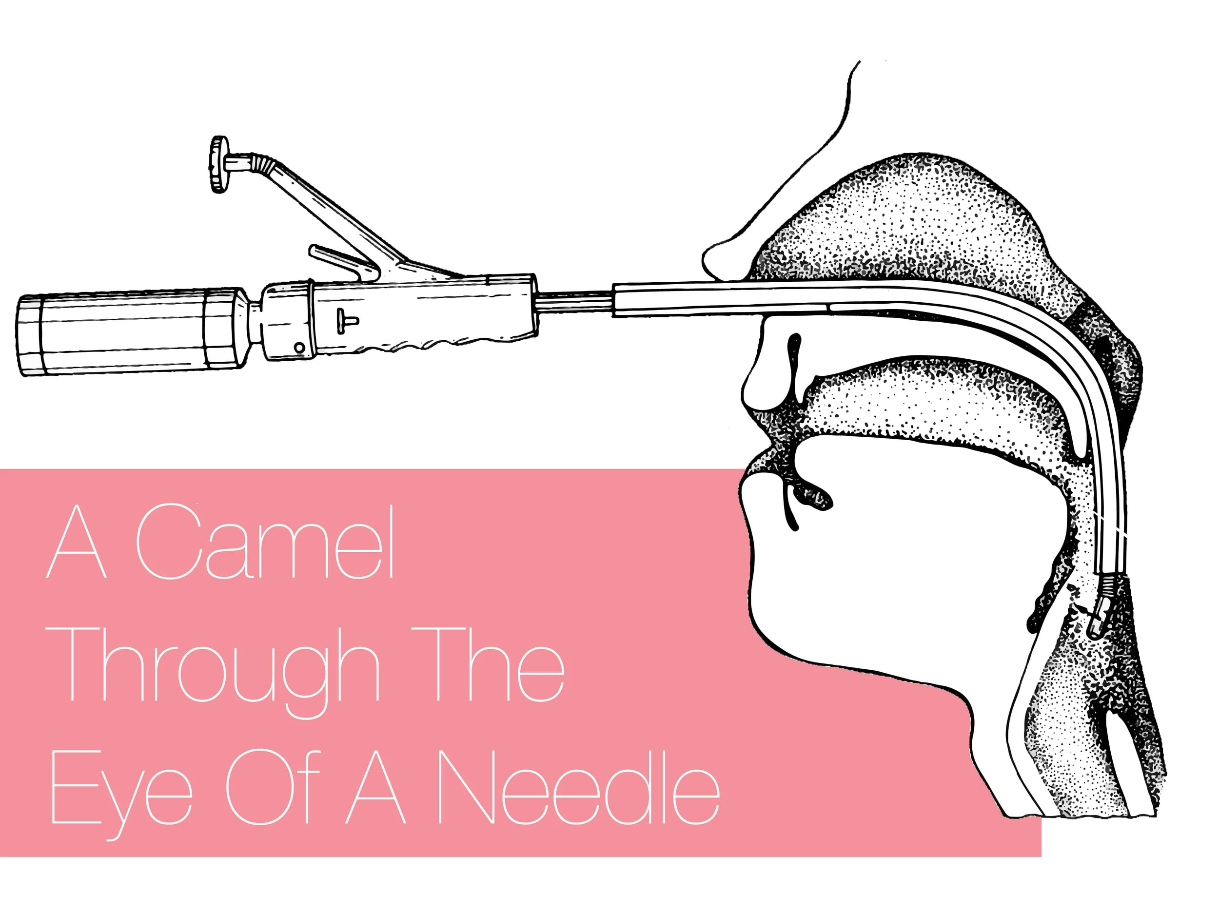
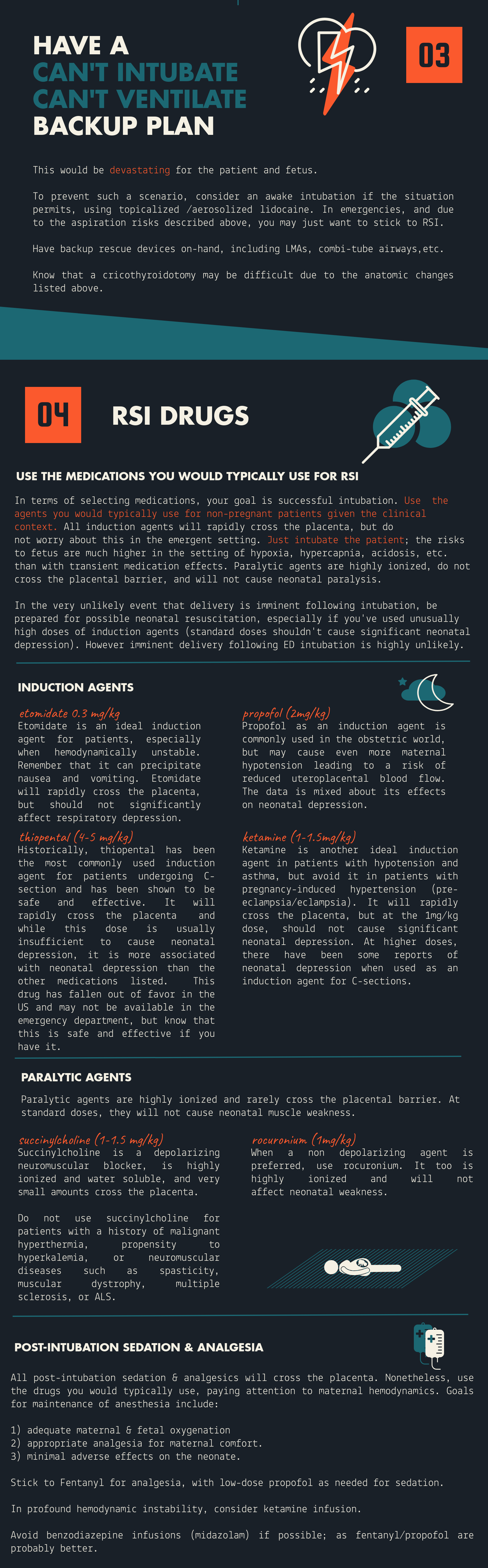
Awake intubation can sound imposing but simply means the patient is still breathing on their own. It is mostly just a matter of using topical lidocaine instead of paralyzing, and sedating the patient a bit to tolerate it. It will almost always require some sedation – ketamine procedural sedation works very well as the patient’s protective reflexes will be intact (until we topicalize) as will their respiratory drive.

It does not take long! Just spray lido instead of pushing NMBA. This is the key concept. If time is really a factor, I atomize the larynx, push ketamine, and then reload and spray more lidocaine as I do laryngoscopy; everything else is just like every other ED intubation.

Glycopyrrolate is nice but if it’s not handy, not worth a delay.

I find nebulizing doesn’t add much, mostly just gets the mouth. I still nebulize if I can get it set up quickly while prepping everything else (and it can help tolerate the atomizer).

Small touches of propofol might help relax the ketamine-sedated patient as well, including spontaneous/dissociated movements and tightly closed mouths. Dexmedetomidine might not be fast enough for ED intubations.

I usually use hyperangulated VL (eg Glidescope S3) – we are usually doing this for predicted difficult intubation, and now not optimizing intubating conditions. Fiberoptic requires a fair amount of skill and time. One of the main things that demystified awake intubation for me is it is a medication choice; it doesn’t always mean awake-fiber optic.

In non-COVID times, I would keep the nasal cannula on at 5-15lpm to keep the patient as oxygenated as possible, which is even better than during RSI because they’re still breathing, now with extra oxygen.

The paradox of awake intubation is that we take the patients we predict to be the most difficult anatomically, and then don’t optimize intubating conditions (no NMBA). Part of the beauty of awake intubation is that we also gain a ton of information even if unsuccessful without losing much; if I get a partial view in non-NMBA circumstances I can make a judgment call about proceeding to paralysis (ie RSI) or calling for help, etc.

Sedation-only or ketamine-only intubation can sound like a good idea but neither makes sense to me. It takes a lot of sedation to knock out protective airway reflexes to allow laryngoscopy, i.e. enough to impair respiratory drive. Topicalization is not hard with atomizers. Similarly, ketamine keeps the airway reflexes intact, which is why it is so safe for procedural sedation, so hard to imagine that laryngoscopy won’t be an issue.

Seth Trueger, MD, MPH

Assistant Professor of Emergency Medicine

Department of Emergency Medicine

Northwestern University


How To Cite This Post:

[Peer-Reviewed, Web Publication] Bigach, P. Whipple, T. (2021, Aug 20). Awake Intubation. [NUEM Blog. Expert Commentary by Trueger, S]. Retrieved from http://www.nuemblog.com/blog/awake-intubation


Other Posts You May Enjoy

Posted on August 23, 2021 and filed under Airway.

Hanging Injuries

Written by: Vytas Karalius, MD, MPH (NUEM ‘22) Edited by: Nery Porras, MD (NUEM ‘21) Expert Commentary by: Kevin Emmerich, MD, MS

Written by: Vytas Karalius, MD, MPH (NUEM ‘22) Edited by: Nery Porras, MD (NUEM ‘21) Expert Commentary by: Kevin Emmerich, MD, MS


Today’s post was inspired by the near-hanging of young gentleman who ended up passing away due to complications related to his near-hanging. His parents decided to donate his organs to Gift of Hope, allowing the passing of his life to extend the lives of others. While we hope to never see cases like these, they are an inevitable part of our job as emergency medicine physicians. As with most rare and complex pathology, preparation and knowledge can help us with the management of these cases when things often get chaotic. Lastly, as emergency medicine physicians who see the sequelae of mental illness daily in their EDs, I encourage us all to advocate for better funding and access to mental health care in the United States.

Hanging Injury

Terms/Classification [1]

  • “Hanging” is used to describe a death from a form of strangulation that involves hanging from the neck.

  • “Near-hanging” is a term for patients who have survived an attempted hanging (or at least long enough to reach the hospital).

  • “Complete hanging” defines when a patient’s legs are fully suspended off the ground and the patient's bodyweight is fully suspended by the neck.

  • “Incomplete hanging” defines when some part of the patient’s body is still on the ground and the body's full weight is not suspended off the ground.

  • “Judicial hanging” classically refers to victims who fell at least the height of their body.

Epidemiology:

  • Hanging is the 2nd most common form of successful suicide in the US after firearms

  • Accounts for 23% of >34,500 suicides in 2007

  • In the jail system, hanging is the most common form of successful suicide

  • Increasing incidence in US

  • Risk Factors: male, aged 15-44 years, history of drug or alcohol abuse, history of psychiatric illness

Pathophysiology of Injury:

Spine/Spinal Cord:

  • When the drop is greater than or equal to the height of the victim, as in a judicial hanging, there will almost always be cervical spine injury.

  • The head hyperextends, leading to fracture of the upper cervical spine ("hangman's fracture” of C2) and transection of the spinal cord.

  • Cervical injuries are in non-judicial hangings are rare. [2] One retrospective case review of near-hangings over a 10-year period found the incidence of cervical spine fracture to be as low as 5%. [3]

Vascular:

  • The major pathologic mechanism of death in hanging/strangulation is neck vessel occlusion, not airway obstruction. [1,4]

  • Death ultimately results from cerebral hypoxia and global ischemia.

  • There are two mechanisms by which this happens:

    • Venous: The most implicated cause of death is actually venous obstruction. Jugular veins are superficial and easily compressible. Obstruction of venous outflow from the brain leads to stagnant hypoxia and loss of consciousness in as little as 15 seconds.

    • Arterial: The risk of damage to the major arterial blood flow to the brain (such as carotid artery dissection) is rare, but should suspected in patients. [4]

Cardiac:

  • Carotid body reflex-mediated cardiac dysrhythmias are reported, and likely a minor mechanism of death.

Pulmonary:

  • Airway compromise plays less of a role in the immediate death of complete hanging/strangulation. However, it is a major cause of delayed mortality in near-hanging victims. [1,4]

  • Significant pulmonary edema occurs through two mechanisms:

    • Neurogenic: centrally mediated, massive sympathetic discharge; often in association with serious brain injury and a poor prognostic implication.

    • Post-obstructive: strangulation causes marked negative intrapleural pressure, generated by forceful inspiratory effort against extra-thoracic obstruction; when the obstruction is removed, there is a rapid onset pulmonary edema leading to ARDS.

  • Aspiration pneumonia later sequela of near-hanging injury.

  • Airway edema from mechanical trauma to the airway, which can make intubation difficult.

  • Tracheal stenosis can develop later in the hospital course.

Other Injuries:

  • Hyoid bone fracture

  • Cricoid or thyroid cartilage injury [5]

Physical Examination:

  • "Ligature marks" or abrasions, lacerations, contusions, bruising, edema of the neck

  • Tardieu spots of the eyes

  • Severe pain on gentle palpation of the larynx (laryngeal fracture)

  • Respiratory signs: cough, stridor, dysphonia/muffled voice, aphonia

  • Varying levels of respiratory distress

  • Hypoxia

  • Mental status changes

Early Management/Stabilization:

  • ABCs as always

  • Early endotracheal intubation may become necessary with little warning.

  • Patients who are unconscious or have symptoms such as odynophagia, hoarseness, neurologic changes, or dyspnea require aggressive airway management.

  • If ETI unsuccessful, consider cricothyroidotomy; if unsuccessful, percutaneous trans-laryngeal ventilation may be used temporarily.

  • Judicious and cautious fluid resuscitation - avoid large fluid volume resuscitation and consider early pressors, as fluids increases the risk/severity of ARDS and cerebral edema.

  • Monitor for cardiac arrhythmias.

  • The altered/comatose patient should be assumed to have cerebral edema with elevated ICP.

Imaging/Further Testing:

  • Chest radiograph

  • CT brain

  • CT C-spine

  • CTA head/neck

  • Can consider soft-tissue neck x-ray, if CT not immediately available

Further Management:

  • In patients with signs of hanging/strangulation, there should be a low threshold to obtain diagnostic imaging/testing as discussed above.

  • Expect pulmonary complications early.

    • They are a major cause of delayed mortality in near-hanging victims, as stated above.

  • Early intubation and airway management are important.

  • Non-intubated patients with pulmonary edema may benefit from positive end-expiratory pressure ventilation.

  • Patients with symptoms of laryngeal or tracheal injury (e.g. dyspnea, dysphonia, aphonia, or odynophagia), should undergo laryngobronchoscopy with ENT. [4,6]

  • Tracheal stenosis has been reported during the hospital course. Address cerebral edema from anoxic brain injury, using strategies to reduce intracranial pressure or seizure prophylaxis. [4]

  • Address vascular complications seen on CTA and coordinate intervention with the appropriate specialty at your institution.

  • Therapeutic Hypothermia

    • There is some evidence for therapeutic hypothermia in those with cardiac arrest from hanging injury [7,8] and those who are comatose from hanging injury. [9-11] While the evidence is weak, in the absence of better evidence, it is reasonable to consider hypothermia treatment in all comatose near-hanging victims. [1,12,13]

  • When suicide is suspected, evaluate patients for other methods of self-harm (e.g. wrist lacerations, self-stabbing, ingestions). It is also important to consider drug and alcohol intoxication. [4]

Disposition:

  • Admit critically ill patients to the appropriate ICU-level care.

  • Admit patients with abnormal radiologic or endoscopic imaging to the appropriate service and level of care.

  • Even if the initial presentation is clinically benign, all near-hanging victims should be observed for 24 hours, given the high risk of delayed neurologic, airway and pulmonary complications. [14]

  • Observe asymptomatic patients with normal imaging.

  • Psychiatry/Crisis Team consult on all suspected intentional cases.

  • Emphasize strict return precautions as well as education about possible delayed respiratory and neurologic dysfunction when discharging patients.

  • Some patients may require transfer to a trauma center if the required services are not available at the initial receiving facility. [1]

Prognostication:

  • GCS 3/GCS 3T is a predictor of very poor outcome, [15-19] but there is mixed evidence on the GCS as a predictor of outcomes in GCS scores greater than 3, especially with regard to neurologic intactness. [3,19]

  • Recovery of patients with neurology symptoms is unpredictable. [4]

  • Patients presenting with cardiac arrest have a very poor prognosis, and might be the strongest predictor of poor prognosis. [4,8,16,18,20]

  • Other predictors of poor clinical outcome include:

    • Anoxic brain injury or cerebral edema on head CT [3,19]

    • Prolonged hanging time [18]

    • Cardiopulmonary arrest [8,11,19]

    • Cervical spine injury

    • Hypotension on arrival


Expert Commentary

We’ve all certainly been involved with a patient with reported hanging injury at some point in our time in the ED. They are usually unimpressive if a person does it as more of a gesture rather than a true suicide attempt. When they are unfortunately done “correctly,” they usually result in a trip to the morgue instead of the ED. When the swiss cheese holes align and a true hanging attempt results in a serious but not fatal presentation, things can get quite hairy. I’ve been a part of one such case, and will never forget it. Here are my two cents.

Airway

This should undoubtedly be treated as a predicted difficult airway, not only due to likely cervical spine trauma, but also possibly due to airway edema. Get your ducks in a row for this unless this patient is crashing in front of you. Get your consultants/help (if available), preoxygenate, airway adjuncts open and ready, backup airway supplies if your first plan fails. Most importantly, have a plan and discuss this with your team beforehand. Don’t be afraid to take an awake look with a hyperangulated video laryngoscope, especially if this patient presents with stridor. Ketamine can be your friend here. This should be an airway that you do not undertake without a scalpel, finger, and bougie ready just in case. I like to draw a line on the patient’s skin overlying the cricothyroid membrane beforehand.

Trauma

Self explanatory, but don’t be stingy here. Light this patient up from head to pelvis, including the neck angiogram. Document a repeat neuro exam every time you move this patient.

Overdose/psych

Don’t forget your Tylenol and salicylate levels, EKG in this suicide attempt. If you feel the need to add the useless urine drug screen, I suppose this is fine as well.

Kevin Emmerich, MD, MS

Emergency Medicine Physician

Methodist Hospital

Gary, Indiana


How To Cite This Post:

[Peer-Reviewed, Web Publication] Karalius, V. Porras, N. (2021, Aug 9). Hanging Injuries. [NUEM Blog. Expert Commentary by Emmerich, K]. Retrieved from http://www.nuemblog.com/blog/hanging-emergencies


Other Posts You May Enjoy

References

1. Walls RM, Hockberger RS, Gausche-Hill M. Rosen's emergency medicine : concepts and clinical practice. Ninth edition. ed. Philadelphia, PA: Elsevier; 2018.

2. Aufderheide TP, Aprahamian C, Mateer JR, et al. Emergency airway management in hanging victims. Ann Emerg Med. 1994;24(5):879-884.

3. Salim A, Martin M, Sangthong B, Brown C, Rhee P, Demetriades D. Near-hanging injuries: a 10-year experience. Injury. 2006;37(5):435-439.

4. Tintinalli JE, Stapczynski JS, Ma OJ, Yealy DM, Meckler GD, Cline DM. Tintinalli's emergency medicine: a comprehensive study guide. 9th. ed. New York: McGraw-Hill Education; 2019.

5. Tugaleva E, Gorassini DR, Shkrum MJ. Retrospective Analysis of Hanging Deaths in Ontario. J Forensic Sci. 2016;61(6):1498-1507.

6. Hackett AM, Kitsko DJ. Evaluation and management of pediatric near-hanging injury. Int J Pediatr Otorhinolaryngol. 2013;77(11):1899-1901.

7. Hsu CH, Haac B, McQuillan KA, Tisherman SA, Scalea TM, Stein DM. Outcome of suicidal hanging patients and the role of targeted temperature management in hanging-induced cardiac arrest. J Trauma Acute Care Surg. 2017;82(2):387-391.

8. Kim MJ, Yoon YS, Park JM, et al. Neurologic outcome of comatose survivors after hanging: a retrospective multicenter study. Am J Emerg Med. 2016;34(8):1467-1472.

9. Jehle D, Meyer M, Gemme S. Beneficial response to mild therapeutic hypothermia for comatose survivors of near-hanging. Am J Emerg Med. 2010;28(3):390.e391-393.

10. Lee BK, Jeung KW, Lee HY, Lim JH. Outcomes of therapeutic hypothermia in unconscious patients after near-hanging. Emerg Med J. 2012;29(9):748-752.

11. Hsu CH, Haac BE, Drake M, et al. EAST Multicenter Trial on targeted temperature management for hanging-induced cardiac arrest. J Trauma Acute Care Surg. 2018;85(1):37-47.

12. Borgquist O, Friberg H. Therapeutic hypothermia for comatose survivors after near-hanging-a retrospective analysis. Resuscitation. 2009;80(2):210-212.

13. Sadaka F, Wood MP, Cox M. Therapeutic hypothermia for a comatose survivor of near-hanging. Am J Emerg Med. 2012;30(1):251.e251-252.

14. McHugh TP, Stout M. Near-hanging injury. Ann Emerg Med. 1983;12(12):774-776.

15. Kao CL, Hsu IL. Predictors of functional outcome after hanging injury. Chin J Traumatol. 2018;21(2):84-87.

16. La Count S, Lovett ME, Zhao S, et al. Factors Associated With Poor Outcome in Pediatric Near-Hanging Injuries. J Emerg Med. 2019;57(1):21-28.

17. Martin MJ, Weng J, Demetriades D, Salim A. Patterns of injury and functional outcome after hanging: analysis of the National Trauma Data Bank. Am J Surg. 2005;190(6):836-840.

18. Matsuyama T, Okuchi K, Seki T, Murao Y. Prognostic factors in hanging injuries. Am J Emerg Med. 2004;22(3):207-210.

19. Nichols SD, McCarthy MC, Ekeh AP, Woods RJ, Walusimbi MS, Saxe JM. Outcome of cervical near-hanging injuries. J Trauma. 2009;66(1):174-178.

20. Gantois G, Parmentier-Decrucq E, Duburcq T, Favory R, Mathieu D, Poissy J. Prognosis at 6 and 12months after self-attempted hanging. Am J Emerg Med. 2017;35(11):1672-1676.

Posted on August 9, 2021 and filed under Airway, Critical care, Trauma.

Intubating the Pregnant Patient in the ED

Written by: Priyanka Sista, MD (NUEM ‘20) Edited by: Steve Chukwulebe, MD (NUEM ‘19) Expert Commentary by: Samir Patel, MD

Written by: Priyanka Sista, MD (NUEM ‘20) Edited by: Steve Chukwulebe, MD (NUEM ‘19) Expert Commentary by: Samir Patel, MD


Expert Commentary

Tip for #1 - While 3-5 minutes of 100% oxygen is ideal to achieve denitrogenation, in an emergency 8 vital capacity breaths (maximal inhalation and exhalation) with a high FiO2 source is sufficient in a cooperative patient.

Tip for #2 - Airway edema is even worse in preeclamptic patients, and Mallampati scores acutely worsen DURING labor. Don’t bother with direct laryngoscopy and go straight to the video laryngoscope if it’s available.

Tip for #3 - In this scenario, the ideal LMA or supraglottic airway is one that includes a port for passage of an OG tube. Your pregnant patient in the ER with increased aspiration risk is not likely to be NPO for 8 hours like they are for anesthesiologists before surgery.

Tip for #4 - The rapid sequence dose of rocuronium is 1.2 mg/kg. You can immediately reverse rocuronium with sugammadex 16 mg/kg if necessary. For cost purposes, succinylcholine is still the best choice unless medically contraindicated.

Tip for #5 - According to ACOG, if cardiac arrest occurs in a woman greater than 23 weeks gestation, and there is no return of spontaneous circulation after 4 minutes of correctly performed CPR, a perimortem c-section should be performed with the goal of delivering the fetus by the fifth minute.

Samir Patel.PNG

Samir K. Patel, MD

Assistant Professor

Northwestern University Feinberg School of Medicine

Department of Anesthesiology


How To Cite This Post:

[Peer-Reviewed, Web Publication] Sista, P. Chukwulebe, S. (2021, Jan 18). Intubating the pregnant patient in the ED. [NUEM Blog. Expert Commentary by Patel, S]. Retrieved from http://www.nuemblog.com/blog/intubating-the-pregnant-patient.


Other Posts You May Enjoy

Posted on January 18, 2021 and filed under ENT, Airway.

Physiologically Difficult Intubations

Written by: Samantha Stark, MD (NUEM ‘20) Edited by: Steve Chukwulebe, MD (NUEM ‘19) Expert Commentary by: Seth Trueger, MD, MPH

Written by: Samantha Stark, MD (NUEM ‘20) Edited by: Steve Chukwulebe, MD (NUEM ‘19) Expert Commentary by: Seth Trueger, MD, MPH


It’s the first few minutes of your shift, and the paramedics roll by your workstation with your first patient, a young woman clutching an inhaler and breathing with every accessory muscle in her body. You direct them to your resuscitation room and they inform you that she has a history of asthma and is having an attack; she’s too exhausted from breathing to verify this, but it seems true. You quickly get her on BiPAP, which mildly improves her work of breathing, but as she becomes drowsy, you obtain a VBG showing a climbing CO2 of 45. You realize that it’s time to intubate this patient, and as you get set up, you collect your thoughts and quickly review everything you’ve heard about intubating asthmatics.

Obstructive Airway Disease

First, remember that asthma is an obstructive airway disease, meaning that there are two main processes to worry about during and after intubation:

  • Auto PEEP

  • Hypotension secondary to increased intrathoracic pressure from auto PEEP

*Note: auto PEEP is caused by breath stacking in a patient whose expiration is impaired (such as asthma or COPD) – the ventilator initiates a breath before there’s time for full exhalation, and this leads to progressively more volume retained in the lungs, increasing the risk of barotrauma. This can also lead to increased intrathoracic pressure, in turn decreasing preload to the heart and thus causing hypotension.

How to optimize the intubation:

  • As you already have this patient on BiPAP, try to preoxygenate as much as possible with this mode of positive pressure

  • Consider attempting delayed sequence intubation with ketamine

    • It will maintain the patient’s respiratory drive and may help with BiPAP synchrony and anxiolysis

    • It serves as a bronchodilator

  • Use rocuronium for paralysis

    • It will last longer than succinylcholine, and initially help with vent synchrony

    • *Note: remember to fully sedate the patient after intubation, as they won’t tell us that they’re not sedated during their prolonged paralysis

  • Decrease the dead space and resistance of your vent by using the largest endotracheal tube feasible

  • Frequently reassess the ventilator to ensure that breath stacking is not occurring:

    • Low respiratory rate to allow for exhalation

    • Higher tidal volumes of 6-8 cc/kg IBW

    • Decreased I:E ratio (at least 1:3, may very well need to be longer)

You’ve successfully intubated this patient, and now the lab pages you to let you know that there is a patient in the waiting room with a bicarb of 9. When the patient is wheeled back, his marked tachypnea and work of breathing makes you think he may need to be intubated as well. But he’s so acidotic, and you’re sure you’ve hear something about intubating acidotic people…

Metabolic Acidosis

What you’ve heard is that if you intubate a severely acidotic patient, you’ve killed them. There are two reasons for this:

  • It’s very difficult to keep up with their minute ventilation

  • There is a transient increase in pCO2 with paralysis (this is normally inconsequential, but in the decompensating acidotic patient, can lead to cardiovascular collapse)

How to optimize the intubation:

  • Optimize cardiovascular status as much as possible beforehand

  • Bolus fluids

  • Consider starting pressors pre-intubation, or having push-dose phenylephrine or epinephrine on hand during intubation

  • Match the patient’s minute ventilation

  • Ensure adequate pre-oxygenation, using NIPPV

  • However, even if oxygenation is not an issue, BiPAP should be used to assess the minute ventilation the patient is maintaining on their own, to help determine what is needed post intubation

  • Using delayed sequence technique with ketamine as the induction agent and a short acting paralytic like succinylcholine could theoretically help to avoid apnea as much as possible

  • Once intubated, the patient’s pre-intubation minute ventilation (respiratory rate and tidal volume) MUST be matched on the ventilator

  • Don’t be surprised to see higher tidal volumes of 8 cc/kg IBW

As you’re sitting down to catch up on notes, a nurse gets your attention to let you know that there is an altered, febrile, tachycardic patient with a pressure of 65/40 tucked away in a bed at the back of the ED that you should probably see right away. As it turns out, this patient needs to be intubated as well.

Shock

As mentioned above, increased intrathoracic pressure from PPV results in decreased venous return to the heart, leading to decreased preload. This obviously has the potential to be quite detrimental to a patient with shock.

How to optimize the intubation:

  • Optimize cardiovascular status as much as possible beforehand

  • Fluid resuscitation and vasopressors started prior to intubation

  • Have push dose pressors available at the bedside should they be needed

  • Induction agents:

    • Avoid propofol as it has a propensity to cause hypotension

    • Use etomidate or ketamine

    • Ketamine has been shown to be more hemodynamically stable than etomidate

    • Also, the body should prioritize cerebrovascular blood flow in shock, therefore if etomidate is used, consider decreasing the dose to minimize hemodynamic effects

At this point, you’re too tired to write any notes, so you decide to sit down and, given how your shift has been going so far, do some reading about patients that are dangerous to intubate or difficult to manage on the vent. The first topic you come across is pulmonary hypertension.

Pulmonary Hypertension

Mechanical ventilation is dangerous in these patients due to their inability to tolerate decreased preload, increased afterload, or really any alteration in their tenuous hemodynamics. Unfortunately, in patients with pulmonary hypertension but also systemic hypotension, IV fluids can over-distend the right ventricle and make things worse. There’s not a super reliable way to tell if these patients will be fluid responsive or not; most would suggest a small fluid bolus challenge to see how they respond. There may or may not be time for this prior to intubation, but if there is time, it’s probably worth a try.

How to optimize the intubation:

  • Can consider pre-medication with fentanyl:

    • Thought to blunt the hypertensive response to laryngoscopy, similar to head-injured patients

    • In theory, this prevents increased afterload in the pulmonary vasculature

  • Induction agent:

    • Consider etomidate

    • Theoretically should have less of an effect on preload than propofol

    • Additionally, less of an effect on afterload than ketamine

  • Ventilator settings:

    • Closely monitor plateau pressures to keep them less than 30 cm H2O, to avoid drops in preload due to increased intrathoracic pressure

    • Consider placing an arterial line for frequent ABG checks

    • Both hypercapnia and hypoxia can cause vasoconstriction (increasing afterload in the pulmonary vasculature)

Two days later, while you’re following up on some of your prior patients, you note that the patient in septic shock that you intubated a couple of days ago now has ARDS, and it seems that the inpatient team is having some difficulty managing her on the vent.

ARDS

While this is an area of active research and there are different strategies and methods for helping to improve these patients’ oxygenation, the main thing to remember from the perspective of managing the ventilator is the lung protective strategy:

  • Tidal volume 6 cc/kg IBW

  • Plateau pressure less than 30 cm H2O

  • Minimum PEEP of 5 cm H2O (and remember that these patients may often need significantly higher PEEP) 


Expert Commentary

Thank you for this review of intubating sick patients - intubating complex physiology is arguably one of the most dangerous things we can do, but there are some straightforward, concrete steps we can take to do it as safely as possible.

For me, the first step is to consider every ED intubation potentially dangerous. Maximize resuscitation (IV fluids; pressors if needed, always ready) and optimize preoxygenation to provide the biggest possible safety net. It’s much more CBA than ABC.

Every patient we intubate in the ED has potential to crump: the sympatholysis from sedation will reduce endogenous catecholamines, and the switch to positive pressure ventilation impairs preload.

Every intubated patient needs post-intubation sedation. I generally default to a fentanyl drip and modify from there (eg add propofol if BP tolerates; add ketamine if not). Do not remove sedation for hypotension; do not use pain as a pressor. That is torture and it is bad. Sedate the patient adequately and if that means more resuscitation (fluid, blood, pressors, etc) then do that too. Do not torture patients to maintain BP.

The easiest tactic to ensure post-intubation sedation is to think of RSI as 3 medications: NMBA, induction agent, and post-intubation sedative. I should not be surprised that I will need post-intubation sedation shortly after intubation.

Perhaps the biggest lesson in ARDS management and prevention in recent years is that nearly everyone potentially benefits from lung protective ventilation, i.e. 6 ml/kg *ideal* body weight. I’ve changed my default tidal volume to 400-450ml (it was 550-600 when I was in med school). Otherwise, ventilation (minute ventilation, or CO2 management) is all about adjusting respiratory rate (my default is 16-18, not 12) as the patient’s height usually does not change in the ED.

Special situations: asthma patients don’t have a big enough tube to exhale properly. Pay special attention, make sure they have sufficient time to exhale (and they may the one group that may benefit from *not* being on 6 ml/kg IBW. Perhaps even more importantly, unlike many other situations, intubation does not fix asthma; it makes it even harder to manage, as even the largest ET tubes are, by definition, smaller than the patient’s natural airway. Maximize NIV and other management options (eg epinephrine) if at all possible.

Acidosis is tough and the key is maximizing ventilation before and after intubation. These patients may need absurd-seeming respiratory rates and regardless of how hypercarbic they are, acidosis does not make patients taller so there is no reason to adjust tidal volume.

Pulmonary hypertension is complex and scary. Prepare beforehand, and work with your intensivists and other relevant specialists.

The most important part of airway management is preparation – not just in the ED, but learning as much as I can beforehand.

Seth Trueger.PNG

Seth Trueger, MD, MPH

Assistant Professor of Emergency Medicine

Department of Emergency Medicine

Northwestern University expert commentator


How To Cite This Post:

[Peer-Reviewed, Web Publication] Stark, S. Chukwulebe, S. (2020, Oct 5). Physiologically Difficult Intubations. [NUEM Blog. Expert Commentary by Trueger, S]. Retrieved from http://www.nuemblog.com/blog/physiologically-difficult-intubations


Other Posts You May Enjoy

References

  1. Ebert TJ, Muzi M, Berens R. Sympathetic responses to induction of anesthesia in humans with propofol or etomidate. Anesthesiology. 1992;76:725-33.

  2. Van Berkel MA, Exline MC, Cape KM, et al. Increased incidence of clinical hypotension with etomidate compared to ketamine for intubation in septic patients: a propensity matched analysis. Journal of Critical Care. 2017;38:209-214.

  3. Dalabih M, Rischard F, Mosier JM. What’s new: the management of acute right ventricular decompensation of chronic pulmonary hypertension. Intensive Care Med. 2014;40(12):1930-3.

  4. Hemmingsen C, Nielson PC, Odorico J. Ketamine in the treatment of bronchospasm during mechanical ventilation. Am J emerg Med. July 1994;12(4):417-420.

  5. Eames WO, Rooke GA, Wu RS, Bishop MJ. Comparison of the effects of etomidate, propofol, and thiopental on respiratory resistance after tracheal intubation. Anesthesiology. June 1996;84(6):1307-11.

  6. Gragossian A, Asp A, Hamilton R. High Risk Post Intubation Patients. www.emdocs.net/ high-risk-post-intubation-patients/ June 2017

  7. The Acute Respiratory Distress Syndrome Network. Ventilation with lower tidal volumes as compared with traditional tidal volumes fo acute lung injury and the acute respiratory distress syndrome. N Engl J Med. 2000;342:1301-1308.

  8. NIH NHLBI ARDS Clinical Network. Mechanical Ventilation Protocol Summary. www.ardsnet.org/files/ventilator_protocol_2008-07.pdf

  9. Marino, Paul L. 2009. The Little ICU Book. Wolters Kluwer Health. Philadelphia, PA.

  10. Arbo, John E. 2015. Decision Making in Emergency Critical Care: An Evidence-Based Handbook. Wolters Kluwer Health. Philadelphia, PA.

Posted on October 5, 2020 and filed under Airway.

Intubation Positioning: Beyond Sniffing

Screen Shot 2019-01-30 at 1.01.19 PM.png

Written by: Katie Colton, MD (NUEM PGY-4) Edited by: Charles Caffrey, MD (NUEM Alum ‘18) Expert commentary by: Andrew Pirotte, MD


The patient

On a recent Thursday night in a single-attending ED, we received a call that a patient was several minutes out with respiratory distress.  He had been enjoying his routine post-dinner cocaine insufflation and became dyspneic, per bystanders. We could hear yelling in the back of the ambulance and EMS reported that they were only able to get an oxygen saturation, which was about 70%. 


The scene

Two minutes later, EMS rushed in with a patient who looked to be in his mid-50s who was clearly struggling to breathe; it required 6 people to transfer him to our cart.  He was altered, hypoxic, approximately 500 pounds, and needing an airway in the near future.  While there are many considerations for the difficult airway, what are particular positioning options that may increase the chances of success in this patient?


“Positioning is 90% of the battle”

Beyond the technical difficulties posed by the morbidly obese patient, there are physiologic differences that complicate their oxygenation and ventilation.  Due to the weight of the chest and larger abdomen they will have a decreased functional residual capacity and total lung capacity.  Supine position can complicate pre-oxygenation, endotracheal intubation (ETI), and cause hypotension in these patients.  

Many providers are still trained almost entirely in ETI with a supine patient, but there is growing evidence that a head-up position can improve pre-oxygenation and facilitate ETI.

Some authors advocate for an aggressive ramped position, using either a pre-formed foam ramp or a stack of pillows or blankets, like in the pictures below.  I would argue that unless you have a stack of pillows at the ready and a number of spare hands this technique may be difficult in the less-controlled setting.

(Simoni 2005)

(Simoni 2005)

sniff2.png

So then what?

Newer studies – and anecdotal experience - are showing good results with upright intubation through simple manipulation of the head of the bed.  One example of this is the back-up head-elevated position as seen below[2]. First, by brief placement of the patient into Trendelenburg, the patient is brought all the way to the top of the bed (1 in image), and then the back of the bed is ramped up to at least 30 degrees above the horizontal (2 in image), with the head placed into the “sniffing” position with a towel roll (3 in image).[2] In their retrospective analysis, Khandelwal et al. found a lower rate of intubation-related complications as compared to a supine cohort, 9.3% vs 22.6%.[2]

(Khandewal 2016)How to place the patient in the advised back-up, head-elevated position.

(Khandewal 2016)

How to place the patient in the advised back-up, head-elevated position.

A team from IU showed improved intubation success with head of bed elevation in both a simulated [4] and ED setting [3].  This approach allows the patient to be positioned during preoxygenation.  Redundant tissue falls away from the face and chest, improving the patients ability to breathe for themselves and the ease of BVM, if needed.  Khandelwal et al found lower risk of aspiration and esophageal tube placement. For every 5 degree increase in head of bed angulation above the horizontal, Turner et al found increased likelihood of first pass success with ETI.

(Turner 2017)

(Turner 2017)

In short, consider using the bed to your advantage in these difficult patients.  It takes time to overcome habits but there is good evidence for changing up your positioning plan.


Case Conclusion

The patient’s head of the bed was ramped up to 45 degrees. Utilizing rapid sequence intubation, the resident took one look with a size 4 Macintosh acquiring a Grade II view, and was able to place an 8-0 tube. A follow-up chest x-ray showed appropriate placement and frank pulmonary edema.  The patient was treated for pulmonary edema and admitted to the ICU.


Expert Commentary

As with all clinical excellence, the devil is in the details.  Skillful airway management requires attention to detail, notably patient positioning.  This case and review serve as a reminder that close attention to setup and positioning can help enhance successful airway management.

Positioning is critical to airway success.  Particularly in the setting of higher body-mass index patients, optimized positioning is a critical step to safe and successful airway management.  As suggested by the review, simply placing the patient in supine positioning is not optimal and should be avoided if possible.  Improved positioning can be achieved in several ways, but often the most straight-forward is by raising the head of the bed, or stacking towels and pillows.  This position is often referred to as the “sniffing” position. 

The sniffing position refers to bringing the sternal notch and the ear into the same plain (see blog post image).  This positioning not only improves ergonomics for the clinician, but provides enhanced laryngoscopy and endotracheal tube delivery success.  In addition, sniffing positioning compliments and enhances other airway optimization strategies. 

One significant benefit of the sniffing position is preventing collapse of soft tissue and occlusion of the airway.  The relief of redundant tissue with the sniffing position likely improves high-quality mask ventilation, as the tissue collapse into the posterior oropharynx is less prominent.  Physiological benefits of sniffing position also include decreased lung atelectasis and improved delivery of oxygen during airway preparation. Considering these points, utilization of the sniffing position (rather than supine positioning), vitally strengthens the airway management pathway.

Positioning remains crucial to optimized airway delivery. The sniffing position does not require expensive equipment or great skill; it is a straight-forward, useful, and impactful strategy to enhance airway management.  As emergency airway management continues to evolve, much focus has been on enhancing laryngoscopy.  In addition, there have been great strides in technology and monitoring equipment.  But even with the best equipment and technology, simple strategies such as optimizing positioning can lead to high-yield results. 

Special thanks to Dr. Jordan Kaylor and Dr. Matthew Pirotte

andrew_pirotte.png
 

Andrew Pirotte, MD

Department of Emergency Medicine, University of Kansas Hospital

Clinical Assistant Professor, University of Kansas Medical Center


How to Cite This Post

[Peer-Reviewed, Web Publication] Colton K, Caffrey C (2019, February 4). Intubation Positioning: Beyond Sniffing [NUEM Blog. Expert Commentary by Pirotte A]. Retrieved from http://www.nuemblog.com/blog/intubation-positioning.


Other Posts You May Enjoy


Resources 

  1. RF Simoni et al. Tracheal Intubation of Morbidly Obese Patients: A Useful Device. Brazilian Journal of Anesthesiology, 2005; 55: 2: 256-260

  2. N Khandelwal et al. Head-Elevated Patient Positioning Decreases Complications of Emergent Tracheal Intubation in the Ward and Intensive Care Unit. Anesth Analg. 2016 Apr;122(4):1101-7.

  3. Turner JS et al.  Feasibility of upright patient positioning and intubation success rates at two academic emergency departments. Am J Emerg Med. 2017 Feb 5. pii: S0735-6757(17)30100-6.

  4. Turner JS et al. Cross-over study of novice intubators performing endotracheal intubation in an upright versus supine position. Intern Emerg Med. 2016 Jun 14.


Posted on February 4, 2019 and filed under Airway.