The ED Guide to Neuroimaging: Part 1

Emergency Department Neuroimaging

Written by: Justin Seltzer, MD (NUEM PGY-1) Edited by: Andrew Cunningham, MD, (NUEM PGY-3) Expert commentary by:  David Rusinak, MD


Neuroimaging, mainly using CT, has become an indispensable part of our emergency diagnostic process, but, all too often we rely on radiologists to interpret what we ordered. The goal of this multi-part blog is as follows:

  • To cover the basics of how to look at a CT brain and quickly identify life threat

  • Review the literature supporting the major ED indications

  • Discuss special considerations, such as when to use contrast, angiography, or MRI instead.


Systematic Reading of a CT Brain

The first portion of this blog will focus on how to read a CT brain quickly with a focus on life threats.

The classic mnemonic, “Blood Can Be Very Bad,” is a pathology oriented, step-wise method to look for blood, cistern changes, and alterations to the brain parenchyma, ventricle appearance, and bony anatomy.  Applying this approach to each image cut individually can help reveal subtle findings that would otherwise be easily missed by quick scrolling.

Blood:

Blood can collect both intra-axially (parenchymal) and extra-axially (outside the parenchyma).

Figure 1

  • Classically, spontaneous intra-axial bleeding originates in deep structures such as the basal ganglia and thalamus (Figure 1).

    • In the setting of trauma, intra-axial bleeding is often ipsilateral or directly contralateral to the injury site (coup-contrecoup) but can be anywhere

    • Inferior frontal and anterior temporal lobes are high risk for traumatic contusions due to close proximity to bone (Figure 2)


Figure 2

 
  • Extra-axial bleeding is defined by location: mainly subdural, epidural, subarachnoid, and intraventricular hemorrhages. The patterns for these are well known and readily identified, however below are some key points on extra-axial bleeding.

    • Finding chronic subdural hematomas can be difficult as older blood and grey matter are similar appearing

    • Mass effect, abnormal appearing brain folds on that side, and use of coronal reconstructions can help identify

    • Subarachnoid hemorrhage becomes difficult to see within hours to days but acutely is often observed well in the cisterns (see below)

    • Be careful not to mistake choroid or pineal calcifications for hemorrhage

    • Don’t forget about scalp hematomas


Cisterns:

The cisterns are not ventricles but rather outpouchings of the subarachnoid space. When evaluating a CT brain the following, certain cisterns have clinical relevance for potential herniation syndromes, layering of subarachnoid blood, and/or the significant structures that run through them. Figures 3-5 show the locations of the major cisterns described below.

Figure 3

Figure 4

  • Suprasellar: Located in the area of the sella turcica, forms a pentagon/star shape

    • Classic location of subarachnoid hemorrhage due to proximity to circle of Willis

    • Obliteration associated with downward transtentorial (i.e. uncal) herniation or due to severe elevated ICP

  • Perimesencephalic cistern: A group of interconnected basal cisterns surrounding the midbrain (mesencephalon), important location of subarachnoid hemorrhage, may see effacement (reduction or loss) with tonsillar herniation

    • Interpeduncular: Located in the area of the cerebral peduncles

    • Quadrigeminal: Classically forms a W shape, obliteration associated with upward herniation

    • Ambient and crural: Connections between quadrigeminal and interpeduncular cisterns

  • Cerebellopontine: Located between anterior cerebellum and lateral pons, synonymous with area of cerebellopontine angle

  • Cisterna magna: Located between the cerebellum and medulla, receives fourth ventricular CSF outflow (Figure 4)

  • Prepontine: Located at the anterior aspect of the pons

Figure 5

Brain parenchyma:

CT allows for gross evaluation of the major structures as well as a differentiation of grey and white matter by Hounsfield units. The focus here is major parenchymal disruptions.

  • Mass lesions, mass effect, midline shift: Because of the fixed nature of the skull, mass lesions of any type easily exert pressure on the surrounding tissue (mass effect) that can result in increased ICP, midline shift, and herniation.

    • Midline shift is measured in millimeters of displacement of the septum pellucidum at the level of the foramen of Monro from the midline of the skull

  • Ischemic changes: depending on the size of the involved territory and duration, may be subtle or obvious density or architectural changes.

    • Early signs of infarction: reduced grey-white matter distinction and loss of insular hyperdensity

Ventricles:

The ventricular system is where CSF is produced and the route by which it travels into the subarachnoid space. The lateral ventricles drain via the foramina of Monro to the third ventricle, which then drains via the cerebral aqueduct (aqueduct of Sylvius) to the fourth ventricle and then to the cisterna magna and the rest of the subarachnoid space via the median and lateral apertures (foramina of Magendie and Lushka, respectively). Figures 3-5 also show the locations of the major ventricles.

  • Interruption of ventricular CSF flow will cause proximal ventricular dilation that helps localize the level of obstruction

  • If unsure between hydrocephalus and atrophy, dilation of temporal horns of the lateral ventricles can be helpful as it occurs in hydrocephalus involving the lateral ventricles but not with hydrocephalus ex vacuo


Bones:

Intimate knowledge of bony anatomy is not essential fracture evaluation. However, it is crucial that the bony anatomy be viewed with a dedicated bone window. Skull and facial fractures can be subtle and the presence of blood, especially an epidural hematoma, may help localize them. As noted above, soft tissue findings such as scalp hematomas are important to rule out as well.


Key Learning Points and Conclusions

  • A systematic approach is essential to avoid missing significant findings, especially with complex neuroimaging—remember “Blood Can Be Very Bad”

  • Immediately look for: blood anywhere (don’t forget the scalp!), effacement of major cisterns, mass effect/midline shift, enlarged ventricles (temporal horns), skull fractures

  • Older blood, such as a chronic subdural hematoma, can be hard to find and may require different cuts or inference from mass effect or effaced sulci

  • Signs of infarction may be subtle (more on this later)


In the next installation, we will discuss the major indications for CT brain and the utility of CT for these indications.


Expert Commentary

Overall, this is a very nice approach to head CT interpretation.  The classic mnemonic, “Blood Can Be Very Bad,” is not something I’ve heard of before, but it works.  Let’s take each search item in turn.


Blood

A helpful way to think about intracranial hemorrhage is to consider the causes of hemorrhage and the most common location for each pathology.  Common causes include trauma, stroke (hypertensive or hemorrhagic conversion of a venous or arterial infarct), neoplasm (primary or secondary), vascular (aneurysm, AVM, dural AV fistula), and spontaneous (anticoagulation, amyloid angiopathy, vasculitis).  If you consider the location of each of these pathologies, the hemorrhage will typically be primarily in this location.  A tumor, for example, will cause a parenchymal bleed, a ruptured aneurysm will cause subarachnoid hemorrhage, an AVM will result in a parenchymal bleed, etc.  Often with parenchymal bleeds additional imaging, vascular and MRI, as well as follow up imaging will be necessary to determine the underlying cause.

A correction is that subacute hemorrhage, not chronic, has a density similar to gray matter.  Chronic subdural hemorrhages are usually very hypodense and easy to detect on CT. So, from a practical perspective, a patient experiencing headaches from subarachnoid hemorrhage that is greater than 3 or 4 days old may be occult by CT.  This underscores the role of lumbar puncture and vascular imaging in working up patients with headaches.

Another important concept to keep in mind is window and level when interpreting CTs. Different substances (air, metal, bone, blood, fat, etc) have different and defined densities.  The pathologies associated with each of these substances (fractures, edema in the setting of stroke, etc) can be better seen by adjusting the window and level settings.  This can be done manually or, typically, PACS viewers have preset brain, bone, lung and soft tissue windows that can be displayed by pressing different numbers on the keypad.  Subtle subdural hemorrhages are often only seen with the appropriate window and level that allows distinction of the hemorrhage from the overlying calvarium.


Cisterns

The blood vessels course through the cisterns, so these must be scrutinized for the presence of hemorrhage secondary to a ruptured aneurysm in a patient presenting with an atraumatic headache.  The cisterns are also effaced in the setting of mass effect. Mass effect may be from a space occupying lesion; such as a tumor, abscess, or hemorrhage; or from diffuse cerebral edema with generalized brain swelling.  Often the absence of something (i.e. patent basal cisterns) can be harder to detect than the presence of something, like hemorrhage.  It is, therefore, important to examine the basal cisterns on each case to get comfortable with their normal variation of appearance so that their absence, such as in diffuse cerebral edema, is not missed.


Brain parenchyma

Subtle changes in parenchymal density can be difficult to detect.  It is important to get acquainted with ideal window and level settings to uncover subtle parenchymal changes.  Also comparison with prior imaging, if available, is necessary to determine the chronicity of parenchymal findings.  Understanding where a physical exam finding localizes intracranially can also be very useful- aphasia or left upper extremity weakness localize to very different locations, for example.  Lastly, always look at the vessels in the subarachnoid space to identify hyperdense thrombus in the setting of a suspected stroke.


Ventricles

Distinguishing volume loss from ventricular dilatation takes experience to understand the variation of normal across the entire age spectrum.  If hydrocephalus is suspected, determining if it is obstructive or communicating can help to understand the underlying cause. The temporal horns are the most elastic portion of the ventricles and dilate first in the setting of hydrocephalus. 


Bones

Depressed skull fractures and easy to see on routine bone windows.  Things get complicated when subtle non-displaced fractures mimic normal sutures or if the fracture involves the skull base/temporal bones.  It is probably not within the normal ED physician’s scope of practice to have a detailed knowledge of skull base anatomy, but if a skull base fracture is suspected (loss of hearing, hemorrhage in the external auditory canal, facial nerve paralysis, etc) it is important or order the proper test for further evaluation, like a temporal bone CT.  A helpful tip is to look for subtle foci of intracranial air and soft tissue swelling which may direct you to a subtle fracture.    

Screen Shot 2018-04-20 at 1.09.34 PM.png

David Rusinak, MD

Assistant Professor of Radiology, Northwestern Medicine


 How to cite this post

[Peer-Reviewed, Web Publication]  Whipple T,   Gappmeier V (2018, April 23 ). Demystifying the Hand Exam.  [NUEM Blog. Expert Commentary by Rusinak D ]. Retrieved from http://www.nuemblog.com/blog/neuroimaging


Posts you may also enjoy


References

1. Shaw AS, Prokop M. Computed Tomography. In Grainger & Allison's Diagnostic Radiology, 6th Edition (2016).

2. McKetty MH. The AAPM/RSNA physics tutorial for residents. X-ray attenuation. RadioGraphics, 1998; 18(1):151-163

3. Cadogan M. CT Head Scan. Life in the Fast Lane. Retrieved from https://lifeinthefastlane.com/investigations/ct-head-scan/.

4. Nadgir R, Yousem DM. Approach and Pitfalls in Neuroimaging. In Neuroradiology: The Requisites, 4th Edition (2017).

5. Mehta A, Jones BP. Neurovascular Diseases. In Grainger & Allison's Diagnostic Radiology, 6th Edition (2016). Chapter 62, 1456-1496.

6. Jones J. Subarachnoid Cisterns. Radiopaedia. Retrieved from https://radiopaedia.org/articles/subarachnoid-cisterns.

7. Nadgir R, Yousem DM. Cranial Anatomy. In Neuroradiology: The Requisites, 4th Edition (2017).

8. Skalski M, Dawes L. Cerebral herniation. Radiopaedia. Retrieved from  https://radiopaedia.org/articles/cerebral-herniation.

9. Baron EM, Jallo JI. TBI: Pathology, Pathophysiology, Acute Care and Surgical Management, Critical Care Principles, and Outcomes. In Brain Injury Medicine: Principles and Practice, 2nd Edition (2012). Chapter 18: 265-282.

10. Nadgir R, Yousem DM. Head Trauma. In Neuroradiology: The Requisites, 4th Edition (2017).

11. Waxman SG. Ventricles and Coverings of the Brain. In Clinical Neuroanatomy, 28th Edition (2013).

12. Nadgir R, Yousem DM. Neurodegenerative Diseases and Hydrocephalus. In Neuroradiology: The Requisites, 4th Edition (2017).

13. Galliard F, Jones J. Intraventricular haemorrhage. Radiopaedia. Retrieved from https://radiopaedia.org/articles/intraventricular-haemorrhage


Demystifying the Hand Exam

Screen Shot 2018-03-05 at 8.31.59 AM (1).png

Written by: Terese Whipple, MD (NUEM PGY-2) Edited by: Victor Gappmaier, MD, (NUEM PGY-4) Expert commentary by:  Aviram Gialdi, MD, MS


The human hand is a fascinatingly intricate arrangement of pulleys, tendons, muscles, and nerves that work together in a complex system to perform daily tasks. It is often difficult to visualize the various paths that the tendons and muscles take.  It can also make a thorough hand exam difficult to perform with proficiency. This post will review the clinically relevant anatomy of the hand, and apply it to both a screening exam and detailed exam with maneuvers used in the diagnosis of common hand injuries.


This screening exam can be used in the case of a fracture/dislocation at or proximal to the wrist, or in a general trauma to ensure that there has not been a nerve injury – from the cervical spine, through the brachial plexus, and into the extremity.

Basic Screening Exam:  

Vascular

To examine the vascular supply of the hand the examiner should palpate the radial pulse and check digital capillary refill.  Using a finger pulse oximeter is a useful adjunct for evaluating perfusion; anything below 95 in a traumatized limb/digit raises concern.

Neuro

The radial, median, and ulnar nerves each have sensory and motor functions that should be evaluated. 

 Radial (C5-C8):

Img 1. Sensory innervation of hand

  • Motor: Extend the wrist. If too painful due to injury, then extension of the thumb IP joint may be substituted.
  • Sensory: Test the dorsal webspace between the thumb and index finger

Median (C5-T1):

  • Motor:
    •  Recurrent motor branch of the median nerve: Have the patient attempt opposition (bringing the thumb tip across to the small finger tip)
    • Anterior interosseus branch of the median nerve:  Make an OK sign by having the patient touch the tip of the thumb to the tip of the index finger

Img 2. Correct OK Sign

Img 3. Incorrect OK Sign

  •  Sensory: Palmar surface of the index finger or thumb

Ulnar (C8-T1):

  • Motor: Test by having patient spread fingers against resistance
  • Sensory: Palmar aspect of the little finger


Check the individual digital sensory nerves to any finger by testing the radial and ulnar sides of each digit

If the patient can perform each of the above functions and has intact sensation, as well as good cap refill and pulses, they have passed the basic screening exam and are “neurovascularly intact.”


Now for a more detailed exam, which should be used when a patient comes in with a specific hand complaint or if there is concern for muscle or tendon injury.

A thorough musculoskeletal (MSK) exam should include:

  • Inspection
  • Palpation
  • Range of Motion (ROM)
  • Nerve/Vascular assessment
  • Muscle/tendon exam
  • Specific maneuvers


Detailed Hand Exam:

Inspection

Inspect the hand for evidence of:

Img 3. Mallet finger

  • Asymmetry
  • Lacerations/abrasions: Any skin break over a joint (eg : fight bite) may look innocent, but actually provides a route for inoculation of the joint with infection and can be serious.
  • Inflammation:  Can be acute from recent injury/infection or chronic from inflammatory states such as RA.
  • Atrophy: Think critically about the location of the atrophy, is it diffuse or does it fit one nerve distribution? For example, carpal tunnel syndrome may produce atrophy in the thenar muscles supplied by the median nerve. Ulnar nerve entrapment at the elbow (Cubital tunnel syndrome) could cause hypothenar muscle wasting and intrinsic wasting (most visible at first dorsal interosseous, along dorsal radial border of the index metacarpal).
  • Any evidence of traumatic deformity such as unusual angulation or rotation. You should always check alignment of the fingers in flexion and extension. Sometimes abnormal rotation will only be visible when making a fist, when one finger crosses over/under the next
    • Any alteration to the normal cascade of the fingers (one finger that is not flexed/extended to match the position of the others) may represent a tendon injury
    • Mallet finger: A flexed DIP with inability to actively extend due to rupture of the terminal extensor tendon of the digit. (Img. 3)
    • Boxer’s fracture: May have a “dropped knuckle sign” where the fracture of the metacarpal shaft causes a “disappearance” of the metacarpal head  (Img. 4)
 

Img 4. Dropped knuckle sign


Palpation

Img 5. Scaphoid Tubercle

It can be difficult to visualize all of the bones in the hand and wrist in order to palpate them correctly. However, there are a few that emergency medical providers should know in order to catch the most common and consequential injuries.

The scaphoid is technically part of the wrist, however it is usually part of a screening hand exam for anyone with a fall onto a hand. It can be palpated in 3 places:

Img 6. Anatomic snuffbox

  • Scaphoid tubercle   
  • The waist of the scaphoid can be palpated in the anatomic snuffbox 
  • The proximal scaphoid can be palpated on the dorsal wrist in the soft spot between the tendons of the 3rd and 4th compartment of the wrist, just distal to Lister’s tubercle 

Img 7. Lister's Tubercle

Img 8. Proximal Scaphoid

Previous studies have demonstrated that tenderness at the scaphoid tubercle is actually more sensitive than the anatomic snuffbox (95% v. 85%) in diagnosing scaphoid fracture. When palpating the anatomic snuffbox you can maximize the surface area that you are palpating by having the patient move their hand into ulnar deviation and thumb abduction.

Range of Motion

Test range of motion both passively and actively in each joint. Passive ROM gives you information about the joint.  You may feel clicking, catching or crepitance. Active ROM provides information about nerve function, muscle strength, joint congruity/stability, and tendon integrity.

[Insert aforementioned neurovascular exam here]

Muscle/tendon exam

A full muscle/tendon exam doesn’t need to be a part of every exam in the Emergency Department, we don’t have the time. However, if there is an injury that makes you concerned about the integrity of deep structures in the hand, wrist, or forearm, knowing the course and function of each muscle and tendon is useful.  Theoretical cases have been included to provide context.

Case 1:

A patient sustained a deep laceration to his right volar forearm from a glass bottle during an altercation at a bar. The sensory exam in the hand is normal, but function is abnormal.  In addition to the usual laceration care, you want to ensure all of the underlying tendons from the extrinsic muscles are intact. You need to check the finger and wrist flexors. Most of these are innervated by the median nerve, with the exception being the Flexor Carpi Ulnaris and the Flexor Digitorum Profundus to the small and ring fingers, which are innervated by the ulnar nerve.

Img 9. Flexors of forearm

  • Flexor Pollicis Longus (FPL): Test by asking patient to flex thumb at the IP joint (AIN)
  • Flexor Digitorum Profundus (FDP): Test by asking patient to flex DIP joint of index or middle finger while stabilizing PIP of the same digit
  • Flexor Digitorum Superficialis (FDS): Test by asking patient to flex PIP while examiner holds all the other digits in extension (this blocks FDP and completely isolates the FDS)
  • Flexor Carpi Ulnaris and Flexor Carpi Radialis: Test by asking patient to flex the wrist and palpate tendon/muscular contraction


In summary, to test the extrinsic flexors:

  • Flex thumb IP joint
  • Stabilize PIP and have patient flex each DIP in succession
  • Hold remainder of fingers in extension, ask patient to flex each PIP in succession
  • Volar flex wrist


Case 2:

The same patient presents again after a bar fight, this time sustaining a deep laceration to his dorsal forearm.  You want to ensure all of the underlying tendons from the extrinsic muscles are intact. You need to check the extensors.  These muscles are all innervated by the radial nerve and are separated into six compartments.

First Dorsal Wrist Compartment

  • Abductor Pollicis Longus and Extensor Pollicis Brevis: Ask the patient to bring their thumb out to the side (abduct) and palpate the tendons along the radial border of the wrist

Img 10. Extensors of forearm

2nd Dorsal Wrist Compartment

  • Extensor Carpi Radialis Longus (ECRL) and Extensor Carpi Radialis Brevis (ECRB): Have the patient make a fist and extend against resistance

3rd Dorsal Wrist Compartment

  • Extensor Pollicis Longus: Place hand flat on table and lift thumb off the table

4th Dorsal Wrist Compartment (the MCP joint extensors of the fingers)

  • Extensor Digitorum Communis and Extensor Indicis Proprius (EIP): Test by straightening individual fingers at the MCP. The EIP can be isolated by extending index finger with the rest of the fingers closed in a fist

Img 11. Extensors of forearm 

5th Dorsal Wrist Compartment

  • Extensor Digiti Minimi: Extend small finger with the rest of the fingers closed in a fist

6th Dorsal Wrist Compartment

  • Extensor Carpi Ulnaris: Extend and ulnar deviate wrist

In summary, to test the finger extensors:

  • Abduct the thumb, then place on table and lift thumb off
  • Extend fingers against resistance at MCP
  • Make fist and extend wrist against resistance
  • Ulnar deviate fist
  • Extend index finger from closed fist
  • Extend small finger from closed fist

Case 3:

The same unfortunate patient returns after yet another bar fight. His other two lacerations are well healed, but now he has sustained a deep laceration to his right palm. This time you need to check the intrinsic muscles and tendons of the hand. These are innervated by the median and ulnar nerves and are also separated into compartments.

Thenar muscles: both median and ulnar nerve innervation

Img 12. Intrinsic muscles of hand

  • Abductor Pollicis Brevis, Opponens Pollicis, Flexor Pollicis Brevis (median n.): Ask the patient to touch thumb and small finger tips together so the nails are parallel
  • Adductor pollicis (ulnar n.): Have the patient hold paper between thumb base and radial side of 1st finger.  Try to pull the paper away and see if they can hold it.  When the adductor muscle is weak the thumb flexes at the IP joint to grab the paper (Froment’s sign)


Interosseus and Lumbrical: ulnar nerve innervation

Interosseus testing

  • Lumbricals: Flex MCP and straighten IP
  • Interosseus: Adduct and abduct the fingers.  Place the hand flat on table to eliminate interference by extrinsic extensors, hyperextend middle MCP, and move finger from side to side.


Hypothenar muscles: ulnar nerve innervation (difficult to isolate, especially in an injured patient)

  • Abductor Digiti Minimi: Test by abducting small finger
  • Opponens Digiti Minimi: Function to bring small finger towards thumb

In summary, to test the intrinsic muscles of the hand:

  • Touch small finger to the thumb so the nails are parallel
  • Pinch paper between thumb and radial side of index finger in the first webspace
  • Flex MCP and straighten PIP
  • Place hand flat on table, hyperextend at MCP, adduct and abduct each finger
  • Spread fingers against resistance, (also abducts the 5th finger and tests the hypothenar muscles)


-------------------------------------------------------------------------------------------------------------------

Special tests:

There are several tests that can be used to examine for common and important injuries.

Case 4

You are working at a ski clinic in Lake Tahoe as part of an elective rotation. A patient presents after a fall backwards onto his R hand while holding his ski pole.  He has pain in his thumb, especially on the ulnar aspect of the MCP joint.

  • Most likely diagnosis: Skier’s thumb/Gamekeeper's thumb, a rupture of the ulnar collateral ligament (UCL)
  •  Evaluation: Test UCL integrity. Hold thumb metacarpal with one hand, and fully extend thumb MCP and apply gentle radial deviation force to see if there is laxity or pain. Test again at 30 degrees of MCP flexion. Test other thumb as a reference (people vary widely in baseline joint laxity).


Case 5

A patient reports that he was playing pickup basketball, got a finger snagged on the opposing player’s shirt, and felt pain when the player pulled away suddenly.  Now he has difficulty flexing the fingertip.

  • Most likely diagnosis: Jersey finger, a rupture of the FDP tendon from the distal phalanx.
  • Special test:  Hold the patient's MCP and PIP in full extension and ask patient to flex at the DIP. If the FDP is intact the patient will be able to flex at the DIP.  The PIP must be held in full extension to isolate FDP function. 

Case 6

The same patient presents 6 months later, again playing pickup basketball, but this time he got his finger jammed on the ball going up for a rebound.  Now he cannot fully extend it at the tip.

  • Most likely diagnosis: Mallet finger, an avulsion of the extensor digitorum from the distal phalanx.
  • Special test: Hold the middle phalanx of affected finger to isolate DIP and ask patient to actively straighten DIP. If the patient cannot, then the test is positive for Extensor Digitorum injury.  You can also passively extend the tip and see if patient is able to hold it there or if it returns to the flexed position.


Case 7

A patient presents with a deep laceration to the dorsum of his 3rd finger, over the middle phalanx. He appears to be able to extend and flex the finger easily, however as an astute ED physician, you are concerned about occult tendon injury.

Screen Shot 2018-04-14 at 3.40.35 PM.png
  • Most likely diagnosis: Central slip injury, a rupture of the central band of the extensor mechanism causes the lateral bands to slide ventrally, preventing extension of the PIP and extension of the DIP.
  • Special Test: Elston’s test. Passively flex the PIP to 90 degrees to relax the lateral bands. Have patient try to extend the finger and provide counter force on middle phalanx. When the patient tries to extend the PIP test the tension at the DIP: If DIP is floppy the central slip is intact, but if the DIP becomes taut then central slip is injured.

Expert Commentary

Evaluating hand trauma requires understanding the anatomy and the functions associated with that anatomy.  Having a systematic approach helps, as pain, bleeding, intoxication, and fear can affect the upper extremity exam.   It is good practice to start by evaluating for deformity, color change, and wounds.  Ask the patient to make a fist and then open it, which can help direct you to the problem area.  Test wrist flexion and extension.  Evaluate extension of each finger.  Evaluate flexion and extension of the thumb IP.  Evaluate FDP and FDS of each finger.  Test OK sign, fingers crossed (index and middle), spread fingers wide and hold them out against resistance.  Test gross sensation in each fingertip, and on the back of the hand.  If you do this every time, you are unlikely to miss a substantial injury. From the findings of the general hand exam you can then focally test any trouble areas.

Determining adequate perfusion, often via clinical exam (color, temperature, turgor, etc) is critical.  Using a pulse oximeter on an injured finger can help identify threatened digits before the clinical ischemia or venous congestion becomes obvious.  Doppler exam of each digital vessel is another useful evaluation tool. 

Always consider the proximal to distal nature of the anatomy, and tailor the focal exam based on the level of injury.  For example, if a patient presents with an injury at the wrist, testing finger flexion will not give any information about the median nerve.  The level of injury helps guide what additional components of the exam you need to perform to get the full picture, as laid out in the different discussions between cases 1 and 3 above.

The sensory exam is often challenging, especially in traumatized fingers.  Pain can be distracting, and edema can cause sensory changes.  Gentle sharp sensation (pin-prick) testing is a useful adjunct to the digital sensory exam, especially if only one side is injured and you are trying to clarify whether the digital nerve is intact.  Also, if a patient had a tourniquet placed in the field, they may present with an abnormal sensory exam (or even functional exam, depending on duration of ischemia) even if all structures are intact.

And, although this may be an obvious reminder, always document a thorough sensory exam before ever administering local anesthesia. 


Screen Shot 2018-04-14 at 3.45.33 PM.png


Aviram Giladi, MD, MS

The Curtis National Hand Center, MedStar Union Memorial Hospital


 How to cite this post

[Peer-Reviewed, Web Publication]  Whipple T,   Gappmeier V (2018, April 16). Demystifying the Hand Exam.  [NUEM Blog. Expert Commentary by Giladi A ]. Retrieved from http://www.nuemblog.com/blog/hand-exam


Posts you may also enjoy


References

• Ghane, MR et al. How trustworthy are clinical examinations and plain radiographs for diagnosis of scaphoid fractures. Trauma Mon Nov 2016. 21(5): e23345.

• Giuglae, J et al. The palpable scaphoid surface area in various wrist positions. Journal of Hand Surgery. 1 Oct 2015. 40(1): 2039-2044. 

• Netters Orthopedic Clinical Exam. Ed: Cleland, Joshua A., PT, DPT, PhD; Koppenhaver, Shane, PT, PhD; Su, Jonathan, PT, DPT, LMT. Third Edition. Copyright 2017

• Lin, M.  Quick Tip: Elston’s Test for the Finger.  Jul 29 2013. ALiEM.

• Bookman, A. A., von Schroeder, H. P., & Pham, A. G. (2010). The Wrist and Hand. In Fam’s Muskuloskeletal Exam and Joint Injection Techniques (pp. 29–43). Mosby.

• Seiler, JG. (2002). Essentials of Hand Surgery (pp 23-48). Lippincott Williams & Wilkins.


Approach to Nail Trauma

Screen Shot 2018-03-05 at 8.26.14 AM.png

Written by: Gabby Alzadeh, MD (NUEM PGY-3) Edited by: Jim Kenny, MD, (NUEM PGY-4) Expert commentary by:  Matt Levine, MD


Why nails are important

  • Nail injuries may have significant associated functional and cosmetic morbidity
  • The nail bed provides adherence and support for the nail
 
Nail_anatomy diagram 2.png
 

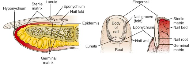
Nail Anatomy

  •  Nail bed: overlies the cortex of the distal phalanx and lies directly beneath the nail plate
  •  Eponychium: the skin that covers the proximal end
  •  Hyponychium: the skin edge at the distal nail margin
  • Cuticle: an outgrowth of the eponychium that provides a seal between the proximal nail fold and nail plate
  • Germinal matrix: the proximal portion of the nail bed responsible for nail formation and begins 7 to 8 mm under the eponychium; the distal end of the germinal matrix is the lunula


Subungual Hematoma

  • A simple subungual hematoma is not an indication to remove the nail. Trephination is not indicated if the hematoma encompasses only 25%, there is no significant pain, or if injury was over 24 hours ago, as the blood likely clotted and will not flow out.
  • Blood under the cuticle proximal to the nail is a clue that there is a deeper injury and usually the nail should be removed if there is significant pain.
  • There is controversy regarding treatment of subungual hematomas and whether simple trephination is enough or whether inspection of the nail bed for injury is required.
  • It was suggested that for subungual hematomas involving more than 50% of the nail bed, the nail should be removed given the risk of nail bed laceration.
    • This was based on an initial study in 1987 that found that 16/27 patients with hematomas >50% had associated nail bed lacerations that required repair
    • This study did not follow up with patients and did not have a control group, so long term outcomes are unknown.
    • However, subsequent studies have shown that if there is no other significant finger tip injury, treatment by trephinating alone provides a similar good cosmetic and functional result.
  •  If you don’t have a trephinator, what else can you use?
    •  Heated paper clip
    • 23-gauge 1-inch needle: Hold the needle over the hematoma, avoiding the lunula, twist and rotate the needle back and forth like a drill; no pressure needed.
    •  Number 11 scalpel (slower, more painful, larger hole and better drainage)
    •  Insulin syringe needle (29-gauge)
  •  What if there is a fracture underneath?
    • Though there is a risk of turning the fracture into an open fracture, consider still performing the procedure if the injury is painful.
    •  You can consider antibiotics if trephination is pursued, though there is no data.
    •  It is always important to obtain an x-ray with any traumatic injury.

Nail Bed Repair

Suture the nail bed if a large subungual hematoma is associated with an unstable or avulsed nail. Good outcome depends on maintaining the space under the cuticle where the new nail will grow out from (the germinal matrix). If this area scars down a new nail will not grow. 

Figure 1

Figure 1

  •  If the nail is only partially avulsed or loose, especially at the base, lift the nail slightly to assess the nail bed.
  • If the nail is completely transected, it is best to remove the entire nail to suture the nail bed. In this case, suture the proximal and lateral nail folds first for better approximation prior to repairing the actual nail bed.
  • A sturdy needle (3-0 or 4-0) is needed to suture the nail back in place. Before replacing the nail and suturing it back in place, you can poke a hole through so the needle and suture can pass more easily.
  • A study in 2008 used dermabond for nail bed laceration repair showed similar follow up cosmetic and functional outcomes; using dermabond took about 1/3 of the time. It was a small study with only 40 patients and repair was done by orthopedic residents, but definitely a consideration
    •  The key to success is achieving hemostasis and making sure you have a dry field before dermabond application
  • Another method to secure the nail in place is the figure 8 stitch (Figure 1, 2) proposed by hand surgeons 
 

Figure 2


Protecting the exposed nail bed is essential, which can be done with the nail itself (wash well beforehand with normal saline), with the sterile aluminum foil from the suture pack, or with a piece of vaseline gauze. The nail should be reinserted under the eponychium to protect the open space for nail growth. Consider a hand surgeon consult if the nail bed is extensively lacerated or if part of the nail bed is lost, as the patient my need a matrix graft.


Discharge Instructions:

  • Tell the patient to return for a wound check 3-5 days post repair. Replace any non-adherent material that was inserted into the proximal nail fold. Afterwards, the patient should perform dressing changes every 3-5 days.
  • Sutures that were used to reattach the nail should be removed in 2 weeks.
  • Nails grow at a rate of 0.1 mm/day and it takes approximately 6 months for a new nail to grow.
  • Instruct the patient to avoid any trauma or chemical irritants to the healing nail.

Tips:

Figure 3

  • Always use absorbable suture to repair the nail 
  • Use a large suture and sturdy needle when suturing the nail back in place; consider dermabond as an option
  • Use a finger tourniquet to maintain a bloodless field (Figure 3)
  • Digital blocks are key
  • Clean the nail bed prior to repair; clean the nail very well before replacement
  • If possible use the avulsed nail to protect the exposed nail bed and maintain the space for a new nail to grow
  • Repair the proximal and lateral nail folds first

Expert Commentary

Dr Ahlzadeh has presented a nice review of the approach to nail trauma and some useful techniques.  While there are seldom formal lectures during residency training dedicated to nail trauma, it is something we regularly see and treat in the ED, so it is important that we do this well.  It is well within the scope of practice of Emergency Medicine to be the primary providers for most nail injuries.

The subungual hematoma is probably the most common nail injury encountered in Emergency Medicine.  Traditional dogma directed the provider to remove the nail to repair underlying injuries in the presence of >25-50% hematoma.  This recommendation left many of us scratching our heads. We would go through the elaborate procedure of removing the nail by dissecting the nail away from the underlying nailbed, meticulously repairing a nailbed laceration if required, and then stenting the eponychial fold open, hoping that a normal nail would regrow without deformity despite the trauma from the injury and the procedure itself.  It seemed like we were introducing a lot of trauma to the nail bed and eponychial fold for a theoretical (non evidence based) benefit.  The more we did this, the more many of us would ask ourselves, “What if we just left this alone?”

Today it seems the tide has turned.  It is now well accepted practice to leave a nail in place in the setting of a subungual hematoma as long as the nail is intact and laying flat on the nailbed, regardless of the percentage size of the hematoma.  This makes intuitive sense.  An anatomically intact nail lying flat upon the nailbed should lead to the flat healing of an underlying laceration.  A common practice to ensure that the nail remains flat after discharge is to trephinate nail so that pressure from the underlying hematoma does not elevate the nail off of the nailbed after discharge.

Nail trephination is one of the more rewarding procedures in Emergency Medicine.  Patients present with throbbing pressure from the tense subungual hematoma and typically get immediate improvement once the nail is trephinated.  Many techniques have been described. Dr Ahlzadeh even refers to a “trephinator”.  I am not actually sure what this device even is.  Regardless, this procedure should be quick and easy.  No anesthesia is necessary.  I suggest keeping it simple.  Rather than heating a needle or paper clip, use a cautery device.  In our ED we have a cordless disposable plastic cautery device that is the size of a magic marker.  It has a single button and the tip becomes orange with heat when the button is pressed so you know it is hot and ready to go.  Warn the patient that they will feel some heat and possibly see or smell a bit of smoke but it will be brief and not painful (unless you are too forceful, in which case it will be hot and painful!).  Pick the location in the center of the hematoma.  Repeatedly very lightly touch the hot cautery tip to the nail over the center of the hematoma until you are through the nail.  You will know you are through when a drop of blood comes out of the hole you have trephinated.  At that point you can stop and gently express whatever blood you can.  That’s the whole procedure.  It should take mere seconds. When the patient gets over the initial anxiety of the procedure they typically realize they feel better. 

Nails that are deformed or elevated off of the nail bed are not good candidates for trephination. These nails should be removed for nailbed repair so that the nailbed heals flat.  Don’t even bother unless your digital block is highly effective.  We do not have commercial finger tourniquets in our ED so I like to make my own professional looking tourniquet.  I get a sterile glove is about the size (or a half size smaller) than would fit the patient.  I cut the very tip off the finger of the glove that corresponds with the patient’s injured finger.  Then I put the glove on the patient and roll the cut glove finger proximally and voila, you have both a tourniquet and a clean field.  Now you are ready to get comfy and repair the nail bed according to Dr Ahlzadeh’s techniques!  While a sturdy needle is needed to puncture through a nail, I still find the nail often deforms the needle. So instead of piercing the nail with my needle, I once again use my trusty cautery device to make holes in the nail that my needle will easily pass through without deformity.

Screen Shot 2018-04-08 at 5.24.00 PM.png


Matt Levine, MD

Assistant Professor, Northwestern Emergency Medicine


 How to cite this post

[Peer-Reviewed, Web Publication]  Alzadeh G,  Kenny J  (2018, April 8). Nail Trauma.  [NUEM Blog. Expert Commentary by Levine M ]. Retrieved from http://www.nuemblog.com/blog/nail-trauma. 


References

  • Batrick N. Treatment of uncomplicated subungual hematoma. Emerg Med J 2003;20:65.
  • Bowen WT, Slaven EM. Evidence-based management of acute hand injuries in the emergency department. Emergency Medicine Practice  EB Medicine. 2014;16(12):1-28. http://www.ebmedicine.net/media_library/files/1214%20Hand%20Injuries
  • Guthrie, Kane. “Minor Injuries 001.” Life in the Fastlane. <http://lifeinthefastlane.com/minor-injuries-001/>.
  • Hedges, Jerris, James Robers. “Methods of Wound Closure.” Clinical Procedures in Emergency Medicine, 6th ed. Philadelphia: Elsevier/Saunders, 2014.
  • Roser SE, Gellman H. Comparison of nail bed repair versus nail trephination for subungual hematomas in children. J Hand Surg [Am]1999;24:1166–70.
  • Strauss E, Weil W, Jordan C, Paksima N. A prospective, randomized, controlled trial of 2-octylcyanoacrylate versus suture repair for nail bed injuries. J Hand Surg Am. 2008;33(2):250-253.


Ocular Ultrasound: From Floaters to Fogginess

Screen Shot 2018-01-25 at 10.42.52 AM (3).png

Written by: Steve Chukwulebe, MD (NUEM PGY-3) Edited by: Michael Macias, MD, (NUEM Graduate 2017, US Fellow UC San Diego) Expert review by:  John Bailitz, MD 


The Case

A 60 year old male with a history of hypertension presents to the emergency department with three days of intermittent floaters in his right eye.  Concurrently, he also notes that vision in the right eye has become progressively blurred, first starting at the base of his visual field and now advancing up towards the center of his vision. He denies any trauma to the area as well as any other neurological complaints.


Ocular Exam

Motor: PERRL, EOMI,

Superficial Exam: Clear conjuctiva, left eye cataract

Visual Acuity: OD 20/25, OS 20/40 values corrected for a distance of 10 feet from the chart

Visual Fields: Decreased in the lower quadrants of the R eye to confrontation


The Differential Diagnosis for Floaters

  • Retinal detachment (RD)

  • Posterior vitreous detachment (PVD)

  • Vitreous hemorrhage

  • Intraocular foreign body

  • Posterior uveitis/vitreous inflammation

  • Migraine w/ aura

A bedside ultrasound obtained the following image:

 
eye.gif


The patient was found to have a retinal detachment and was admitted for definitive management under ophthalmology.


 Acute Vision Loss and Floaters

Floaters, often described by patients as lines, circles, dots, cobwebs, and other shapes, are common as part of the degenerative process of the vitreous body.  While in the chronic setting they are thought to be related to condensation of the vitreous collagen fibers, new onset floaters in patients 50 years or older have been related to PVD in 95% of cases. Of patients with vitreous floaters and/or flashes as a consequence of PVD, the incidence of RD is 14% [1]. If the PVD is complicated by vitreous hemorrhage, the incidence of RD rises to 70%.

From a population perspective, the incidence of RD ranges between 6.2-17.9 per 100,000 people with the highest rates occurring in the age group 60-69 [2].  Additionally, patients with history of myopia or uncomplicated cataract surgery have a significantly increased risk of developing RD compared to the general population.  It is also important to note that there is a 3-33% chance of bilateral involvement [3].


Using the Ultrasound for Detection of Retinal Detachment

On ultrasound, RD appears as a hyperechoic rippled (or undulating) line/membrane in the posterior to lateral globe.  A recent paper in Annals of Emergency Medicine reviewed 78 articles and ultimately included three studies (or 201 eyes) in a meta-analysis to evaluate the performance of emergency physicians at identifying RD through ultrasound [4].  Though the 95% confidence intervals for sensitivity and specificity range from 60%-100% in the three studies, each study boasted high accuracy to diagnosing RD.  Furthermore the receiver operating characteristic curve for the three studies had an excellent summary area of 0.97, suggesting that bedside ocular ultrasonography is an accurate tool in an emergency physician’s arsenal when a fundoscopic exam is technically challenging.


Keys to Successful Evaluation for Retinal Detachment with Ultrasound
 

Differentiating between RD and PVD - Keep the gain down:

Since the eye is usually a homogeneous fluid filled structure, it provides a great acoustic window for ultrasonography.  Too much posterior retrobulbar acoustic enhancement decreases the observer’s ability to visualize pathology in the vitreous body.  It can be difficult to see a difference between RD and PVD on ultrasound since both may present as a wavy membrane in the posterior orbit.  In this case, it is important to remember that the retina is a highly reflective surface and should still be seen as a thick or stiff undulating membrane with reduced ultrasonographic gain [5]. Another trick is when asking the patient to move their eye, the PVD membrane appears to be more mobile and dynamic, as if it is moving with the motions of the vitreous, while a RD membrane tends to retain its slow oscillation.


Ultrasound both eyes not just the affected one:

Though RD commonly occurs with the classic story of painless monocular vision loss, described as if a “curtain” or a “shade” is being pulled over their eye, in this case the patient actually presented with better visual acuity in his affected eye.  Remember that there is a 3-33% likelihood that a RD can be developing in the other eye.


Artifacts in the vitreous:

Artifact appears as bright echogenic material in the vitreous body that disappear when the patient is asked to move their eye.  However, if a persistent hyperechoic object is seen in the body, or there is shadowing or reverberations associated with the object, this is concerning for a foreign body or vitreous hemorrhage.


Using Tegaderm bandage over the eye:

This technique provides a few advantages.  One, it allows the ultrasonographer to apply as much gel as needed over the eye without the worry of getting it into the patient’s eye.  It is important to note that any pressure on the eye should be avoided if there is any suspicion for globe rupture or foreign body.  By applying a generous amount of gel over the eye and stabilizing the hand by placing a finger on the forehead or bridge of the nose, it is possible to stabilize the probe without having it make any contact with the eyelid.  This technique also allows for easier clean up after the exam, again focusing on preventing any pressure to the eye.


Conclusion

Bedside ultrasound is a useful tool for rapidly diagnosing RD and getting the patient seen by an ophthalmologist emergently.  However, if there is a high enough clinical suspicion for RD and ultrasound is negative, it is still important for the patient to receive a dilated fundoscopic exam by an ophthalmologist in a timely manner.


Expert Commentary:

Thank you for sharing this outstanding case describing one of the most straightforward and useful clinical ultrasound (CUS) applications. Few ED shifts go by when I do not need to perform an Ocular US exam. As mentioned, CUS is helpful for the evaluation of patients with suspected PVD, RD, Vitreous Hemorrhage, and Intraocular Foreign Body, but also for the assessment of patients with other ocular injuries or increased intracranial pressure. New clinicians may sometimes not perform an ocular US due to the perceived difficulty of preparing for, and safely performing the exam.

Pro-Tips on Preparing and Performing Ocular CUS Quickly and Safely

A physical examination of any sensitive structure such as the eye begins by first simply explaining the exam thoroughly to the patient. The explanation also provides a nice review for junior trainees in the room and even for the clinician sonographer! So take the time to explain the ocular ultrasound just as you would a Tonopen measurement or Slit Lamp exam.

Tegaderms are important for any patient who may not be entirely reliable or with suspected traumatic injury. However, for the experienced clinician sonographer with a reliable non-trauma patient, covering the orbit with a Tegaderm is not always necessary. Getting every artifact producing air bubble out of the way during the Tegaderm application may prove difficult. And patients may not appreciate the eye lash and brow waxing, as well as sticky residue left behind by the Tegaderm. As long as the patient agrees to keep their eyes completely until all gel is removed, the likelihood of gel getting into the eye is low. If cooperation is at all an issue, then I ask the patient their preference. From having the technique performed without a Tegaderm on myself countless times by students, and performing clinically on hundreds of patients, gel contamination is rare, and minor eye irritation even less so.

Next, recline the patient to a 45 degrees or even completely supine position. Then place a rim of gel on the clean linear probe. Ask the patient to close both eyes completely but not forcefully. Perform the exam from the head of the bed, first stabilizing your hand on the patient’s forehead, then gently placing the probe over the closed lid. With any soft tissue or musculoskeletal exam, always start with the normal side first to relearn your anatomy and optimize your settings. In particular, according to the As Low As Reasonably Achievable (ALARA) Principle, utilize the lowest Mechanical Index (MI) possible when performing ocular ultrasound. This minimizes the theoretical risk of damage to the delicate retinae from excess ultrasound energy. If you do not know how to adjust MI, then just select the Ocular preset on your machine. If you have no ocular preset, but a patient who desperately needs the ultrasound, at the very least be sure to minimize the ultrasound exam duration.

As you finish examining each eye, remind the patient to keep both eyes closed until all gel is removed. Have an assistant gently wipe any gel from the eyelid using gauze to lift the gel completely from the lid and lashes. For most CUS applications, white cotton napkins found in just about every ED work the best for gel removal. Paper towels simply smear gel, chucks are too expensive, and clothe towels hard to locate in a busy ED. For the ocular exam stick with gauze to lift all the gel away and give the patient one after you are finished just in case. With brief but adequate preparation and explanation, ocular CUS is a safe and effective technique to rapidly rule in emergent ocular pathology!


Screen Shot 2018-03-05 at 10.12.42 AM.png

John Bailitz, MD

Associate Professor of Emergency Medicine

Program Director, Northwestern Emergency Medicine


FOAMed Resources

1. Just in Time Learning

         a. Jacob Avilla’s 5 Minute Sono; http://5minsono.com/vids/

         b. ACEP Sonoguide https://www.acep.org/sonoguide/smparts_ocular.html

 2. Detailed Learning:

         a. Introduction to Bedside Ultrasound iBook Volume 2 Chapter 16. https://itunes.apple.com/us/book/introduction-to-bedside-ultrasound-volume-2/id647356692?mt=11

          b. Ultrasound of the Week: Ocular Ultrasound https://www.ultrasoundoftheweek.com/tag/ocular/


Posts you may also enjoy


 How to cite this post

[Peer-Reviewed, Web Publication]  Chukwulebe S,  Macias M  (2018, March 19). From Floaters to Fogginess.  [NUEM Blog. Expert Review by Bailitz J ]. Retrieved from http://www.nuemblog.com/blog/ultrasound-in-RD. 


References

  1. Lumi X, Hawlina M, Glavač D et al. Ageing of the vitreous: From acute onset floaters and flashes to retinal detachment. Ageing Research Reviews. 21:71-77. 2015.

  2. Mitry D, Charteris DG, Fleck BW, Campbell H, Singh J. The epidemiology of rhegmatogenous retinal detachment: geographical variation and clinical associations. British Journal of Ophthalmology. 94(6):678-684. 2009.

  3. Gupta OP, Benson WE. The risk of fellow eyes in patients with rhegmatogenous retinal detachment. Current opinion in ophthalmology. 16(3):175-8. 2005.

  4. Vrablik ME, Snead GR, Minnigan HJ, Kirschner JM, Emmett TW, Seupaul RA. The diagnostic accuracy of bedside ocular ultrasonography for the diagnosis of retinal detachment: a systematic review and meta-analysis. Annals of emergency medicine. 65(2):199-203.e1. 2015.

  5. Schott ML, Pierog JE, Williams SR. Pitfalls in the Use of Ocular Ultrasound for Evaluation of Acute Vision Loss. The Journal of Emergency Medicine. 44(6):1136-1139. 2013.


5 Palliative Care Myths DEBUNKED!

Written by: Hashim Zaidi, MD (NUEM PGY-3) Edited by: Alison Marshall, MD, (NUEM Graduate 2017) Expert review by:  Eashwar B. Chandrasekaran, MD MSc


1) “Palliative care is only for people close to death”

Image Credit: “Reconceptualizing palliative care as a continuum of support”Wang, David H., MD. Published March 31, 2017. Volume 69, Issue 4. Pages 437-443.

End-of-life care is only one of the many components of palliative care. Palliative care consists of interdisciplinary efforts to decrease the physical, spiritual, and psychological stress related to an incurable or chronic illness. Sound familiar? We do this daily in the emergency department (ED) for a variety of patients with acute on chronic conditions, many of whom would benefit from early palliative care involvement. Despite multiple studies illustrating the benefit of early palliative intervention, palliative care has traditionally been offered late in one’s disease course, thus creating the misperception that palliative care is synonymous with hospice, or end-of-life care. Specifically, hospice is a benefit and program for patients whom providers believe are prognostically likely to live less than 6 months if the disease is to run its usual course and are philosophically inclined with not pursuing further life-prolonging interventions. Many conditions and disease states could benefit from the model proposed below, with palliative care offered in tandem with life-prolonging care in order to ease symptom burden and improve quality of life. As someone’s chronic illness starts to worsen with concern that their time may be getting shorter, palliative care can then transition to planning for end-of-life, including discussions about enrolling in hospice. Within the landscape of modern disease shifting towards long term management of chronic disease, emergency medicine and palliative care are beginning to share increasing common ground. Many of these patients present to the ED on a regular basis, making the emergency department a unique access point for initiation of early palliative services.

2) “Palliative care discussions are really just about establishing code status.”

            As mentioned previously,  palliative care accomplishes much more that just what happens when a patient has cardio-respiratory compromise. The exact actions performed at the very end of life are not as significant as identifying the values that inform patients and surrogate decision making. It is better to avoid jumping into a goals of care discussion with questions such as “Would your loved one like everything done?” or more gore filled specifics of “Do you really want us beating on their chest or shocking them with electricity to try and keep them alive?”  No one would like to approach the context of their goals for treatment and care framed as a series of end of life checkboxes and procedures. Rather, palliative care providers stress the importance of identifying what’s important to patients and families with the worsening of their disease progression, in order to fill the gap between expectations and reality.

“‘Hello I’m Dr. Emergency please fill out this checklist of invasive procedures that you think sound appealing for your loved one.”

While it is important for us as ED providers to understand how a patients’ wishes relate to medical interventions, we must remember that collaboration and the development of mutual trust are needed to provide excellent care. Trust is achieved through verbal communication style and through non-verbal cues such as sitting with the patient and/or family and giving them time to communicate their wishes without haste or decision-steering conversation. Attempts should be made to identify broad goals that are important to the patient and family. Building on those principles, providers should then propose appropriate goals of treatment based upon expectations.

Screen Shot 2018-01-26 at 10.38.05 AM.png


Once trust and rapport are developed and the goals of care identified, providers should elaborate on a plan as well as timeline just as you do with all ED patients. If your patient's goals are more aligned with comfort and aggressive symptom control at the end of life, future discussions should establish code status as well as discussion of initiating hospice services be it as an inpatient or outside the hospital. . When speaking specifically of code status, a “Do Not Resuscitate” (DNR) specifically comes into play when the patient’s heart stops beating, even though it is often misconstrued as a theme for withholding treatment of any kind at the end of life. Thus, discussions for withholding interventions at the end of life should include discussion of not only code status, but also of discussion issues such as artificial nutrition/hydration and use of antibiotics. If appropriate, most emergency departments have a case-management based approach to initiate a hospice referral. Often, this is done in concert with the admitting team and/or palliative care providers to guarantee a smooth transition and often may require the patient to be admitted for setting up these services. Future directions will look to establish formalized care patterns for the initiation of hospice services in the ED to reduce hospitalizations for individuals at the end of life.

If the patient’s or family’s goals align with ensuring they live as long as possible or continue for hope of curative treatments, then it would be prudent still to offer your perspective of prognosis and document this discussion for future providers that may come in communication with the patient in the future. This certainly does not preclude trying to get palliative care involved with the patient as they can continue to follow the patient even if he is pursuing life-prolonging therapy in anticipation of a change in disease progression in the future.

3) “A patient who is DNR or DNR/DNI should not be admitted to the ICU”

The rough translation of this statement is “if we can’t offer everything in terms of intensive care then the only option is no critical care at all.”

“Wait a minute- if I can’t intubate the patient then how do I get a plateau pressure? Where are we on the CVP monitoring?” - ICU MD

“Wait a minute- if I can’t intubate the patient then how do I get a plateau pressure? Where are we on the CVP monitoring?” - ICU MD

For the emergency physician, identifying the broad goals of care in a criticallyi ill patient helps to delineate what specific interventions should and should not be done in the ED and furthermore during their inpatient stay. Identifying that CPR and intubation aren’t in line with the patient’s goals is critical first step if that intervention could happen in the ED. ICU level of care is not determined by this, but rather based upon the amount of intensive nursing care which will be required for that patient as well as frequency with which medications must be administered. Often times if patients are pursuing life-prolonging therapy, a short ICU stay is necessary to further discussions with inpatient ICU and palliative care providers as they de-escalate use of technology at the end of life and transition to aggressive symptom control. In the end, most ICU doctors have an informal fellowship themselves in palliative care and feel comfortable with admitting these patients knowing that their goals and their disease trajectory may change acutely.  It is important however to be explicit with specific discussions that have already taken place and what decisions may have been made based upon these discussions as to help the accepting service provide better care for these patients.

4) “Having a productive goals of care conversation takes hours; I don’t have that kind of time in the ED”

Meaningful discussions happen between patients and providers every day in the ED on a minute to minute basis. Having a productive and efficient goals of care conversation should be no different. These are critical communication skills that allow us as providers to understand the values of our patients and ensure that they understand their trajectory of care.  ACEP’s Simple Palliative Toolkit includes a framework for a 5-minute goals of care conversation in order to facilitate  meaningful dialogue in a time sensitive manner. Like any procedure, it requires patience, preparation and practice to accomplish well. The initial conversation should focus on defining the current situation and establishing the values of the patient. We’ve found the following question stems useful (shout out to Dr Justin Morgenstern who taught us this and so much more during his EM:RAP segment on Dyspnea in the Palliative Care Patient):

  1. What do you understand about your condition?
  2. How do you normally spend your days?
  3. How much has that changed over the last month?
  4.  What is most important to you (your loved one) right now?

Validate the patient and their loved ones continuously; acknowledge their challenges to date and what they have accomplished to get to that point. Utilizing this value framework will allow you to consider treatment options from the patient’s perspective in the current medical context. Discuss these options and then recommend a course of action based on both your medical knowledge and your understanding of the patient’s values. Finally, remember that emergency medicine is a team sport--get your colleagues from social work and pastoral care involved early, so that they can assist in the established care pathway.

5) “When discussing goals of care for a patient with a dire prognosis, I should avoid partiality in treatment options.”

This myth goes back to the 5 minute goals of care conversation. After establishing wishes and mapping out care goals broadly, it is imperative care providers take the role of adviser rather than host in terms of care. It is key to understand that this does not mean to be irreverent or negligent of a patient or family's wishes. Rather, when the goals of care and treatment have been established and the rapport developed, this is the time you give your professional opinion about the best course of action. If, with the appropriate patient with an incurable disease, you feel the patient would benefit from increased palliative measures and less invasive care you need to communicate that to the patient and family. Connecting their broad reaching goals with actionable palliative interventions such as pain control, dyspnea control, and secretion control will help build the sense that the providers are not “doing nothing” simply. With concerns and care issues validated you can build on issues and also discuss critical invasive interventions that would not help the overall prognosis and lead to prolonged suffering such as intubation, mechanical ventilation, and central line placement. As an ED provider, it is imperative we have discussions about interventions which could directly change the patients trajectory in the hospice such as the procedures detailed above, but more chronic life-prolonging therapy is best guided by the patient’s primary oncologist or in concert with their primary care physician. My recommendation is to make sure you have done due diligence to try to get these individuals in discussion if possible as to have appropriate follow up for future conversations.


Regardless of the patients chronic healthcare conditions, each visit to the ED is an opportunity to assess for palliative care needs

Regardless of the patients chronic healthcare conditions, each visit to the ED is an opportunity to assess for palliative care needs

Talking to the oncologist first often reveals a foundation of previous care goals that can be built upon. Instead of saying, “I’m going to talk to your oncologist about stopping chemotherapy and getting you the palliative care that I think is better for you at this stage,” this would be better communicated by saying, “I know you talked about what care and comfort issues were most important to you as this disease progresses with Dr. X. I talked to them as well. I wanted to revisit this issue and talk more about working to make you actively comfortable moving forward with or without cancer therapies.”


While these “myths” being debunked likely didn’t leave any physicians in shock or awe, it’s important to realize many of the underlying concepts get lost in the chaos that is the emergency department. Remember the ED sets the pace and tone for a lot of care delivered in a hospital system and establishes a patients trajectory through the greater hospital system. By taking a step back and remembering the principles and benefits of early palliative care, a substantial impact can be imparted on patients and their families  that will increase patient satisfaction, and provide better resources that align with their wishes.

Screen Shot 2018-01-26 at 11.14.52 AM.png

Expert Review by Eashwar B Chandrasekaran MD MSc

Assistant Clinical Professor Indiana University SOM Dept of EM

Associate Hospice Medical Direct IUH Ball Memorial Hospital


 How to cite this post

[Peer-Reviewed, Web Publication]  Zaidi H,  Marshall A  (2018, March 12). 5 Palliative Care Myths Debunked.  [NUEM Blog. Expert Review by Chandrasekaran E ]. Retrieved from http://www.nuemblog.com/blog/palliative-care. 


Posted on March 5, 2018 and filed under Palliative Care.

Open Globe Injury

Screen Shot 2018-01-25 at 9.31.41 AM (1).png

Written by: Justine Ko, MD (NUEM PGY-1) Edited by: Hashim Zaidi, MD (NUEM PGY-3)  Expert commentary by:  Rehan Hussain, MD


00001.jpg

Expert Commentary

Hi Drs. Ko & Zaidi,

Thank you for this great review on emergency management of open globe injuries. The presentation of open globe injuries can vary widely depending on the mechanism of injury, and so can the final visual outcome. For obvious ruptured globes with peaked pupils or extruding intraocular contents, I am glad you emphasized that it is best to avoid any manipulation of the eye to prevent further extrusion of contents or increase the risk of infection. Cover the eye with a shield, start topical and systemic antibiotics, and consult ophthalmology so they can arrange surgery in a timely manner. The patient must be kept NPO to avoid delaying surgery unnecessarily. Definitely don’t check the eye pressure if you already know it is a ruptured globe.

For the less obvious ruptured globes, which may sometimes mimic a corneal or conjunctival abrasion, it is imperative to perform a Seidel test carefully. This involves placing a wet fluorescein strip (not the pre-made drop) over the suspected entry site, looking at it under the cobalt blue light of the slit lamp, and checking to see if a stream of aqueous fluid is coming out of the injury site (it can be either quick or a slow stream). Fluorescein staining in absence of a stream of fluid indicates that only an abrasion is present. When in doubt, consult ophthalmology for confirmation. Other slit lamp findings that may be present include subconjunctival hemorrhage, chemosis, corneal abrasion, hyphema, flat anterior chamber, iris defects, foreign body in the anterior chamber, or traumatic cataract.

For imaging workup, CT of the ORBITS is preferable, as it allows for 1 mm sections that increase the likelihood of detecting small intraocular foreign bodies (IOFB) that might be missed with a standard head CT. MRI should be avoided as it is slow, costly, and can cause movement of metallic IOFBs, causing further damage to the eye. Gentle ultrasound over the closed eyelids could be used in absence of availability of CT, but I prefer to avoid it since puts pressure on the eye.

The patient and family members are often times distraught over the injury, and appropriate counseling is an essential part of the encounter. They frequently ask if they will be permanently blind. I avoid making any predictions on the final visual outcome, as it is difficult to predict and can lead to either unmet expectations or unnecessary anxiety. I say that the goal is to save the eye at this time, and we will follow closely to see how the vision turns out. Sometimes patients do very well if the extent of injury is not severe, but on other occasions patients require multiple surgeries to correct associated retinal detachment, vitreous hemorrhage, or lens dislocation. If appropriate antibiotic therapy is not initiated, there is an increased risk of developing endophthalmitis, which portends a poor visual prognosis.

Screen Shot 2018-01-26 at 11.43.21 AM.png

Rehan Hussain, MD

Vitreoretinal Surgery Fellow,  Bascom Palmer Eye Institute, Univeristy of Miami


Posts You May Also Enjoy


How to cite this post

[Peer-Reviewed, Web Publication]  Ko J,  Zaidi H  (2018, Feb 26). Open Globe Injury.  [NUEM Blog. Expert Commentary By Hussain R]. Retrieved from http://www.nuemblog.com/blog/globe-rupture. 


Resources

  1. Alteveer, J and Lahmann B. 2010. “An Evidence-Based Approach To Traumatic Ocular Emergencies.” Emergency Medicine Practice, 12(5): 1-24. 2017.

  2. “Emergency Management of Traumatic Eye Injuries | 2001-07-01 | AHC Media: Continuing Medical Education Publishing.” 2017. Accessed October 6. https://www.ahcmedia.com/articles/71714-emergency-management-of-traumatic-eye-injuries.

  3. Guluma K, Lee JE. Ophthalmology. In: Gausche-Hill M, Hockberger R, Walls R, eds. Rosen’s Emergency Medicine: Concepts and Clinical Practice. 9th ed. Philadelphia, Pa: Elsevier, Inc. 2018:790-819.e3.

  4. Bord, Sharon P., and Judith Linden. 2008. “Trauma to the Globe and Orbit.” Emergency Medicine Clinics of North America, Ophthalmologic Emergencies, 26 (1): 97–123. doi:10.1016/j.emc.2007.11.006.

  5. “Trauma: Open-Globe Injuries.” 2015. American Academy of Ophthalmology. November 4. https://www.aao.org/pediatric-center-detail/open-globe-injuries.

  6. Harlan JB, Pieramici DJ. Evaluation of patients with ocular trauma. Ophthalmol Clin N Am. 2002;15:153-161. (Review article)

  7. Ahmed, Y, A M Schimel, A Pathengay, M H Colyer, and H W Flynn. 2012. “Endophthalmitis Following Open-Globe Injuries.” Eye 26 (2): 212–17. doi:10.1038/eye.2011.313.

  8. Arey, Mark L., V. Vinod Mootha, Anthony R. Whittemore, David P. Chason, and Preston H. Blomquist. 2007. “Computed Tomography in the Diagnosis of Occult Open-Globe Injuries.” Ophthalmology 114 (8): 1448–52. doi:10.1016/j.ophtha.2006.10.051.

Post Partum Hemorrhage in the ED

Screen Shot 2018-01-24 at 6.14.19 PM.png

Written by: Spenser Lang, MD (NUEM PGY-4) Edited by: Michael Macias, MD, (NUEM Graduate 2017)  Expert commentary by:  Annie Dude, MD


Introduction

A 26-year-old female G3P3 arrives via ambulance with heavy vaginal bleeding after having a precipitous home delivery of her third child. EMS reports a “pool of blood,” and en route to your facility she continued to bleed briskly.

Post partum hemorrhage (PPH) is a common and dangerous complication of child birth. According to CDC estimates, hemorrhage is the most common cause of maternal death in both developed and developing countries. About 2 out of every 100 births occur either at home, pre-arrival to the hospital or in the ED. With a trend towards home births and free standing delivery centers increasing dramatically in recent years, emergency physicians need to be able to recognize and treat this life threat.

The official diagnosis of PPH is volume-based, however this information is not easily obtainable in the emergency department.  A more reasonable approach is to treat the PPH patient the same way you would a traumatic hemorrhage. Allow the patient’s vitals and visualized hemorrhaging to guide the aggressiveness of your resuscitation. Remember, a pregnant woman has ~40% extra circulating blood volume and can cope with a higher amount of blood loss than her non-pregnant counterpart.

Following an algorithmic approach as detailed below is essential to management of these patients.


Management

1. Notify an obstetrician.

  • Having the obstetrician on board early will allow for mobilization of definitive treatment such as trans-arterial embolization and/or laparotomy should physical maneuvers, tamponade & uterotonics fail

 2. Resuscitate.

  • As with any resuscitation, begin with the simple ABC algorithm, addressing any issues as they are identified. Patients should be placed on 15 L of oxygen via face mask regardless of their saturation (if hemorrhage is significant, this will dramatically increase their blood oxygen levels via dissolved O2). If a patient is hemodynamically unstable, early administration of blood products should be considered over large volume crystalloid.

3. Obtain adequate access.

  • Two large bore IVs will be necessary if aggressive resuscitation is needed. Consider an intraosseous line early if difficulty obtaining access. Send type and screen, CBC, coagulation panel and fibrinogen. Keep coagulopathy on your differential (Thrombin). This should be done in concert with step 2 described above.

4. Source control

  • By far, the most common cause of PPH is uterine atony (Tone).  Therefore the first action taken should be physical maneuvers to improve tone. A bimanual uterine massage can be useful in stimulating uterine contractions. At the same time, one can evaluate for retained products of conception (Tissue). If tissue is felt, try to sweep out as much as possible while taking care to avoid uterine perforation. Note that this should be done with an empty bladder therefore a foley catheter should be placed prior to attempting massage.


  • If the patient continues to bleed briskly, an effort can be made via balloon tamponade of the uterus, with a foley catheter (or, if available, a Bakri) with ~ 150 ml normal saline injected into the balloon. If the uterus is firm and bleeding continues it is reasonable to assess the genital tract for lacerations of the vaginal wall or cervix (Trauma). Cervical lacerations, ideally, should be repaired by an experienced obstetrician as this can have implications on future fertility. However, a vaginal laceration can be repaired just as a perineal laceration repair, taking care to approximate anatomy with absorbable sutures.

5. Administer uterotonics

  • It is reasonable to begin uterotonic therapy in conjunction with uterine massage in a briskly bleeding patient. Oxytocin is first line and 10 U can be given immediately IM or as an infusion at 10 - 40 mU/minute to achieve and maintain uterine contractions. If hemorrhage is refractory to massage and oxytocin, continue pharmacotherapy for ongoing bleeding with Carboprost (Hemabate - 0.25 mg IM) and Misoprostol (cytotec – 1000 mcg per rectum). 

Summary

Hemodynamically unstable PPH patients should be resuscitated like any other severely hemorrhaging patient. Utilizing a step wise approach as described above will help you maintain control of the situation. Notify an obstetrician. Resuscitate. Obtain adequate access. Source control. Administer uterotonics.

If the patient remains unstable, a myriad of other options exist with your interventional or obstetrical colleagues, including uterine artery embolization, ligation of uterine/internal iliac arteries, or hysterectomy.


Expert Commentary


This case outlines a common presentation of a postpartum hemorrhage in the ED.


A few points:

  1.  While the patient in this scenario had just delivered, postpartum hemorrhage can occur days or even weeks following delivery/hospital discharge. Delayed postpartum hemorrhages are often caused by infection or retained products of conception, so if a patient is stable enough to perform a bedside scan, looking for retained products (which will show as echogenic on ultrasound) can be helpful. Ultimately, a patient with retained products is likely going to need to go to the OR for a D &C, so this is another reason to call OB/Gyn early.
  2. In the case of delayed postpartum hemorrhage, realize a patient may have been sent home following delivery with a fairly low hemoglobin, and may not have much reserve even given that pregnant and recently postpartum women have higher circulating blood volumes. She may have also lost a lot of blood prior to presentation, either that day or slowly over the past days/weeks.
  3.  When interpreting DIC labs, remember that fibrinogen levels are higher in pregnant, as compared to nonpregnant, women. Thus, a ‘normal’ fibrinogen level may still represent a significant decrease. Most protocols for blood product resuscitation in the case of a postpartum hemorrhage involve replacement of clotting factors and fibrinogen along with packed red blood cells as DIC is fairly common with large volume blood losses.
  4. There are two goals of performing a bimanual exam: fundal massage and clot evacuation. Uterine atony will not improve if there is a large volume of clot in the uterus, so be aggressive about clearing these clots out.
  5. Uterine massage can be quite painful; if possible give either IV or IM narcotic (morphine, fentanyl, or dilaudid) prior to starting.
  6. Cervical lacerations and complex perineal lacerations often need to be repaired in the OR, either for better visualization or better pain control, often with either a spinal or epidural to help keep the patient comfortable and still. Packing the vagina with kerlix gauze while waiting for OB/Gyn can be one strategy to reduce bleeding.
  7.  A good way to determine ongoing blood loss is to have someone weigh the chux with the blood on it (1 gram = 1 ml).

One other note: the box with risk factors for postpartum hemorrhage also include parity (the more
babies a woman has had, the higher the risk
), a macrosomic fetus, and polyhydramnios.

Annie Dude, MD PhD

Maternal-Fetal Medicine Fellow, Northwestern Obstetrics & Gynecology


Posts You May Also Enjoy


How to cite this post

[Peer-Reviewed, Web Publication]  Lang S,  Macias M  (2018, Feb 12). Post Partum Hemorrhage in the ED.  [NUEM Blog. Expert Commentary By Dude A]. Retrieved from http://www.nuemblog.com/blog/post-partum-hemorrhage. 


References

  1. MacDorman, M. F., Mathews, T. J., & Declercq, E. (n.d.). Trends in out-of-hospital births in the United States, 1990-2012.
  2. MacDorman, M. F., Mathews, T. J., & Declercq, E. R. (2012). Home births in the United States, 1990-2009. Hyattsville, MD: U.S. Dept. of Health and Human Services, Centers for Disease Control and Prevention, National Center for Health Statistics
  3.  Marx, J. A., Hockberger, R. S., Walls, R. M., & Adams, J. (2002). Rosen's emergency medicine: Concepts and clinical practice. St. Louis: Mosby.
  4.  Prevention and management of postpartum haemorrhage - RCOG. (n.d.). Retrieved July 23, 2016, from https://www.rcog.org.uk/globalassets/documents/guidelines/gt52postpartumhaemorrhage0411.pdf
  5. Anderson, J. M., MD, & Etches, D., MD. (n.d.). Prevention and Management of Postpartum Hemorrhage. Retrieved July 27, 2016, from http://www.aafp.org/afp/2007/0315/p875.html

Journal Club: Do Emergency Physician Opioid Prescribing Practices Impact Long-Term Opioid Use?

Screen Shot 2018-01-25 at 10.11.06 AM.png

Written by: Jon Andereck, MD (NUEM PGY-3) Edited by: Rachel Haney, MD, (NUEM Graduate 2017) Expert
commentary by:  Seth Trueger, MD


Introduction

Figure 1. Opioid Pain Reliever Sales, Related Treatment Admissions, and Related Deaths from 1999-2010 (CDC).

Over the past two decades, rates of opioid prescribing in the United States have skyrocketed, with the total amount of opioids distributed quadrupling from  1999 to 2010 (see Fig. 1). [1]  Rates of opioid related unintentional overdoses and deaths have risen in concurrent fashion, from 4,030 in 1999 to 14,800 in 2008. Public health experts, policy makers, and physicians have slowly come around to acknowledging the epidemic of opioid abuse now facing the country.

Much like in other care settings, there has been an increasing push to reduce the volume of opioids prescribed from the ED. The exact role of Emergency Department (ED) opioid prescriptions in this epidemic has been difficult to quantify. Among people aged 10-29, EDs represented 12% of opioid prescriptions and ranked as the third most common setting for which opioids were prescribed. [2]

Indeed, nearly 4 in 5 heroin users reported prior exposure to non-medical prescription pain relievers, and prior exposure to narcotic pain medications carried a 19-fold increased risk of future heroin use. [3] A study from 2014 estimated that as many as 13.8% of patients discharged from ED's across the country in 2010 were written a prescription for opioid pain medications, up from 11% in 2005. [2] Still, a large amount of uncertainty persists about the true impact of prescribing habits of Emergency Physicians (EP's) on the incidence of narcotic abuse. This study helps shed some light on how EP prescribing practices impact long-term narcotic use.


Study

Barnett ML, Olenski AR, Jena AB. Opioid-Prescribing Patterns of Emergency Physicians and Risk of Long-Term Use. N Engl J Med. Feb 16 2017. 376(7): 663-673.

Study Design

Retrospective analysis.

Population

Medicare Beneficiaries who visited any ED in the US from Jan 1, 2008 – Dec 31, 2011 who had not received an opioid prescription in the preceding 6 months and who were not admitted to the hospital on the index ED visit studied. Patients with cancer or on hospice were also excluded.

Measurement Protocol

Using Medicare Part D data, the authors calculated the morphine equivalents dispensed both in the 7 days following the index ED visit and any further opioid prescriptions over the following 12 months.

Treating EPs were categorized as either high-intensity or low-intensity opioid prescribers based on comparison with their peers at the same hospital. The authors calculated the percentage of patients that filled an opioid prescription after seeing any provider in a given hospital, and then divided providers into quartiles of rates of opioid prescribing within their own hospital. Physicians in the top quartile were designated high-intensity prescribers; those in the bottom quartile were designated low-intensity prescribers.

Outcome Measures

The primary outcome of interest was rate of long-term opioid use among patients in the 12 months following a visit in which they were seen by either a low-intensity or high-intensity opioid prescriber. Long-term use was defined as at least 180 days of opioids supplied in the 12 months after the initial ED visit, excluding the first 30 days following the ED visit.

Secondary measures included rate of hospital encounters possibly related to the adverse effects of opioids in the 12 months following the index ED visit. The authors also measured repeat ED visits at 14 and 30 days for the same primary diagnosis to assess for possible undertreated pain.

Results

Long-term opioid use was significantly higher among those treated by high-intensity prescribers, with an odds ratio of 1.3 (p<0.001) and an absolute rate of 1.51% as compared to 1.16% in the low-intensity group. The authors calculate a number needed to harm of 48 patients receiving an opioid prescription to lead to one excess long-term opioid user.

Long-term opioid use increased in a stepwise fashion for patients treated by physicians in each quartile of prescribing intensity (Fig 1).

A total of 377,629 patients were included in this retrospective analysis; 215,678 were seen by low-intensity EPs and 161,951 were seen by high-intensity EPs. Characteristics of each patient population were similar, though several of these were significant given the large sample size (see Table 1).

Over three times as many patients seen by a high-intensity prescriber were discharged with an opioid prescription than those seen by a low-intensity prescriber (24.1% vs. 7.3%), though there was no difference between the two groups in the median dose of morphine equivalents per prescription.

Figure 1

Table 1

In the secondary analysis, the authors found a small but significant increase in rates of opioid-related encounters (OR 1.03, p = 0.02) as well as ED visits for fall or fracture (OR 1.07, p < 0.001) for patients treated by high-intensity prescribers. In contrast, there was no difference in rates of hospital encounters for non-opioid related complaints. Additionally, rates of short-term ED visits for the same chief complaint were no different at 14 or 30 days for patients treated by either low- or high-intensity prescribers (See Table 3).


Table 3


Interpretation

This study does an impressive job of looking at an important but poorly understood issue in the field of emergency medicine – how do prescribing practices of physicians affect long-term opioid abuse their patients?

This retrospective study design is limited in that it was not randomized-controlled, but it was the most logical design to answer the question at hand. The number of patients included was certainly adequate to detect a meaningful difference. The study was limited by the fact that only Medicare beneficiaries were studied, in part because this was the most accessible database for such a large retrospective study. However, it therefore excludes many in the 19-39 age range in which long-term abuse potential is highest. Designing a randomized-controlled trial to attempt to answer this question would be difficult as it would require standardization of discharge prescriptions; few physicians would be amenable to ceding their right to determine the analgesic plan for their own patients.

The primary outcome of interest – long-term opioid use among patients seen by either type of provider – demonstrated that there is a correlation between high-intensity prescribers and long-term opioid use among patients they see. The authors calculate a number needed to harm (NNH) of 48 among patients prescribed opioids on discharge. This means that for every 48 patients given a discharge prescription for an opioid analgesic by a high-intensity prescriber, one will go on to use opioids long-term (as defined by this study) that could have been avoided if the patient had been seen by a low-intensity prescriber.

One of the most interesting results from this study is not even the question the authors set out to answer, but is the difference in opioid prescribing rates between high-intensity and low-intensity prescribers. This difference was over three-fold (7.3% to 24.1%) and represents an extraordinarily wide practice variability that underscores the lack of standard practice for opioid use. However, this variation was only in the number of prescriptions written and not for the amount of morphine equivalents per prescription as demonstrated in Fig. S3 above.

The counter-measure of pain control adequacy is an important one. The authors attempt to address whether patients treated by low-intensity providers had inadequate analgesia at home. While this question was not directly answered by the study, a surrogate measure of return visits to the ED with the same chief complaint demonstrated no significant difference between the two groups, which suggests but does not prove that there was no meaningful difference in analgesia between the groups.

Is this article practice changing? Perhaps. It does provide evidence that there is a correlation, small though it may be, between prescribing practices of EPs and long-term opioid use of patients. There are also small but significant differences in complications from the opioids given by high-intensity prescribers. The study further highlights the profound variability among EPs regarding their opioid prescribing practice, which I argue is an area to target for improvement especially without any known deficiency of pain treatment by doing so. We can all strive to only prescribe opioids that are truly necessary to treat acute pain, and this article serves as further motivation that over-prescribing can in fact cause our patients direct harm.


Take Home Points

  •  Variability of opioid prescribing within departments is large
  •  Opioid prescribing patterns do have an impact on long-term opioid use
  •  Fewer opioids do not lead to worse pain control, at least as measured by the return rate to the ED

Expert Commentary

This is a great overview of an impactful article. While ultimately there will always be some variation in opioid prescribing (by chance, some physicians will likely see more patients with more painful conditions), this paper suggests that the prescriber variability is high and its not due to chance. Regardless, my takeaway here is that there does seem to be a dose-response to opioid prescribing in the ED and longer term opioid use.

Both the sheer scale and physicians’ role in the opioid epidemic is startling and a number of factors are at play. Years of focus on oligo-analgesia were likely a mix of genuine concern for undertreating pain but unfortunately also driven by those with specific financial interests. Similarly, the increasing focus on patient satisfaction/experience and even with the link to payments, I suspect physicians are too quick to shift blame to others and we need to prescribe more responsibly. While most emergency department opioid prescriptions are short (75% are for 20 pills or fewer [4]), as Barnett and others have shown, a startling fraction of ED patients receive opioid prescriptions. And to borrow from Lewis Nelson, everyone who is addicted to opioids had to have had a first exposure.

Of course there are no easy answers – plenty of patients we see in the emergency department are in substantial pain, and we do not have a lot of tools. But it is not hopeless. My approach to pain management is similar to patients with URIs and antibiotics: sometimes we cut corners (“they’re just here for a z pack / Norco prescription”) and underestimate how satisfied our patients can be being taken care of by a physician who cares and explains things. Some of the things I focus on:

  1.  Acknowledge the patient’s pain: just because I’m not writing for a ton of opioids doesn’t mean I don’t believe they’re not in pain
  2.  Set realistic goals: I don’t have a silver bullet to make pain go away. My goal is to make their pain manageable, not gone.
  3.  Emphasize our priorities: Our main goal in the ED is to make sure there isn’t anything dangerous causing the patient’s symptoms.
  4.  The door isn’t shut when I discharge the patient: “The good news is you don’t need an MRI now but you do need to follow up with your primary doctor over time who will keep an eye on your symptoms and help determine if you do eventually need more testing or to see a specialist.”
  5. Information, information, information: what concerning symptoms to look for at home, when to call your doctor, when to come back to the ED.
  6.  What to do for symptoms: What works well for this? I find a lot of patients are very happy to hear me thoughtfully say “what works really well for this is prescription-strength ibuprofen.” I’ve also had a lot of success with lidocaine patches. Anecdotally they seem to work well, but more importantly, I think it demonstrates to the patient that we’re paying attention and being thoughtful (especially as I need to explain that sometimes insurance doesn’t cover them well but there are over the counter versions so here is how to approach that…)

Incidentally I rarely prescribe or co-prescribe benzodiazepines as we have some data they aren’t very helpful (e.g. Friedman [5]) and that we vastly underestimate their harms, particularly when patients take both opioids and benzos (e.g. Sun [6]).

We have a tight needle to thread between oligoanalgesia and the opioid epidemic, but right now I think it’s clear that the pendulum has swung too far. I don’t think we can nor should stop using opioids altogether (yet) but we can be thoughtful and careful as we care for our patients.

Seth Trueger MD MPH

Assistant Professor of Emergency Medicine, NUEM


Posts You May Also Enjoy


How to cite this post

[Peer-Reviewed, Web Publication]  Andereck J,  Haney R  (2018, Jan 29). Journal Club:  Do Emergency Physician Opioid Prescribing Practices Impact Long-Term Opioid Use? [NUEM Blog. Expert Commentary By Trueger S]. Retrieved from http://www.nuemblog.com/blog/opioids. 


Resources

  1.  “Vital Signs: Overdoses of Prescription Opioid Pain Relievers --- United States, 1999-2008.” Morbidity and Mortality Weekly Report. Centers for Disease Control and Prevention. Nov 4, 2011. 60(43): 1487-1492. < https://www.cdc.gov/mmwr/preview/mmwrhtml/mm6043a4.htm#fig2>. 
  2.  Cantrill SV, Brown MD et al. Clinical Policy: Critical Issues in the Prescribing of Opioids for Adult Patients in the Emergency Department. From the American College of Emergency Physicians Opioid Guideline Writing Panel. Ann Emerg Med. 2012. 60:499-525.
  3.  Muhuri PK, Gfroerer JC, and Davies MC. Associations of Nonmedical Pain Reliever Use and Initiation of Heroin Use in the United States. Center For Behavioral Health Statistics and Quality. CBHSQ Data Review. August 2013.
  4. Hoppe JA, Nelson LS, Perrone J, Weiner SG. Opioid Prescribing in a Cross Section of US Emergency Departments. Ann Emerg Med. 2015 Sep;66(3):253-259.
  5. Friedman BW, Irizarry E, Solorzano C, Khankel N, Zapata J, Zias E, Gallagher EJ. Diazepam Is No Better Than Placebo When Added to Naproxen for Acute Low Back Pain. Ann Emerg Med. 2017 Aug;70(2):169-176.
  6. Sun EC, Dixit A, Humphreys K, Darnall BD, Baker LC, Mackey S. Association between concurrent use of prescription opioids and benzodiazepines and overdose: retrospective analysis. BMJ. 2017 Mar 14;356:j760. doi: 10.1136/bmj.j760.


Vertigo: A hint on the HiNTs exam

Screen Shot 2018-01-25 at 11.37.47 AM.png

Written by: William LaPlant, MD (NUEM PGY-2) Edited by: Mitali Parmar, MD, (NUEM PGY-4) Expert
review by:  Phillip Chang, MD


What is the HiNTs Exam?

The HiNTs Exam is a screening tool for distinguishing a central cause of vertigo from an acute peripheral vestibulopathy (APV), such as vestibular neuritis. The reason for stratifying is obvious (early intervention for central processes, prevention of adverse outcomes), but the degree of difficulty in correctly stratifying a patient is not.  


Why is this important?

A normal neurological exam cannot accurately exclude a central process; 10% of patients with a cerebellar infarct, usually in the medial branch of the PICA, will have isolated vertigo without other associated deficit. [1] In the HiNTs exam derivation paper, only 51% of those with a central process causing vertigo had a neurological sign on exam.  [2] While risk factors, including age, can be helpful in risk stratification, there is again no definitive cutoff that can eliminate the risk of a central process.


How to perform the HiNTs exam:

Notably, it can only be used if the patient is currently symptomatic, and is meant for continuous, not intermittent, symptoms. This means in conditions such as benign paroxysmal positional vertigo (BPPV) where symptoms are not continuous, HiNTs exam should NOT be performed.

It is comprised of three components: head impulse, nystagmus and skew

Head Impulse:

How to correctly perform the head impulse test with demonstrations of a negative (A) and positive (B) finding [1].

Ask the patient to relax his/her head and maintain his/her gaze on your nose. Gently move the patient’s head to one side, then rapidly move it back to the neutral position. The patient may have a small corrective saccade. The head impulse test is positive (consistent with peripheral vertigo) if there is a significant lag with corrective saccades. If you can see the correction, it is abnormal. Compare this to the contralateral side; a difference in the speed of correction should be noted. Video for Good Technique


The horizontal head impulse test is consistent with peripheral vertigo if it is positive in one direction only. If there is a lag in corrective saccades in both directions, it may be concerning for central vertigo. This test can also be performed in the vertical plane.  A lag in corrective saccades in the vertical plane is always suspicious for a central etiology for vertigo.


Pitfalls: The patient must be awake and cooperative. This is essentially an awake “doll’s eye” that requires conscious fixation on an object. Patients who are mentally impaired, unable to fixate, or sedated cannot do this maneuver. Likewise, anxious patients who are unable to relax their neck are unable to do this procedure adequately. False negatives often result from an inexperienced practitioner being too gentle with the head thrust due to fear of causing neck injury.


Contraindications: Any patient that has head trauma, neck trauma, an unstable spine, or neck pain concerning for arterial dissection. This maneuver may extend the injury. In addition, one should avoid this in patients with known severe carotid stenosis as it may embolize unstable plaque. An acceptable alternative is assessing for ocular dysmetria.


Nystagmus:

Note if nystagmus is present in primary gaze (i.e. looking straight ahead) and/or in lateral gaze. What direction is the fast component? If the nystagmus is worse looking in one direction, with the fast component present in that same direction on contralateral gaze, it is unidirectional and reassuring for peripheral vertigo. If, for example, a patient has right-beating (fast direction to the right) nystagmus with rightward gaze and leftward gaze, this is unidirectional right-beating nystagmus.

Bidirectional nystagmus, I.E fast component to the right with rightward gaze and to the left with leftward gaze, is concerning for a central process, as is vertical nystagmus or pure torsional nystagmus.


Skew: 

Again have the patient maintain his/her gaze on your nose. Alternate covering each of the patient’s eyes. A positive result will be the deviation of one eye while it is being covered, followed by correction after uncovering it.


Interpretation:                    

If the HiNTs exam is entirely consistent with peripheral vertigo (positive head impulse test, unidirectional and horizontal nystagmus, negative test of skew), then, according to the derivation paper, it is 100% sensitive and 96% specific for a peripheral cause of vertigo. [2]  While this sounds appealing, there are some caveats:

  • ALL findings must be present for the HiNTs exam to be invoked. If, for example, the patient has horizontal rightward nystagmus with right gaze and no skew, but has no findings on impulse test, you cannot invoke the HiNTs exam to clear the patient

  • In the derivation study, it was performed by one practitioner who was a neuro-ophthalmologist using specialized equipment to measure skew.

  • The study has not yet been validated by a large external group, let alone a large external group of emergency medicine providers. We do not know the sensitivity of the test in this population. As such, while useful in stratifying patients and documenting low risk patients, it would be prudent to err on the side of caution with moderate risk patients given the limited evidence of the sensitivity and specificity of the HiNTs exam in the hands of emergency medicine providers.


Should l get imaging, and if so, what?

CT scan has low sensitivity (16%) for acute infarction in the posterior fossa, so unless you are only concerned for bleed (CT non con) or dissection (CTA), evaluating the patient with just a CT brain essentially commits the patient for an MR stroke rule out. [1] Notably, even initial MRI (48 hours from symptom onset) is falsely negative in 12%, so a patient with a concerning story and exam may require repeat imaging. [1]

As such, there are three possible pathways for these patients: no imaging in the lowest risk, CT vs CTA for patients with concern for bleed or dissection, or MRI +/- CT for ischemic stroke rule out.

Bonus Facts

In the HiNTs derivation paper, patients presenting with central vertigo were more likely to have headache or neck pain (38% vs 12%), a neurological sign on exam (51% vs 0%) including truncal instability (the inability to sit upright unassisted [34%]. [2]


Documentation

 Here is an example of one way to document the HiNTs exam. As a default, a negative exam is used. Please feel free to use and adapt it.

Head impulse test: loss of fixation with corrective saccades when head turned to the ***

Nystagmus: unidirectional, horizontal ***-beating nystagmus

Skew deviation: grossly absent


Posts You May Also Enjoy


How to cite this post

[Peer-Reviewed, Web Publication]  LaPlant W, Parmar M  (2018, Jan 15). Vertigo: A hint on the HiNTS exam. [NUEM Blog. Expert Review By Chang P]. Retrieved from http://www.nuemblog.com/blog/hints. 


References

1.        Nelson J a, Viirre E. The clinical differentiation of cerebellar infarction from common vertigo syndromes. West J Emerg Med. 2009;10(4):273-277.

2.        Kattah JC, Talkad A V., Wang DZ, Hsieh YH, Newman-Toker DE. HINTS to diagnose stroke in the acute vestibular syndrome: Three-step bedside oculomotor examination more sensitive than early MRI diffusion-weighted imaging. Stroke. 2009;40(11):3504-3510. doi:10.1161/STROKEAHA.109.551234.


Clinical Concept: Managing first trimester vaginal bleeding in the ED

Screen Shot 2017-12-18 at 7.38.39 AM.png

Written by: Priyanka Sista, MD (NUEM PGY-2) Edited by: Spencer Lang, MD, (NUEM PGY-4) Expert
Commentary by
:  Natasha Wheaton, MD


Introduction

Vaginal bleeding in a pregnant patient occurs frequently (20-40% of all 1st-trimester pregnancies) and these patients present to the Emergency Department often. Patients in their 1st trimester (prior to 12 weeks gestational age) may not have initiated prenatal care and may not have a definitive intrauterine pregnancy (IUP). In fact, they may not be aware that they are pregnant at all. In patients with 1st trimester bleeding, consideration of ectopic pregnancy should be a priority for the emergency physician. Ectopic pregnancies, while rare, are responsible for the greatest morbidity and mortality in early pregnancy. When ectopic pregnancies are diagnosed - or even strongly suspected - management should involve consultation with an OB/GYN, either for emergent surgical management or medical management. The vast majority of 1st trimester bleeding, however, falls into a spectrum of “it’s too early to tell.” The differential of early bleeding with an undetermined pregnancy location ranges from completed abortions to benign subchorionic hemorrhages, with multiple options in between. This degree of uncertainty is often emotionally difficult to absorb for both patients and clinicians. Having a thorough management approach to 1st trimester bleeding, clearly communicating the uncertainty in prognosis, and providing accurate anticipatory guidance can better protect patients, ensure appropriate follow-up, and may even offer reassurance.  


Diagnostic algorithm for vaginal bleeding in early pregnancy (< 12 weeks gestation age)

1.     Is the patient hemodynamically stable?

If not, you should begin resuscitation and consult gynecology early for strong suspicion of ectopic pregnancy. Ensure the patient has 2 large-bore IVs, give crystalloids, check a hemoglobin, type and cross-match blood, and perform a FAST exam to look for free fluid in the abdomen. If the patient is actively hemorrhaging and you cannot wait for a type and cross, transfuse O-negative blood. Administer RhoGam (see below). If you suspect the patient is septic from a septic abortion, give antibiotics (see below).  If you suspect the patient is headed to the OR emergently, order the appropriate pre-op labs.

Before continuing with the rest of the diagnostic algorithm, all pregnant vaginal bleeders will typically receive the following, unless there is rationale not to do so: urine pregnancy test, serum beta-hCG, type and screen, a CBC, and a transvaginal ultrasound.

2.     Confirm an IUP with transvaginal ultrasound (TVUS).

For all intents and purposes, confirmation of an intrauterine pregnancy effectively rules out ectopic pregnancy (with rare exceptions, see heterotopic pregnancy, below).

How do you confirm an IUP? 

With ultrasound:

  • Visualize a gestational sac (dark anechoic area) within the endometrial cavity. Typically visualized around 4-5 weeks gestational age.

  • Visualize a yolk sac (concentric circle, around 5.5 weeks) or fetal pole (6.5 weeks) within the gestational sac.

Measuring the Yolk Sac, a concentric circle within the gestational sac

Ectopic pregnancies secrete hormones that can cause changes in the endometrial lining, which may look like a gestational sac, called a “pseudogestational sac of ectopic pregnancy.” Therefore, an IUP is confirmed when the yolk sac or fetal pole is visualized within the gestational sac.


A and B: Endovaginal views showing an intrauterine pregnancy. The gestational sac (blue) is located within the endometrial stripe of the uterus (yellow) and contains a fetal pole (purple).

3.     What if the TVUS is indeterminate and you cannot confirm an IUP?

If you cannot confirm an IUP by TVUS, then you cannot effectively rule out an ectopic pregnancy – this is deemed a pregnancy of unknown location (PUL). This means the patient may have an early presentation of ectopic pregnancy, may be too early in their pregnancy to visualize an IUP, or may have a nonviable IUP.

With an indeterminate TVUS, consider the serum beta-hCG level. The discriminatory zone is the level of beta-hCG above which one should be able to identify an IUP by ultrasound (for TVUS, the discriminatory zone 1000-1500 mIU/mL; for transabdominal ultrasound, it is 4000-6500 mIU/mL).

  • When patients have beta-hCG levels above the discriminatory zone, presume an ectopic pregnancy until proven otherwise and consult OB/GYN.

  • When patients have beta-hCG levels below the discriminatory zone and an indeterminate TVUS, you still cannot rule out ectopic pregnancy. Patients have presented with ruptured ectopic pregnancies with beta-hCG levels well below the discriminatory zone. If clinical suspicion is high or you identify free fluid in the pelvis, presume ectopic pregnancy and consult gynecology, regardless of beta-hCG level. If there is no free fluid identified and the patient remains stable and well-appearing, they will need close follow-up and monitoring in 48 hours to re-check serum-hCG levels. In normal viable pregnancies, the serum hCG is expected to roughly double every 48 hours in the 1st trimester. In ectopic or nonviable pregnancies, the serum hCG may seem normal, rise less quickly than expected, or in the case of spontaneous abortion, may plateau or fall. It is the ED physician’s responsibility to ensure this patient has good follow-up in 48 hours for serum beta-hCG recheck, as well as to give strict return precautions for signs of a ruptured ectopic pregnancy.

(A word on heterotopic pregnancy: Heterotropic pregnancy refers to pregnancies at two different implantation sites, most typically a combination of an IUP and an ectopic pregnancy. While it was and is quite rare,  (1 per p 30,000 pregnancies), the incidence is increasing with the use of assisted reproduction techniques (ART). An estimated 1.5 per 1000 ART pregnancies are heterotopic. Therefore, patients who used  ART should get a formal ultrasound in the ED, and all women should have close follow-up. )

4.     Is the cervix open or closed? Is the pregnancy viable or nonviable?

When an IUP is confirmed, a patient with 1st trimester vaginal bleeding may be presenting with a spontaneous abortion. Classification of spontaneous abortion is provided in the table below, and requires a pelvic exam to determine whether the cervix is open or closed.

The majority of patients who present to the ED will be diagnosed with “threatened abortion,” which includes bleeding or cramping, a viable pregnancy, and a closed cervix. Approximately 50% of these patients will proceed to have a miscarriage.

Most spontaneous abortions in the 1st trimester will progress to completion without complications. If they do not progress, the patient may require medical or surgical intervention to remove all POC. Septic abortions may occur if infection complicates the pregnancy loss.

What makes a pregnancy nonviable? The following ultrasound findings:  

  • Crown-rump length of > or = 7 mm and no heartbeat.

  • Mean sac diameter > or = 25 mm and no embryo

  • Absence of embryo w/ heartbeat > or = 2 weeks after a scan that showed a gestational sac without a yolk sac.

  • Absence of embryo with heartbeat > or = 11 days after a scan that showed a gestational sac with a yolk sac


Rh immunization

All Rh negative mothers should get 50 mcgs of RhoGam in the 1st trimester when they are less than 12 weeks gestation, based on expert clinical opinion.  This will be good for any subsequent bleeding that occurs in the following 12 weeks. At some institutions, patients get the full dose RhoGam (300 mcgs) regardless of gestational age, but this is only recommended for patients beyond 12 weeks.


Counseling, FAQs, and Anticipatory Guidance

Given that implications of 1st trimester bleeding are often determined with time, it is critically important to establish appropriate follow-up plans for patients. Providing a safe, reassuring presence with clear information and clear instructions in the midst of uncertainty may help ensure appropriate follow-up. Here are some tips to keep in mind:

  • Ask all patients “Is this a desired pregnancy?” Do not make assumptions or judgments about the nuances of a patient’s situation.

  • Patients may mourn and grieve pregnancy loss similar to the loss of a child or family member. This can be true even for undesired pregnancies. We may forget that we need the same level of kindness for patients experiencing a spontaneous abortion as we do for the mother of a patient with fatal GSW injuries.

  • While we use the term “abortion” in a medical context, for most people it is fraught with political and emotional complexity. Please explain the medical term so that a patient does not see the word for the first time as a discharge diagnosis.


Pregnancy of Unknown Location Discharge Instructions:

1.     Patients MUST follow-up in 48 hours to re-check a serum beta-hCG level.

What will happen at that visit?  If there is an inappropriate rise in beta-hCG level, there is concern for ectopic or nonviable pregnancy. When the beta-hCG level is above the discriminatory zone, a repeat TVUS will be obtained to confirm a viable IUP. If there is no IUP determined, there is concern for ectopic or nonviable pregnancy. Gynecology will help in management decisions at that point: they may choose to monitor the hCG levels, or they may give you medications or consider operative management to help facilitate the nonviable pregnancy to its completion.

What will happen if you don’t follow-up? There is risk of the pregnancy growing in a dangerous location outside the uterus, rupturing, and causing life-threatening bleeding into the abdomen. There is also the risk of missed, incomplete, or septic abortions, which can be life-threatening if allowed to progress. Intervention is sometimes needed to help the body terminate an ectopic or nonviable pregnancy to avoid the possibility of life-threatening hemorrhage and/or infection. 

What does a nonviable pregnancy mean? It means an abnormal pregnancy that will not be carried to completion due to some factors that make it incompatible with progression. (See above for diagnostic definitions)


2.     Use Tylenol for abdominal cramping if needed. Stay well hydrated. Have 2 weeks of pelvic rest (nothing in the vagina for two weeks). There is no evidence to prove that pelvic rest improves outcomes or reduces the likelihood of spontaneous abortion. Theoretically, sexual activity may trigger ectopic rupture but there is no evidence to suggest this. However, pelvic rest is widely recommended to eliminate any possible feelings of guilt that patients may have regarding being responsible for a spontaneous abortion. 

Patients with known open cervical os may continue to bleed significantly. Those with closed cervical os may develop an open os in the coming hours/days. Warn the patient that they may pass blood clots or products of conception. If a patient is concerned they passed products of conception, ask them to retrieve it and bring to their doctor’s or to the ED. Given them gloves and a specimen cup. If a patient is bleeding more than 2 pads an hour for more than 2 hours in a row, tell them to return to the ED.


3.     You must immediately return to the emergency department if you have:

  • Increased or persistent abdominal pain

  • Heavy vaginal bleeding (more than 2 pads an hour for 2 consecutive hours)

  • Weakness, dizziness, lightheadedness.


FAQs, and answers to have in your back pocket:

1.     How common is 1st trimester bleeding? 25-33% of pregnancies will have 1st trimester bleeding.

2.     What does this mean regarding the future of this pregnancy? Approximately half of patients with 1st trimester bleeding will have a subsequent spontaneous miscarriage.

3.     Does this mean I’m miscarrying? Unless the patient has an open os or a documented nonviable pregnancy, it is difficult to tell in the ED. It’s okay to say “I don’t yet know,” and “time will tell, which is why follow-up is important.”

4.     What do I need to watch out for?  What activities can I resume or not resume? With any vaginal bleeding in pregnancy, suggest pelvic rest for 2 weeks. Avoid ibuprofen; take Tylenol for pain. Watch out for the ectopic precautions listed above. Otherwise, if you feel up for doing something, it is OK to do it.

5.     How much bleeding is too much? More than 2 pads an hour for more than 2 hours is too much bleeding.

6.     Why did this happen? Miscarriages are common, occurring in 25-33% of pregnancies. More than half the time, miscarriages occur due to an intrinsic genetic or chromosomal abnormality that occurs during fertilization, rather than due to some inheritable trait, or extrinsic factor. For patients who have recurrent miscarriages, there may be structural or extrinsic causes for pregnancy termination, which warrants workup by the gynecologist.

7.     Did I miscarry because I had sex / took Tylenol / went on a run / had a  glass of wine before I realized I was pregnant? Reassure the patients that miscarriage is common and that there’s nothing they did to cause this. Miscarriage happens in up to one-third of all pregnancies.  

8.     Who gets genetic testing and has tissue pathology testing? For a first-time miscarriage, no genetic testing or tissue pathology is warranted. For subsequent miscarriages, products of conception can be sent to tissue pathology, and extrinsic causes of spontaneous abortion can be evaluated by a gynecologist.

9.     What does a miscarriage mean for future pregnancies? Most patients will subsequently have normal future pregnancies., but it is true that having a miscarriage increases the chance of subsequent miscarriages slightly.

10.  When can I start trying to get pregnant again? Patients can ovulate and become pregnant as soon as 2 weeks after miscarriage. Most gynecologists recommend waiting one menstrual cycle so that it is easier to calculate the due date of the next pregnancy, but there is no medical reason to wait to begin trying again. If patients do not want to get pregnant again right away, be sure to use birth control.

11.  Reassure patients that if they are concerned about anything at all, or there is any change in their health, there should be a low threshold for calling a gynecologist on-call or returning to the ED for re-evaluation.


Expert Commentary

This is a comprehensive and well written entry on a very common complaint seen in the ED. Though the basic principles of early pregnancy related vaginal bleeding are relatively straightforward; confirm an IUP, administer Rhogam if indicated and provide anticipatory guidance, there are several nuances that bear mentioning and will improve patient care as well as patient experience.

As stated in the blog, after determining hemodynamic stability, the next step is usually to ensure the pregnancy is developing in the correct anatomic location (ie “find the IUP!”). However, there are some nuances regarding transvaginal ultrasound in early pregnancy. First, it is worth emphasizing that ectopics can exist at truly ANY hcg level (fine, fine except at zero) so don’t let anyone convince you to skip the ultrasound because “the beta is too low”. In fact, in a retrospective study of 730 women seen in the ED with early pregnancy bleeding, a β-HCG of <1,500 mIU/mL more than doubled their chances of having an ectopic pregnancy (Acad. Emerg. Med. 2003;10:119-26). Another study found that 40% of ruptured ectopics had β-HCG < 1,000 mIU/mL, suggesting that those women with ectopics and lower quants may actually have poorer outcomes.

If you are doing your own transvaginal ultrasound (or even when reviewing those done by radiology), don’t confuse a pseudogestational sac for a true gestational sac. Pseudosacs are seen in up to 20% of ectopic pregnancies (Radiology. 1979;133:451–454) and are irregular, heterogeneous and lack a yolk sac. When you are concerned for a pseudosac, be especially vigilant for secondary signs of ectopic including free fluid in Douglas’ pouch posterior to the uterus or abnormal adnexal masses. Finally, remember that while the vast majority of ectopics are tubal, there are multiple other locations where ectopics can implant some of which are particularly difficult to detect on ultrasound. Interstitial ectopics are particularly difficult as they appear to be in the uterus but are in fact within the myometrial tissue rather than in the uterine cavity. Though the exact measurements remain controversial, most agree that a measurement less than 8 mm of myometrium surrounding the gestational sac is abnormal (https://radiopaedia.org/articles/interstitial-ectopic-pregnancy).

Though there is some data suggesting that pelvic exams in the ED often don’t alter management, I still advocate for a pelvic exam in these patients for several reasons. First, it helps quantify the amount of bleeding. There is some data that those with heavy bleeding are more likely to miscarry than those with lighter bleeding (Hasan et al, Obstet Gynecol. 2005 Nov;106(5 Pt 1):993-9). If there are products present at the cervical os, delivery of those with ring forceps may shorten the patients pain and bleeding. An evaluation of the cervix may rule out other causes of bleeding including STIs or trauma. Remember that domestic violence levels peak during pregnancy so be on the lookout for it, GU trauma can be a sign! Finally, a bimanual exam can help risk stratify the patient and aid in your counseling. If the cervix is dilated, that is a nonviable pregnancy and an “inevitable abortion”. The counseling for that woman is significantly different than counseling for one with mild vaginal spotting and a closed cervical os.  

A few notes on the practicalities of performing this physical exam. First, a pelvic can be particularly uncomfortable when pregnant so please take your time and warn the woman before you start. Be ready to collect tissue so you’re not scrambling if you find tissue unexpectedly. Grab ring forceps, a sterile collection cup as well as Fox swabs and an extra Chux to cover your used instruments. If you collect tissue, it may be worth sending to the lab for chromosomal analysis though this is really only indicated in recurrent pregnancy loss.

A word on ART patients. First, know that their dates are exact so if you don’t see a yolk sac when expected (5.5 weeks) or a heartbeat when expected (just under 6 weeks) it’s probably not a viable pregnancy. Also, be highly suspicious of hetero-ectopics in these patients! Their risk is upwards of 2-5% (Fertil Res Pract. 2015; 1: 15.) so don’t be too comforted by a TVUS showing an IUP. I would advocate for early involvement of their REI physicians to ensure close and appropriate early follow up.

As we wrap up, let’s talk numbers. While there is some data showing a 50% miscarriage rate for all-comers to the ED with vaginal bleeding in early pregnancy (< 12 weeks), this number changes significantly depending on what we find in the ED. With a closed cervix, mild bleeding and a TVUS showing a heartbeat, the risk drops to closer to 10%. The heavier the bleeding, the higher the risk. Finally, a subchorionic hemorrhage on ultrasound does increase the risk of miscarriage to closer to 25% though the risk also depends on the size of the hemorrhage (J Obstet Gynaecol. 2006 Nov;26(8):782-4.)

So, what about follow up? As the blog notes, if we have yet to confirm an IUP, these patients need 48-hour follow up for a repeat β-HCG and consideration of an ultrasound. But what if we did find an IUP? If the woman does go on to miscarry, does she need to see a provider? And how long can she bleed before Ob/Gyn would consider offering her surgical intervention (ie D&C) if desired by the patient? Does she have to have a D&C?

In general, I counsel women with documented IUPs that if they begin to bleed significantly more, pass clots or tissue and go on to have more intense cramping they are likely to be experiencing a miscarriage. They do not necessarily need to return to care as many women prefer to miscarry in the comfort of their own homes, as long as there is not excessive bleeding, severe abdominal pain, fainting or other severe symptoms. I warn her (and her partner if present) that the cramping will be significant and she will pass clots and tissue but the pain should not be persistent and/or severe. Finally, I counsel women that she will likely have bleeding for 1-2 weeks but it should slow after several days. If it does not and she continues to have pain and/or heavy bleeding at the 1 week mark she should follow up with a provider for a repeat ultrasound to ensure she does not have retained products of conception and discussion of possible D&C versus continued expectant management.

Lastly, some comments on the words we use in these cases; it matters! Though this is “bread and butter” emergency medicine for us, this day will likely stick in this patient’s, and her families’, minds for the rest of their lives. Do all those things we were taught in medical school when breaking bad news; Sit down, take your time and make eye contact. Say you’re sorry this is happening and you’re sorry there are often no black or white answers in these cases. Don’t use the word abortion and if those words are in her discharge instructions explain why. At the end of my counseling I always tell the woman that it’s not her fault and that there was nothing she could have done to prevent this or prevent an ultimate miscarriage if it does occur. I say “Pregnancy is a very complicated process and it doesn’t always go right. Usually pregnancies that miscarry this early because the baby wasn’t developing normally and would have never developed into a healthy baby”. Be the doctor you would want for your family member in this sensitive, scary and potentially life-altering time!


Screen Shot 2017-12-18 at 8.45.42 AM.png

Natasha Wheaton, MD

Associate Program Director, UCLA Emergency Medicine


Posts You May Also Enjoy


How to cite this post

[Peer-Reviewed, Web Publication] Sista P,  Lang S (2017, Dec 18). Clinical Concept: Managing first trimester vaginal bleeding in the ED.   [NUEM Blog. Expert Review By Wheaton N]. Retrieved from http://www.nuemblog.com/blog/1st-tri-bleed


When Algorithms Fail: Uncommon, Can't Miss Causes of Altered Mental Status

Screen Shot 2017-11-26 at 8.48.16 PM.png

Written by: Justin Seltzer, MD (NUEM PGY-1) Edited by: Quentin Reuter, MD, (NUEM PGY-4) Expert
Review by
: Alexander Lo, MD


Introduction

Undifferentiated altered mental status (AMS) is among the most common and vexing problems faced by emergency physicians. The differential is incredibly broad and complicated by varied clinical presentations ranging from mild confusion to coma. Given this, patient stabilization and rapid diagnosis of life threatening etiologies (i.e. glycemic emergencies) are the initial priorities.[1-4] A full description of the initial diagnostic evaluation has been well summarized here previously by Trinquero, Alkawham, and Fant.[5] 

Problem

Sometimes the initial evaluation will fail to determine the etiology responsible for the patient’s with AMS. This blog is intended to highlight four “can’t miss,” but relatively uncommon, conditions you should consider when standard labs and imaging leave you without a clear diagnosis.  


1. Methanol and Ethylene Glycol

When should I consider this? 

High-risk populations: chronic alcoholics, homeless, and patients with previous ingestions/suicide attempts. 

Classically, methanol is associated with blurred “snow field” visual disturbance and acute renal failure from calcium oxalate crystals.  

However, varied clinical presentations, from mild inebriation/sedation to hyperpnea, seizure, and coma, make clinical diagnosis difficult. Significant ingestions can present with mild symptoms, as the toxic metabolites take time to form. Ethanol co-ingestion competitively inhibits alcohol dehydrogenase, slowing toxic metabolite formation; as a result, chronic alcoholics may show minimal symptoms and have delayed formation of an anion gap. 

How do I confirm this?  

Figure 1

Toxic alcohol levels are usually not available quickly and therefore have limited clinical utility in the emergency department. 

If possible, identify the product ingested. Common sources are automotive antifreeze and de-icing solutions, windshield wiper fluid, solvents, cleaners, and fuels. 

Classically, methanol and EG present with “double gap,” or both an osmolar gap and anion gap metabolic acidosis. As the toxic alcohols are metabolized, the osmolar gap resolves and an anion gap forms as toxic metabolites are created, as shown in Figure 1.  Depending on the timing of ingestion, either gap may not be present at the time of evaluation.  

Figure 2

Strongly consider toxic alcohol ingestion if a large osmolar gap (>50 mOsm/L) is present. An ethanol level should also be checked and factored into the osmolar gap calculation by adding the ethanol level in mg/dl divided by 3.7 to the equation in Figure 2. Late ingestions may show a predominate anion gap metabolic acidosis with little or no osmolar gap.  

Other EG ingestion clues include hypocalcemia, as EG metabolite oxalic acid binds blood calcium to form calcium oxalate, and an elevated lactate, as the EG metabolite, glycolate, can be mistaken for lactate by laboratory instruments. 


How do I treat this? 

If suspicious for toxic alcohol ingestion, treat empirically with fomepizole. 

Emergent dialysis is indicated for severe symptoms, such as new renal failure, hemodynamic compromise, vision changes, or persistent acidosis. 

EG ingestions require serum calcium monitoring and repletion.[6-8] 


2. Chronic salicylate toxicity

When should I consider this? 

High risk populations: elderly or anyone who requires chronic aspirin or salicylate use, especially those with liver or renal impairment. 

Acute toxicity presents classically with GI upset, vertigo, and tinnitus progressing to pulmonary edema, hyperthermia, alerted mental status, coma, and death. It also has a mixed respiratory alkalosis, due to stimulation of the central respiratory centers, and anion gap metabolic acidosis, from the disruption of aerobic respiration. Chronic toxicity mirrors this toxidrome but is more insidious and difficult to recognize as there is no acute ingestion event. 

Remember, salicylates are present in high concentrations in many other over the counter products besides aspirin, including oil of wintergreen and Pepto-bismol. 

An important pitfall is failure to consider chronic salicylate toxicity in patients with pulmonary edema, respiratory distress, and altered mental status. 

How do I confirm this?  

Diagnosis is based on history, presentation, and presence of mixed anion gap metabolic acidosis and respiratory alkalosis. 

Obtaining a salicylate level for altered patients without etiology is recommended, though levels alone are not diagnostic. Toxic levels from chronic ingestion are usually lower than those for acute toxicity. 

Given these diagnostic challenges, a delay in diagnosis is common and results in higher overall morbidity and mortality as compared to acute toxicity.   

How do I treat this? 

Sodium bicarbonate is used to alkalinize the urine and serum to improve excretion.  

Dialysis is indicated for severe symptoms such as altered mental status, severe acidosis, and pulmonary edema, or in those with renal impairment requiring dialysis.[9-12] 


3. Thrombotic thrombocytopenic purpura (TTP) 

TTP is a platelet aggregation disorder that leads to microangiopathic hemolytic anemia (MAHA).  TTP is due to autoimmune destruction of ADAMTS13, a von Willebrand factor multimer cleaving enzyme, which results in abnormal platelet and clot formation, subsequent MAHA and thrombocytopenia, and CNS and renal dysfunction. 

When should I consider this? 

High risk populations: Often idiopathic. Hereditary TTP is a rare disease without specific high-risk groups. Acquired TTP is associated with HIV, malignancy, SLE, transplantation, and many drugs. 

TTP has a high mortality rate (>90%), which drops to 20% with treatment. 

It presents classically with “pentad” of sudden onset of :

  1. Fever
  2. Altered mental status
  3. Thrombocytopenia
  4. Renal failure
  5. Microangiopathic hemolytic anemia

However, this occurs in only up to 40% of cases. A review of 77 TTP patients showed 80% had neurologic sequelae, with 53% having a major symptom (AMS, TIA/stroke, seizure, coma).[13] However, neurologic findings can fluctuate and be transient. Importantly, neuroimaging is often normal. 

Always consider TTP as the cause of thrombocytopenia (<50k) or anemia in an alerted patient, especially if other concerning signs and symptoms of the classic pentad are present. Consideration should not hinge on renal function; the creatinine can be normal or only slightly elevated.  

How do I confirm this?  

Clinical suspicion should be based on presentation and laboratory findings. .  Don’t forget to send a blood smear to evaluate for schistocytes. 

Confirmation of diagnosis in the emergency department is unlikely as laboratory testing is specialized. 

How do I treat this? 

Emergent consultation with a hematologist is necessary. 

In the emergency department, fresh frozen plasma infusions and steroids can be used as a temporizing measure until definitive management (plasma exchange) can be arranged. 

Avoid platelet transfusion unless there is active, severe bleeding, as this can worsen the condition. [14,15] 


4. Non-convulsive status epilepticus

Status epilepticus (SE) is a seizure longer than five minutes or two or more seizures without intervening full recovery. A small subsection of people have a diagnostically challenging variant known as non-convulsive status epilepticus (NCSE), during which persistent cerebral seizure activity is not manifested as muscular convulsions.  

When should I consider this? 

Consider the diagnosis especially in high risk populations such as: known seizure disorder, alcoholics, known intracranial masses or hemorrhage, CNS infections, critically ill patients, toxin exposures, and certain medications (ex. cefepime). 

NCSE itself does not have any specific risk factors beyond those for seizures generally. However, those who experienced convulsive SE (CSE) are at higher risk of subsequently developing NCSE; one study showed 14% of those with CSE had persistent NCSE after termination of convulsions. [16] Similarly, up to 20% critically ill comatose patients have been shown to have epileptiform activity on EEG. [17] 

NCSE presents primarily with altered mental status. The patient may have positive (automatisms, blinking, echolalia, facial twitching, nystagmus, eye deviation) or negative (coma, confusion, lethargy) symptoms. 

NCSE can also occur after a generalized seizure, appearing as a prolonged postictal period; if level of consciousness doesn’t improve after 10 minutes or if mental status remains abnormal after 30 minutes, NCSE should be seriously considered. 

How do I confirm the diagnosis?  

EEG is the only definitive diagnostic test. 

How do I treat this? 

Early anti-epileptic administration is key; the longer SE lasts, the more refractory it becomes to treatment.  

Benzodiazepines are the first line treatment, supplemented with fosphenytoin, phenytoin, or levetriacetam. Valproate may be useful for NCSE as well. Agents such as propofol, phenobarbital, or ketamine may be necessary if the previous drugs are unsuccessful.[18-21]  


Final Thoughts

There are countless etiologies for altered mental status in ED patients.  Defining what is uncommon depends on patient population characteristics.  For example, isoniazid toxicity is common in prison populations because of the higher prevalence of tuberculosis but much rarer among healthier and un-incarcerated communities.  Delirium is relatively common among older adults, often due to infection, but it is far less common among younger patients from infectious or metabolic causes (in younger patients delirium is more likely from toxic causes).  An open mind and deep fund of knowledge provides a strong clinical foundation, but knowing your patient population is key.  Hunting for the proverbial diagnostic ‘zebras’ is irresistible and exciting, but always remember a few fundamental emergency department approaches: 


The emergent (the first 5 minutes): 

  • Airway (and B-C-D) 
  • VS (don’t forget the temp!) 
  • Arrhythmia (telemetry monitor or EMS rhythm strip) 
  • Glucose 


The urgent (the next 30 minutes) 

  • New and old medications 
  • Tox, tox, tox 
  • Labs (shotgun labs) 
  • CT (remember that CT is quite useless for many posterior circulation etiologies) 

One lesson I’ve learned is that people are creatures of habit, so often the patient has done the same thing before.  Reviewing previous visits or talking to family/friends (if available) may be helpful.   

One final thought: If the AMS turns out to result from a toxic overdose of either home medications or easily available substances/chemicals, look for any diagnosis of depression or prescriptions for anti-depressants. If you find either, it’s reasonable to suspect a self-inflicted overdose and now you may have an overt act of self-harm; so order the sitter, document your concerns and communicate this to the admitting team. 


Posts You May Also Enjoy


How to cite this post

[Peer-Reviewed, Web Publication] Seltzer J,  Reuter Q (2017, Nov 27). When Algorithms Fail: Uncommon, Can't Miss Causes of Altered Mental Status.   [NUEM Blog. Expert Review By Lo A]. Retrieved from http://www.nuemblog.com/blog/AMS


References:  

  1. Huff JS. Altered Mental Status and Coma. Tintinalli’s Emergency Medicine: A Comprehensive Study Guide. 8th (2016), Chapter 168.
  2. Initial Diagnosis and Management of Coma. Stephen J. Traub MD and Eelco F. Wijdicks MD. Emergency Medicine Clinics of North America, 2016-11-01, Volume 34, Issue 4, Pages 777-793
  3. Lei C, Smith C. Depressed Consciousness and Coma. Rosen’s Emergency Medicine: Concepts and Clinical Practice. 9th Ed. (2017), Chapter 13. 
  4. Tindall SC. Level of Consciousness. Clinical Methods: The History, Physical, and Laboratory Examinations. 3rd Ed. (1990), Chapter 57. 
  5. Trinquero P, Alkawham L (2016, August 30). The ED Approach To The Comatose Patient [NUEM Blog. Expert Commentary By Fant A]. Retrieved from http://www.nuemblog.com/blog/the-comatose-patient/. 
  6. Nelson ME. Toxic Alcohols. Rosen’s Emergency Medicine: Concepts and Clinical Practice. 9th Ed. (2017), Chapter 141. 
  7. Cohen JP, Quan D. Alcohols. Tintinalli’s Emergency Medicine: A Comprehensive Study Guide. 8th (2016), Chapter 185. 
  8. Mount Sinai Children’s Environmental Health Center. CEHC FACT SHEET: Ethylene Glycol. Retrieved from https://www.mountsinai.org/static_files/MSMC/Files/Patient%20Care /Children/Childrens%20Environmental%20Health%20Center/ethylene%20glycol.pdf. 
  9. Boyer EW, Weibrecht KW. Salicylate (aspirin) poisoning in adults. UpToDate. Updated January 9th, 2017. Retrieved from https://www.uptodate.com/contents/salicylate-aspirin-poisoning-in-adults. 
  10. O’Malley GF. Emergency Department Management of the Salicylate-Poisoned Patient. Emerg Med Clin North Am. 2007;25(2):333-46. 
  11. Temple AR. Acute and Chronic Effects of Aspirin Toxicity and Their Treatment. Arch Intern Med. 1981;141(3 Spec No):364-9. 
  12. Carstairs S. Salicylates. Published in CALL US: The Official Newsletter of the California Poison Control System (Fall 2009). Retrieved from http://www.calpoison.org/hcp/2009/callusvol7no4.htm. 
  13. Page, E. E., Kremer Hovinga, J. A., Terrell, D. R., Vesely, S. K., & George, J. N. (2017). Thrombotic thrombocytopenic purpura: diagnostic criteria, clinical features, and long-term outcomes from 1995 through 2015. Blood Advances,1(10), 590-600.  
  14. Kappler S, Ronan-Bentle S, Graham A . Thrombotic microangiopathies (TTP, HUS, HELLP). Emerg Med Clin North Am. 2014;32(3):649-71. 
  15. Saha M, McDaniel JK, Zheng XL. Thrombotic thrombocytopenic purpura: pathogenesis, diagnosis and potential novel therapeutics. J Thromb Haemost. 2017;Epub ahead of print. 
  16. DeLorenzo, R. J., Waterhouse, E. J., Towne, A. R., Boggs, J. G., KO, D., DeLorenzo, G. A., Brown, A. and Garnett, L. (1998), Persistent Nonconvulsive Status Epilepticus After the Control of Convulsive Status Epilepticus. Epilepsia, 39: 833–840. 
  17. Gaspard, N., Jirsch, J., Hirsch, L. J.. Nonconvulsant Status Epilepticus. April 4, 2017.  uptodate.com
  18. Kornegay JG. Seizures. Tintinalli’s Emergency Medicine: A Comprehensive Study Guide. 8th (2016), Chapter 171. 
  19. Button KC, Mannix R. Neurologic Disorders. Rosen’s Emergency Medicine: Concepts and Clinical Practice. 9th Ed. (2017), Chapter 69. 
  20. Peets AD, Berthiaume LR, Bagshaw SM, Federico P, Doig CJ, Zygun DA. Prolonged refractory status epilepticus following acute traumatic brain injury: a case report of excellent neurological recovery. Crit Care. 2005;9(6):R725-8. 
  21. Gaspard N, Jirsch J, Hirsch LJ. Nonconvulsive status epilepticus. UpToDate. Updated April 4th, 2017. Retrieved from https://www.uptodate.com/contents/nonconvulsive-status-epilepticus 
Posted on November 27, 2017 .

Serotonin Syndrome

Screen Shot 2017-11-11 at 12.28.34 PM.png

Written by: Jacob Stelter, MD (NUEM PGY-3) Edited by: Elizabeth Byrne, MD,  (NUEM Graduate 2017) Expert Commentary by: Patrick Lank, MD


The Case

A 21-yearold male with history of PTSD and depression presented to the ED complaining of “feeling anxious.” When evaluated, the patient was found to be anxious, continuously pacing around the room. His speech was pressured as he tangentially answered questions.  During the interview he stated that he was discharged a few days ago from a psychiatric hospital after he had intentionally overdosed on clonazepam. He then admited that two hours prior to arrival in your ED he took 30 pills of his prescribed sertraline 100mg tabs to “calm his nerves.” He denied any concurrent substance abuse. Despite the stated overdose, he denied any suicidal intent with his ingestion.


Physical Exam:

Vital Signs: T 98.9F oral   HR 137   BP  152/89   RR 20    Sat 99% on room air

General: Awake, alert, anxious-appearing, pacing around the room, mildly diaphoretic

Head: normocephalic, atraumatic

HEENT: PERRL,  EOMI, anicteric sclera, mydriatic pupils b/l, 8mm

Cardiac: tachycardic,  regular rhythm, no MRG

Resp: clear to auscultation, nonlabored, no crackles appreciated

Abd: Soft, not distended, not-tender to palpation, +bowel sounds

Extremities: well perfused, 2+ radial and DP pulses b/l, mildly diaphoretic

Neuro: AAOx3, 5/5 strength in all 4 extremities, +ankle clonus, hyperreflexia on patellar reflexes.

Psych: Cooperative but anxious with pressured and tangential speech., no HI , SI, hallucinations/delusions


Pertinent Labs:

WBC 12.5, Hgb 15.8, electrolytes within normal limits,  LA of 1.7.

Urine drug screen was positive for amphetamines and benzodiazepines. Acetaminophen, salicylate, and ethanol concentrations were below detection limits.


ECG:

Sinus tachycardia, QTc 457, no abnormal morphology of the QRS complexes


Diagnosis?

Serotonin Syndrome due to SSRI overdose.


Serotonin Syndrome

Overview

Serotonin syndrome is a disorder that is precipitated by excess serotonin. It is classically described as having a combination of hyperautonomic hemodynamic changes, neuromuscular derangements, and a change in the patient’s mental status. [1]  Serotonin syndrome is a medical condition seen only in patients with exposure to serotonergic medications and has been more recently appreciated since the advent of serotonergic drugs used to treat depression and anxiety. However, in addition to pharmaceuticals, botanicals and recreational drugs of abuse can also be serotonergic.  Causative substances increase the amount of circulating serotonin in the bloodstream, which leads to an increased propensity to develop serotonin syndrome [2].  This is not a rare diagnosis; there are approximately 7,300 cases per year results in about 100 deaths [1].

Clinical Presentation

Boyer EW, Shannon M. The serotonin syndrome. N Engl J Med 2005;352:1112-20.

The diagnosis of serotonin syndrome will often manifest around the time of a dose increase, addition of another serotonergic agent or an overdose on a serotonergic agent [3]. Serotonin syndrome will typically manifest within 24 hours of exposure to the serotonergic agent [4]. Symptom presentation  can be separated into three main categories: cognitive, autonomic and neuromuscular. Cognitive symptoms range from restlessness and anxiety to agitation and altered mental status [2].  Autonomic signs and symptoms include tachycardia, mydriasis, hyper- or hypotension, hyperthermia and diaphoresis [2]. Neuromuscular symptoms are also usually observed, ranging from akathisia to muscle rigidity, hyperreflexia and myoclonus [2].  Serotonin syndrome can be a life-threatening condition if not promptly recognized and treated as it can progress to seizures or shock [1]. 

Boyer EW, Shannon M. The serotonin syndrome. N Engl J Med 2005;352:1112-20.

Treatment

The most important initial step to the treatment of serotonin syndrome is to remove exposure to the offending agent. Use of activated charcoal to assist with gastrointestinal decontamination can be considered in the setting of a recent ingestion [1].  Following this, the cornerstone of therapy of serotonin syndrome is symptom based supportive care, with benzodiazepines being one of the most important components of treatment [1, 2]:

  • Obtain an ECG to evaluate the QTc and QRS durations.
  • Begin IV fluid rehydration and resuscitation.
  •  If the patient is hyperthermic, especially greater than 40 ºC, begin active external cooling.
  • Antipyretics are not useful for the treatment of hyperthermia secondary to serotonin syndrome. This is because the hyperthermia is due to increased muscle activity and not secondary to change in the hypothalamic temperature set point.
  • Benzodiazepines improve numerous symptoms of serotonin syndrome, especially agitation and are one of the most important elements of treatment.
  • Use easily titrated medications like nitroprusside or esmolol for severe hypertension.
  • If needed, vasopressors should be used for refractory hypotension.
  • A recent study has suggested the potential utility of dexmedetomidine to treat serotonin syndrome (5).
  • Cyproheptadine, a serotonin antagonist, has long been proposed anecdotally for the treatment of serotonin syndrome.  However, multiple studies suggest that it does not change outcomes and may only help temporarily control symptoms (6).

Unless the clinical features are very mild, these patients will likely need monitoring in an ICU setting for at least 24 hours after being initially managed in the Emergency Department (ED).  As always, especially in the case of an intentional overdose, contacting the local Poison Control Center is a valuable resource in helping to treat these patients.

What about Neuroleptic Malignant Syndrome?

One of the disorders that can present similarly to serotonin syndrome is neuroleptic malignant syndrome (NMS).  Pathophysiologically, NMS is related to an inherited genetic mutation in skeletal muscle and is provoked in the presence of certain neuroleptics [7]. Neuroleptic malignant syndrome often occurs after exposure to drugs that affect the central dopaminergic system, such as haloperidol. There are inherent differences between NMS and serotonin syndrome. Serotonin syndrome tends to present within 24 hours of exposure to the offending agent, whereas NMS will often be more delayed, presenting after 7 days of starting the neuroleptic [4]. Patients with serotonin syndrome will often be agitated and delirious while NMS patients will often have dysphagia, incontinence and increased secretions [4]. NMS will often have extrapyramidal side effects, muscle rigidity and rhabdomyolysis compared to the mydriasis, clonus and hyperreflexia of serotonin syndrome [4].


Return to the Case

As noted above, the patient in this case exhibited all the hallmark signs of serotonin syndrome, including agitation, restlessness, diaphoresis, mydriasis, hyperreflexia and clonus.  In the emergency department, this patient was treated with multiple rounds of lorazepam and boluses of saline which helped to control his symptoms.  This patient was admitted to the medical ICU and monitored for an additional 24 hours.  After his symptoms and vital signs normalized, he was transferred to the floor and received the psychiatric evaluation and treatment that he needed.  His hospital course was uncomplicated and he was transferred to an inpatient psychiatric treatment facility for further care.


Take Home Points

  1. Serotonin syndrome can be a life-threatening condition. Keep a high suspicion for this in patients on serotonergic agents.
  2. The hallmark signs and symptoms of serotonin syndrome are anxiety and restlessness, diaphoresis, mydriasis, clonus, hyperrflexia, tachycardia and hypertension.
  3. Treatment is mainly supportive care and includes intravenous fluids, benzodiazepines titrated to symptom control and blood pressure control as needed.
  4.  Serotonin syndrome is often confused with NMS.  Remember, NMS is in the setting of exposure to neuroleptics and tends to present later on with symptoms of extrapyramidal side effects, muscle rigidity and increased secretions.
  5. Local poison control centers are a great resource in helping manage the treatment of patients with serotonin syndrome and most will require at least 24 hours in an ICU setting after ED diagnosis and management.

Expert Commentary

Dear Dr. Stelter,

Thank you again for yet another exciting tox blog post! This case has a tremendous amount to dissect, but I want to focus on a few things.  For this case, let’s talk about the tox history, the tox physical exam, and the “supportive care” recommendation. Really, this is the definition of bread and butter medical toxicology.

The patient history described above is incredibly common, and that makes sense! Patients with a known history of psychiatric disease have higher risk of having psychiatric emergencies and have access to psychiatric medications. So whether by misunderstanding the purpose of their medication or by intentionally trying to harm oneself, it is common that we as medical toxicologists have to consider the effect a patient’s psychiatric medications have had (or will have) on their current clinical state. So what I want to know out of this patient is what all he could have been exposed to and when. Here are examples of questions I ask:

  •  “When did you take this medication?” (Looking at the clock) “Ok. It’s 3pm. You took it 2 hours ago, so at 1pm you sat down and swallowed 30 pills?” In essence, I want the patient to describe exactly how he was exposed – this can also clue me into his intent.
  • “Did you take anything else?” (Answer is commonly “no” – always dig deeper) “Are you on any other medication? When is the last time you took that medication? When you started to feel sick before you came into the emergency department, did you take anything else to help with your symptoms?”
  •  “This may sound silly to you, but I also care about anything else you are taking. Are you on any vitamins or diet pills or herbals or cleanses? Is there anything you have purchased and taken in the last week which was not food or a prescription medication we have already talked about?”
  • “Do you do any drugs?” I purposely leave the word “illicit” out. I don’t really care about legality. I just want to make them better. “I have seen many patients use all sorts of things that make them feel as sick as you feel now. Is there anything you can think of that you’ve taken which could make you this sick?”

This physical examination gave you your diagnosis! Usually we are clued into these diagnoses based on hyperthermia, which this patient frustratingly did not have. But that “hot tox” differential is something all emergency physicians should be aware of and facile in differentiating between. It includes serotonin syndrome, NMS, sympathomimetic toxicity (including cocaine, amphetamines, MDMA, cathinone derivatives, etc.), anticholinergic toxicity, and severe salicylate toxicity just to name a few.  Although this patient did not have hyperthermia, he did have almost every other finding associated with serotonin syndrome with the most specifically-associated being clonus. Once I see clonus, I take a step back and see if serotonin syndrome fits into the clinical picture.

Finally, I greatly appreciate your description of symptom-focused supportive care in this clinical scenario. As you have displayed, “supportive care” is not super simple. It is nuanced and multifaceted and very frequently life-saving. Before fellowship I was frustrated that there wasn’t necessarily a specific antidote or procedure we could do to reverse toxicity in all poisoned patients. With time, however, I have gained great respect for “supportive care” – regretting when it is not performed adequately and truly appreciating when it is life-saving.

So in this single case you have touched on the key major aspects of the specialty of medical toxicology. I hope some of my points were helpful to you in further appreciating the stellar medical care you provided this sample patient.


Patrick_Lank-04.jpg

Sincerely,

Patrick Lank, MD

Associate Program Director Northwestern Emergency Medicine 

Assistant Professor of Emergency Medicine


Posts You May Also Enjoy


How to cite this post

[Peer-Reviewed, Web Publication] Stelter J,  Byrne E (2017, Nov 13). Serotonin Syndrome [NUEM Blog. Expert Commentary By Lank P]. Retrieved from http://www.nuemblog.com/blog/serotonin-syndrome


References

  1. Boyer EW, Shannon M. The serotonin syndrome. N Engl J Med 2005;352:1112-20.
  2.  LoVecchio F, Mattison E. Atypical and serotonergic antidepressants. In Tintinalli’s emergency medicine: A comprehensive study guide. J Tintinalli (Ed.) (pp. 1219-24). 2016. New York, NY: McGraw-Hill.
  3. Pedavally S, Fugate JE, Rabinstein AA. Serotonin syndrome in the intensive care unit: Clinical presentations and precipitating medications. Neurocrit Care 2014;21:108-13.
  4. Birmes P, Coppin D, Schmitt L, Lauque D. Serotonin syndrome: A brief review. Can Med Assc J 2003;168:1439-42.
  5.  Rushton WF, Charlton NP. Dexmedetomidine in the treatment of serotonin syndrome. Ann Pharmacotherapy 2014;14:1651-4.
  6.  McDaniel WW. Serotonin syndrome: Early management with cyproheptadine. Ann Pharmacother 2001;35:870-3.
  7.  Keck PE, Caroff SN, McElroy SL. Neuroleptic malignant syndrome and malignant hyperthermia: End of a controversy? J Neuropsychiatry Clin NeuroSci 1995;7:135-44.

Posted on November 13, 2017 and filed under Toxicology.

My Brain Hurts: Applying Cognitive Load Theory to Emergency Medicine Handoffs

Screen Shot 2017-10-30 at 10.35.39 AM.png

 Written by: Danielle Miller, MD (NUEM PGY-3) Edited by: Matt Klein, MD,  (NUEM PGY-4) Expert Commentary by: Holly Caretta-Weyer, MD


Introduction:

Have you ever been on a shift in the Emergency Department and thought: “My brain hurts,” or, “If I put one more detail in my brain, my brain may explode?”

Emergency Medicine requires synthesizing large amounts of data in a short amount of time for multiple patients. Think of your last busy post-overnight sign-out, when multiple patient-care-plans needed to be safely communicated from one team to the other. Why are these handoffs so difficult for us? Is there some theory that may illuminate this phenomenon and provide ways to improve?


What is Cognitive Load Theory:

Cognitive load theory (CLT)  was initially developed in the 1980s from research in cognitive psychology that concluded that there are multiple elements that can overload the learner and reduce problem solving (1,4,7).

In cognitive load theory, human memory is divided into three subsystems:

  •  Sensory Memory
  •  Long Term Memory
  •  Working Memory

Sensory memory encompasses brief visual and auditory inputs such as faces/colors/environments, but lasts only 1-2 seconds and generally does not reach consciousness.

Long term memory is a permanent form of memory that utilizes meaningful connections between information elements, and thus can easily be recalled.

Working memory is memory for operating a current task (4). Working memory holds information for a few seconds, with almost all information lost after about 20 seconds, unless the information is rehearsed (1).  Working memory is limited  and can hold no more than five to nine information elements (think of memorizing a phone number, ie. seven plus or minus two)(1).  Working memory is critical to completing a successful handoff, but can easily be overwhelmed.


What Impacts Working Memory:

Working memory is impacted by 3 different types of cognitive load:

  • Intrinsic load
  • Extrinsic load
  • Germane load

Intrinsic load  is the load associated with performing the task itself, such as communicating the names, comorbidities, and plans for the patients at signout.

Extraneous load is the load associated with non-essential aspects of the task, such as what is happening in the environment at signout or what is happening in someone’s personal life that may be affecting the handoff.

Germane load is the load to develop strategies to facilitate learning (3). In general, when the cognitive load associated with a task exceeds the learner’s working memory capacity, performance is impaired (3).


How Does This Apply to Handoffs:

Appreciating the limits of working memory can help identify the challenges of handoffs.

One proposed framework for handoffs is to create a “shared mental model between the giver and receiver” (4). There are multiple factors that prevent the sender and receiver from developing the shared mental model, and this concept is explored through cognitive load theory. These factors can be analyzed by the type of cognitive load that they encompass: intrinsic, extrinsic, germane.  

So What Can We Do to Improve Working Memory and Create Good Handoffs? 

The goal in handoff is to:

  • Manage intrinsic load
  • Reduce extraneous load
  • Optimize germane load (4).

Managing Intrinsic Load in the Emergency Department Handoff:

Intrinsic load  is the load associated with performing the task, or in this case, the load associated with performing the process of a handoff itself.

There are four elements that influence intrinsic load: 

First, the number of information elements influences intrinsic load (4). For example, learning about five patients and their comorbidities during sign-out causes a greater load than two. In the Emergency Department, the number of patients that we must signout is difficult to control, but we could reduce this cognitive load by discharging individuals prior to signout and listing only the critical comorbidities for each patient.

Second, the amount of time available for a task influences intrinsic load (4). For example, the need for rapid handoff and decision making consumes more working memory. Again, this is difficult to control in the Emergency Department, but allowing sufficient time for signout is key.

Thirdly, element interactivity, or how information elements interact with each other, influences intrinsic load (4). The goal, then, during handoff in the Emergency Department would be to attempt to limit uncertainty and give contingencies. For example, one could say, “If the lumbar puncture is unremarkable, I would then consider other causes of mild fever with recent travel such as pulmonary embolism.”  

A fourth determinant that influences intrinsic load is the experience of  the learner (4). As expected, the intrinsic load of a handoff is reduced when learners are more advanced, as the advanced learner has well defined illness scripts that he or she can implement and not bog-down working memory. Again, this cognitive load is difficult to control in the Emergency Department, as we cannot age the learner, but it may be helpful to simply be aware that signing out to someone of different training level requires slightly different information to diminish cognitive load.

Reducing Extraneous Load in the Emergency Department Handoff:

Extraneous load is the load associated with non-essential aspects of the task.  

Extraneous load is influenced by  three components: the physical environment, the personal environment, and how information is presented.

First, the physical environment influences extraneous load during handoffs. It is difficult to control the physical environment and distractions of the ED, but having a culture of limiting interruptions during handoffs may decrease this load.

Secondly, extraneous load is influenced by the personal environment. For example, personal distractions such as home life can increase extraneous load during handoffs. Included in this category is learner fatigue or “burnout,” which is a type of extraneous load that decreases working memory capacity and can affect handoffs (4). Focusing on wellness outside of work may limit this factor.

Thirdly, how information is presented influences extraneous load. For example, when information necessary for the handoff is distributed in space (ie, requiring the sender and receiver to access multiple different databases for information), then extraneous load is increased. Thus, having a cohesive tracking board with all pertinent information in one place, as opposed to having to click through multiple screens for information, would decrease extraneous load during handoffs in the ED. Additionally, combining auditory and visual elements during signout decreases extraneous load.

Optimizing Germane Load in the Emergency Department Handoff:

Germane load  is associated with the load to develop strategies to facilitate learning.  Strategies associated with optimizing germane load include asking clarifying questions and summarizing what has been heard. In the Emergency Department, encouraging clarifying questions during handoff would be a way to improve handoffs. Additionally, having the receiver give a summary to the sender at the end of signout, may also prove useful.


Conclusion:

Cognitive load theory gives a framework for illuminating why handoffs can be challenging in the Emergency Department. We largely use working memory during handoffs, which is a limited and influenced by intrinsic, extrinsic, and germane cognitive loads.  By implementing strategies to manage intrinsic load, reduce extraneous load, and optimize germane load, we can create more successful handoffs.


Commentary

Dr. Miller has done an excellent job of laying out the various factors at play contributing to cognitive load in the Emergency Department and specifically how these factors contribute to making sign-out such a potentially dangerous time.

I want to highlight a few key reasons sign-out conveys such a critical mass of cognitive load and then emphasize some “hacks” to reduce the various contributing factors.

A substantial amount of the critical mass is the product of the nature of the Emergency Department flow, from patient volume to case complexity. I often hear residents say at the beginning of each shift that they want to “work on efficiency.” I would challenge you to take that a step or two further and define the specific type of efficiency in light of considering the various aspects of cognitive load. Do you want to work on your ability to formulate an “if/then” dichotomous decision plan when you walk out of the room to expedite your discharge process? Do you want to further develop your illness scripts by seeing as many patients as possible and discussing where they fit into your bell curve to improve your pattern recognition? Thinking about the various aspects of cognitive load will greatly aid in defining where your efficiency bottlenecks are and how you can improve them, while also improving your knowledge, skills, and personal patient flow for future practice.

Sign-out is the culmination of an entire shift’s worth of cognitive load. It combines both cognitive fatigue from seeing patients and making decisions with physical fatigue from long hours spent working hard and potentially not eating or using the restroom. Cognitive residue from issues outside of work or on cases from previous shifts that you feel could have gone better may also contribute to the cognitive load you experience, and all of this occurs while you try to distill complex patient cases from a crazy shift into concise presentations with understandable dichotomous decision trees for the oncoming team.

I would like to emphasize a few of Dr. Miller’s key points here and add some tips with which I have had or seen significant success.

Tips for reducing cognitive load for handovers:

1.     Be disposition-oriented at all times. If you consistently walk out of the room with a dichotomous decision pathway and know whether the patient will be discharged or admitted, you will have an easier time communicating a concise “if/then” plan to another provider backed up by your decision process. Consistently remaining in this mindset will keep you in “sign-out” mode, especially as you wrap up your shift.

2.     Limit uncertainties (with the caveat that every patient should be reassessed). Providing the oncoming team with decision branch points such as “if the CT is negative, the patient can be discharged” or “this patient will need a sober re-evaluation in two hours at 9:00 AM” makes it easy for them to understand rate-limiting steps. It also allows for you to set time points for reevaluation, and if things are not following the dichotomous decision that was laid out, the receiving team should have a low threshold to take a step back and this should trigger the pursuit of a new pathway. 

3.     Find a system that works for you to reduce your cognitive load and use it! What do I mean by this? Take some of the load in your actual brain and put it into some form of external brain to reference as opposed to having to recall everything. Use old-fashioned paper and pencil with checkboxes for what needs to be done in your decision-tree or using electronic medical record hacks such as the notes section to come back to with the rate-limiting steps when you run your board. Using the EMR to communicate also has the added bonus of letting the nursing staff and others know what the rate-limiting steps for a given patient are and helps relieve some of the cognitive load off of your shoulders.
 

4.     Have a shared mental model for sign-out such as the I-PASS system. The I-PASS system is a mnemonic that stands for conveying the illness severity (for example: stable, watcher, or unstable), patient summary, action items (dichotomous if/then plan), and situational awareness for potential pitfalls and contingency planning for the them. The final S stands for synthesis by the receiver, which can be done for each individual patient, for all patients signed out, or both. By telling the oncoming provider how sick you feel the patient is and their brief presenting history and relevant past history you paint a picture of the patient for the team taking over. Once they have a mental picture of the patient, you give them your dichotomous decision tree and areas where you could foresee this breaking down. Finally, the other team should run the board back after they receive sign-out and ask clarifying questions. This also allows you to get a fresh perspective on cases if you need it. This is the one time we get to really bounce things off one another as a team – take advantage of it!
 

5.     Limit interruptions during handovers. Call the communication system to have them hold your phone calls. Have the other team take traumas and critical patients. Bring your charge nurse into sign-out to expedite workups and dispositions. Do sign-out where you can't hear each other. The key is to limit distractions that take up part of your attentional capacity so that you can efficiently communicate the status and plan for each patient without omitting any important details.
 

6.     Finally, make sure you clear the deck and take care of yourself. Make sure you debrief difficult cases in real-time if you are able to in order to reduce the cognitive residue that this may contribute to the rest of your shift and sign-out. Additionally, make time to eat and use the bathroom on shift to reduce physical stress and improve cognitive processing by taking a short break every hour or two. This allows you to be at your full functional capacity and ensures you are giving your best to each patient and your colleagues once sign=out rolls around.
 

As emergency physicians, cognitive load is everywhere and inherent to our job. How you deal with it will define your “efficiency” on shift and over a long career. Developing functional strategies to manage your cognitive load early will give you the tools you need to have a long and healthy career in emergency medicine.

Holly Caretta-Weyer, MD

Instructor, Department of Emergency Medicine, OHSU                              Education Fellow, Department of Emergency Medicine


Posts You May Also Enjoy


How to cite this post

[Peer-Reviewed, Web Publication] Miller D,  Klein M (2017, Oct 30). My Brain Hurts: Applying Cognitive Theory to Emergency Medicine Handoffs?  [NUEM Blog. Expert Commentary By Caretta-Weyer H]. Retrieved from http://www.nuemblog.com/blog/cognitive-load


References:

  1. Van Merriënboer, Joroen JG, and Sweller, John. Cognitive load theory in health professional education: design principles and strategies. Medical Education. 2009 Dec; Vol 44, 85-93.16.
  2. Wilson EA, Wolf MS. Working memory and the design of health materials: a cognitive factors perspective. Patient Educ Couns. 2009 Mar; 74(3):318-22 
  3. Young JQ, Van Merrienboer J, Durning S, Ten Cate O. Cognitive load theory: implications for medical education. Med Teach. 2014 May; 36(5): 371-84
  4. Young JQ, Ten Cate O, O'Sullivan PS, Irby DM. Unpacking the Complexity of Patient Handoffs Through the Lens of Cognitive Load Theory. Teach Learn Med. 2016; 28(1):88-96.
  5. . Patterson ES, Roth EM, Woods DD, Chow R, Gomes JO. Handoff strategies in settings with high consequences for failure: Lessons for health care operations. International Journal for Quality in Health Care. 2004; 162:125–32.
  6.  Young JQ, O'Sullivan PS, Ruddick V, Irby DM, Ten Cate O. Improving handoffs curricula: Instructional techniques from cognitive load theory. Acad Med. 2017 Mar; DOI: 10.1097.
  7. Sweller, J . Cognitive load during problem solving: Effects on learning. Cognitive Science.1988 Jun;12 (2): 257–285.
  8.  Paas, Fred G. W. C.; Van Merriënboer, Jeroen J. G. The efficiency of instructional conditions: An approach to combine mental effort and performance measures. Human Factors: the Journal of the Human Factors and Ergonomics Society. 1993. 35 (4): 737–743.

Posted on October 30, 2017 and filed under Cognitive Theory.

The ATACH-2 Trial: Blood Pressure Control in ICH

Screen Shot 2017-07-17 at 12.23.29 PM.png

Written by: Jessica Bode, MD (NUEM PGY-3) Edited by: Michael Macias, MD,  (NUEM class of 2017) Expert Commentary by: Andrew Naidech, MD, MSPH


Introduction

Hemorrhagic stroke is the second-leading cause of strokes (behind ischemic strokes). The 30-day mortality from intracerebral hemorrhage (ICH) ranges from 35 to 52 percent, with one-half of these deaths occurring within the first two days[1]. A 2010 systematic review estimated that only between 12 and 39 percent of patients go on to achieve independent function[2]. Somewhat intuitively it has long been thought that aggressive blood pressure (BP) control may improve outcomes by limited hemorrhage expansion.

The INTERACT2 study examined the efficacy of intensive systolic blood pressure (SBP) reduction to a goal of <140 mmHg over the course of 1 hour and within a 6 hour window of symptom onset as measured by death and disability. While initially touted as a positive study further examination of the methodology revealed significant differences in the two arms only with ordinal analysis. When comparing this aggressive intervention to the standard treatment SBP goal of <180, the 2013 study found no significant difference between treatment arms. [3]

The 2016 ATACH-2 study sought to reexamine this question: Does aggressively lowering blood pressure lead to decreased death or disability at 90 days?


Study

Qureshi AI et al. Intensive blood-pressure lowering in patients with acute cerebral hemorrhage (ATACH-2 Trial). NEJM 2016. PMID: 2726234


Study Design

  • Randomized, multicenter, two-group, open-label trial (took place in 110 sites in the US, Japan, China, Taiwan, South Korea and Germany)

Population

  • Age 18-90
  • Intracerebral hemorrhage (< 60 cm3) seen on CT scan with NIHSS score 4 or greater
  •  GCS ≥5 
  •  Presenting within 4.5 hours of symptom onset (initially within 3 hours but later extended)
  • At least one reading of SBP of 180 mmHg or more between symptom onset and the initiation of intravenous antihypertensive treatment

Exclusion Criteria

  • Time of symptom onset not reliably established
  • Previously known AVM, neoplasm or aneurysm
  •  Intracerebral hematoma considered to be related to trauma.
  • lCH located in infratentorial regions such as pons or cerebellum
  •  IVH associated with intraparenchymal hemorrhage and blood completely fills one lateral ventricle or more than half of both ventricles
  • Subject is considered a candidate for immediate surgical intervention by the neurosurgery service
  • Pregnancy or parturition within previous 30 days or active lactation
  •  Any history of bleeding diathesis or coagulopathy
  • Use of warfarin within the last 5 day
  • A platelet count less than 50,000/mm3
  • Known sensitivity to nicardipine
  • Pre-morbid mRS of 4 or greater
  • Subject’s living will precludes aggressive ICU management

Ultimately 8532 patients were screened, 1000 were randomized


Intervention Protocol

Patients were assigned to aggressive (SBP goal <140) or standard (SBP goal <180) arms with BP control achieved via nicardipine or, alternatively, via labetalol or diltiazem. Strict control within these parameters was maintained for 24 hours.

Outcome Measures

Primary

  • Modified Rankin scale of 4-6 (death or disability) at 3 months after randomization

Secondary

  • European Quality of Life–5 Dimensions (EQ-5D) scores at 3 months
  • Visual-analogue scale (VAS) at 3 months
  • Proportion of patients with 33% or more expansion in volume of hematoma on 24h delta scan
  • Safety outcomes (fall in GCS by 2 or increase of NIH Stroke Score by 4)
  • Incidence of serious adverse events within 72 hours

Results

“In conclusion, our results do not support the notion that acute reduction to a target systolic blood pressure of 110 to 139 mmHg in patients with intracerebral hemorrhage is more effective in improving functional outcomes than a reduction to a target systolic blood pressure of 140 to 179 mm Hg.”


Aggressive SBP reduction to a target of 110-139 mmHg did not result in lower rate of death or disability compared to the standard treatment goal of 140-179 mmHg. There was no difference between groups in mRS of 4-6 at 3 months (38.7% in intervention arm, 37.7% in control arm).


Interpretation

 Strengths

  •   Multi-centered trial
  •  Clinically meaningful outcomes

 Weaknesses

  • During the trial, ATACH-2 expanded enrollment from patients who presented within 3 hours to those who presented within 4.5 hours. As the authors acknowledge in the paper, this could mask a time-dependent loss of benefit but they deem this unlikely as subgroup analyses of INTERACT2 data did not demonstrate this phenomenon.
  • Intracerebral hemorrhage grouped as a single entity
  • Primary treatment failure (inability to achieve target BP within 2 hours) was seen in 12.2% of patients in the intensive-treatment group vs just 0.8% in the standard-treatment group. It’s difficult to interpret its impact on treatment effect.
  • 56.2% of patients were Asian making it difficult to generalize to primarily European populations though when subgroups extracted out again no clinically significant benefit to aggressive therapy was found.

The Bottom Line

As mentioned above, ATACH-2 serves as a rebuttal to the purported positive results of the INTERACT2 trial. On further analysis, the interpretation of INTERACT2 results was somewhat flawed and many experts have ultimately come to view it as a negative trial, consistent with ATACH-2. Dr. Rory Spiegel discusses this in more depth here.

As it stands, decisions should be made in conjunction with neurology and neurosurgery at your institution, but the literature at present does not support aggressive BP control and in fact this practice may lead to harm in the form of decreased cerebral perfusion. Standard SBP target of <180 mmHg should probably be maintained.


Expert Commentary

Intracerebral hemorrhage (ICH) is the most deadly form of stroke and has no FDA approved treatment . This is not to say nothing can be done, and the lack of specific therapies has heightened the importance of optimizing the management. Protocols to improve diagnosis, obtain appropriate imaging, obtain subspecialty care, screen for dysphagia, ensure rehabilitation assessments and accredit Stroke Centers are important. Within these broad strokes, hypotheses about which specific management strategies are likely to improve biomarkers and, subsequently, patient outcomes, can be rigorously tested.

Severe hypertension is both a risk factor and a cause of ICH. Hypertension is plausibly linked to growth of the hematoma, a proximate cause of worse patient outcomes. Thus, it is reasonable to test the hypothesis that lowering blood pressure reduces hematoma growth and, subsequently, improves patient outcomes.

INTERACT2 was negative for its primary endpoint of improved odds of good outcome with a dichotomous ordinal scale, while statistically significant for ordinal regression (a method to determine if there is a shift along the score) and health-related quality of life (HRQoL). ATACH-2 found no evidence of overall benefit to rapid blood pressure lowering, and an increased rate of renal failure. There was no evidence that rapid lowering of blood pressure reduced hematoma growth, which makes the potential mechanism of improved outcomes conjectural.

These studies highlight another problem: Outcomes assessment in clinical trials may be too crude to show meaningful differences between groups. Since INTERACT2 and ATACH-2 were designed, NIH has created, normed, and distributed new benchmarks for HRQoL, the Patient Reported Outcomes Measurement Information System (PROMIS) and Neuro-QOL. We have previously validated PROMIS and Neuro-QOL against ordinal scales, and found they highlight facets of HRQoL that are important, but not as well measured by ordinal scales while being more statistically powerful [4].  They are now generally accepted as pivotal outcomes for clinical research [5]. 

Aggressive blood pressure lowering is likely to be of some benefit; a target of 140 – 160 mm Hg systolic is reasonable for patients with systolic blood pressure <220 mm Hg pending the next update of evidence based guidelines. Going forward, accurate assessment of patient outcomes with sensitive, powerful scales will make clinical trials more meaningful, and more likely to be positive.

Screen Shot 2017-10-16 at 6.17.27 PM.png

Andrew Naidech, MD, MSPH

Professor of Neurology (Stroke and Neurocritical Care), Anesthesiology, Medical School Sciences, Neurological Surgery, and Preventive Medicine (Health and Biomedical Informatics), Northwestern University


References

  1. Flaherty ML, Haverbusch M, Sekar P, et al. Long-term mortality after intracerebral hemorrhage. Neurology 2006; 66:1182.
  2. van Asch CJ, Luitse MJ, Rinkel GJ, et al. Incidence, case fatality, and functional outcome of intracerebral haemorrhage over time, according to age, sex, and ethnic origin: a systematic review and meta-analysis. Lancet Neurol 2010; 9:167.
  3. Anderson CS, Heeley E, Huang Y, et al. Rapid blood-pressure lowering in patients with acute intracerebral hemorrhage. N Engl J Med 2013; 368:2355.
  4. Naidech AM, Beaumont JL, Berman M, Francis B, Liotta E, Maas MB, Prabhakaran S, Holl J, Cella D.Dichotomous "Good Outcome" Indicates Mobility More Than Cognitive or Social Quality of Life.  Crit Care Med. 2015 Aug;43(8):1654-9. doi: 10.1097/CCM.000000000000108
  5. Salinas J, Sprinkhuizen SM, Ackerson T, Bernhardt J, Davie C, George MG, Gething S, Kelly AG, Lindsay P, Liu L, Martins SC, Morgan L, Norrving B, Ribbers GM, Silver FL, Smith EE, Williams LS, Schwamm LH.An International Standard Set of Patient-Centered Outcome Measures After Stroke.  Stroke. 2016 Jan;47(1):180-6. doi: 10.1161/STROKEAHA.115.010898. Epub 2015 Nov 24.

Posted on October 16, 2017 and filed under Neurology.

The PESIT Trial: Do All Syncope Patients Need a PE Workup?

pesitgraphic.png

Written by: Alex Ireland, MD (NUEM PGY-2) Edited by: Josh Zimmerman, MD,  (NUEM class of 2017) Expert Commentary by: D. Mark Courtney, MD


Syncope is defined as a transient loss of consciousness and postural tone caused by cerebral hypoperfusion. This chief complaint is familiar to most emergency physicians given it accounts for more than 1-2 million patient visits in the US annually.[1]  The differential diagnosis is broad and crosses multiple organ systems, including, but not limited to, cardiovascular, neurovascular,  and hemorrhagic/hematologic causes.  Pulmonary embolism (PE) is one cardiovascular cause that is considered to be infrequent. This is because when associated with syncope, it is often made more clinically apparent by signs and symptoms such as dyspnea, chest pain, tachycardia, hypotension, and hypoxemia.  The American Heart Association consensus statement on the evaluation of syncope gives little attention to diagnostics for PE, instead focusing on arrhythmia, ischemia, and structural heart disease. [2]  We know from a San Francisco Syncope Rule validation trial that when adverse events occur in the minority of patients with generalized syncope, they are related mostly to cardiac arrhythmias, followed by MI- not PE. [3] Previous studies have cited the prevalence of pulmonary embolism as a cause of syncope in hospitalized patients as low as 1.6%. [4]  But are we underestimating and under-diagnosing this potentially lethal condition? The PESIT study sought to systematically assess the prevalence of pulmonary embolism in patients admitted for syncope. [5] 


Prevalence of Pulmonary Embolism among Patients Hospitalized for Syncope. Paolo Prandoni, M.D., Ph.D., Anthonie W.A. Lensing, M.D., Ph.D., Martin H. Prins, M.D., Ph.D., Maurizio Ciammaichella, M.D., Marica Perlati, M.D., Nicola Mumoli, M.D., Eugenio Bucherini, M.D., Adriana Visonà, M.D., Carlo Bova, M.D., Davide Imberti, M.D., Stefano Campostrini, Ph.D., and Sofia Barbar, M.D., for the PESIT Investigators*. N Engl J Med 2016; 375:1524-1531. October 20, 2016

What was the PESIT trial? 

Figure 1. Inclusion and Exclusion Flowchart

Figure 1. Inclusion and Exclusion Flowchart

    The PESIT trial was a cross-sectional study of patients older than 18 years of age who were hospitalized for a first episode of syncope. This was a multicenter trial, taking place in 11 hospitals (2 academic, 9 nonacademic) in Italy. There were several important exclusion criteria to this study including patients on anticoagulation, pregnant patients, and those with previous syncopal episodes.   Most importantly, however, the study excluded all patients discharged and solely focused on those admitted for an inpatient evaluation (Figure 1). 


    All included patients were subjected to a standardized history aimed at suggesting the cause of syncope and identifying risk factors for pulmonary embolism (Figure 2). They then underwent mandatory chest radiography, electrocardiography, arterial blood gas testing, and routine blood testing, including a D-dimer assay. Further diagnostic workup for causes other than pulmonary embolism was not standardized and varied between patients based on attending physician discretion.

Figure 2. Standardized History Questions

    The presence or absence of PE was assessed with a validated algorithm based on pretest clinical probability and the result of the D-dimer assay. The pretest clinical probability was defined according to the simplified Wells score, which classifies PE as being “likely” or “unlikely” (Table 1). In patients who had a high pretest clinical probability, a positive D-dimer assay, or both, computed tomographic pulmonary angiography or ventilation-perfusion lung scanning was performed.


Outcomes

    The primary outcome of the PESIT trial was to measure the prevalence of pulmonary embolism in patients admitted to the hospital for syncope. Secondarily, the thrombotic burden was assessed by measuring the most proximal location of embolus or the percentage of perfusion defect area.

    In 58.9% (339/560) of patients, PE was ruled out by low pre-test probability and a negative D-dimer. Of the remaining patients, 42.2% (97/229) had a pulmonary embolism. In the entire cohort, the prevalence of PE was 17.3% (97/560). Thus, the authors concluded that nearly one of every six patients hospitalized for a first episode of syncope has a pulmonary embolism (Figure 3, Table 2).

Figure 3. Results Flowchart

Figure 3. Results Flowchart


Strengths and Weaknesses of the Study

    The major strength of the PESIT trial is the systematic approach with which PE's were diagnosed. ALL admitted patients underwent consideration of and testing for PE based on risk factors and pretest probability. Even if, for example, the initial ECG suggested an arrhythmia as the cause of syncope, if they did not rule out for PE, they went on to get a scan. This ensured a complete and thorough investigation. Furthermore, their results were internally valid, as the prevalence of PE was consistently 15-20% across all 11 centers.

    Additionally, the population studied is also fairly representative of those who most emergency medicine physicians would admit to the hospital with syncope. Reasons for admission included trauma, severe comorbidities, failure to identify a cause in the ED, and a high probability of cardiac syncope. While we don’t commonly site the EGSYS score that they used, the components such as palpitations, heart disease, and an exertional component to syncope, are all considerations that factor into our clinical gestalt for admission.

    A glaring problem with this study is the significant variation in diagnostic workup beyond PE. Other potential explanations for syncope were much more likely to be undetermined in those with confirmed embolism (see table 2). Because further workup was left to the discretion of individual physicians, alternative and perhaps more causative causes of syncope may have been under-diagnosed or underreported.

    Furthermore, this study has limited external validity when used to assess our ED population as a whole, given the exclusion criteria of all patients discharged from the ED. When recalculating the prevalence based on all patients who presented to the ED, only 1 in 26 (97/2584 = 3.8%) patients were diagnosed with a PE, far fewer than their conclusive 1 in 6. A major contributing factor is likely age. As expected, the mean age of patients discharged was much younger at 54 years (range 16 to 79). These patients are much less likely to develop PE based on currently available decision rules such as the PERC Rule.

    We commonly use tools such as the PERC Rule or the Wells score to quantify our pretest probability because they include characteristics known to be associated with PE. As seen in the clinical characteristics described in table 2, patients diagnosed with pulmonary embolism had a high prevalence of symptoms commonly associated with PE, such as hypoxemia and tachycardia, or clinical manifestations of DVT. They were also far more likely to have a history of previous venous thromboembolism or to have active cancer. A large proportion of PESIT patients diagnosed with PE presented exactly as we would expect patients with a PE to present. These  data strengthen support for current clinical practice, which for most physicians means only entering the diagnostic pathway for pulmonary embolism when history and physical exam suggests it as a potential diagnosis.

    Lastly, the PESIT trial spends significant effort quantifying radiographic burden of pulmonary embolism across all their positive cases. As mentioned earlier, all patients were evaluated for PE, regardless of clinical gestalt. However, we must remember that degree of radiographic filling defect does not necessarily correlate with clinically significant pulmonary embolism. In 40% of patients with PE, the extent of pulmonary vascular obstruction was at the segmental or subesegmental level or was less than 25% of total lung area. Some of these may have been clinically insignificant, not likely to have caused the syncopal event, and perhaps were discovered incidentally. A better predictor for the severity of PE might have incorporated factors such as heart rate, systolic blood pressure < 100 mmHg, elevated BNP, and elevated troponins, which are used in the simplified PE Severity Index and a subsequently developed composite score to predict mortality from PE. [6,7}


Take Home Message

    In summary, Pulmonary embolism is an important “must-not miss” cause of syncope, but it is likely an uncommon diagnosis in patients who pass out, recover, and appear well – the main way that most patients in the US with syncope present. Overall, 1 in 26 patients who present to the ED with first time syncope may have a PE. A large portion of these may be clinically insignificant and not causative of syncope. Those with symptoms and signs of PE are more likely to have a significant PE and should be evaluated as such. Thus, we should not change our current clinical practice based on the results of the PESIT trial.


Expert Commentary

Internal validity:

Dr. Ireland’s review of the Prandoni study reviews it’s methods and results quite well.  He also points out the important ways in which we as consumers of literature should address a study.  The first is with respect to internal validity (how well the authors measured what they in fact intended to measure).  This takes into account potential for bias, or systematic ways in which error could be introduced into the measurement.  Dr. Ireland does not seem to identify many problems with this study from that standpoint.   In general I agree with this, if the goal of measurement of this study was to exhaustively test all patients who are admitted largely at the discretion of their doctors after a first event of syncope.  If so, then the methods of this paper seem to have resulted in reasonable internal validity.  However if the goal of the paper is to identify the prevalence of symptomatically significant PE among ALL patients with first time PE, then it is quite possible that there is bias due to the methods of inclusion.  Specifically, the fact that only admitted patients were included, and those admitted had a fairly large degree of comorbid conditions may have resulted in a measurement of PE that is higher than what would be expected for in general “syncope.”

External validity:

The second key question for a consumer of medical literature to ask is, simplistically, “are these patients like mine?”  The answer to this is probably no.  The patients included are different than a typical US ED "undifferentiated passing out patient" not just because they are Italian, but also because they had a high prevalence of symptoms to suggest PE such as history of PE/DVT,  or history of malignancy. In the US, we would probably not consider many of these patients to be “undifferentiated syncope.” Rather, we would consider them to be possible symptomatic PE patients and simply test them.  Dr. Ireland points this out in his evaluation of table 2.  It is not a novel finding that many of the patients with these comorbidities, signs, and symptoms went on to have PE when tested (Courtney Annals of EM Annals of Emergency Medicine 2010;55(4):307–315).

Another way to examine generalizability is to examine the course of these syncope patients and ask if this is similar to the course that would be taken in the US. Of the 2584 ED syncope patients in this study, 1867 were discharged. Are we discharging 72% of our syncope patients?  Whether or not we should be is another question.  However,  it is likely that the US ED environment has a much lower threshold for admission for syncope than the Italian setting, similar to the way that the US ED environment has a much lower threshold for testing for PE than the European setting.  Therefore, it is highly likely that, at least to some extent, these Italian syncope patients are more ill than the average US ED syncope patient.  This is supported by the elderly median age in the Prandoni study of 80….meaning half of all their patients were over 80 years of age!  Also note that of the 717 remaining patients not discharged, a further 157 were excluded.  So this sample really is a unique selected group…..making it difficult to generalize this study to the average US ED patient with syncope.  

Dr. Jeff Kline and I explored the possible external generalizability of this report in a re-examination of the PERC dataset which included 7940 patients from 12 US emergency departments, all of whom had  symptoms prompting testing for PE.  Among 466 PE positive patients, 6.6% reported syncope, while among 7474 PE negative patients 6.0% reported syncope (95% CI for difference, -1.7 - 3.0).  This suggested syncope was not a predictor of PE. We also noted in the Prandoni study a mean age of 75 and a high prevalence of main pulmonary artery clot (42%), something we have not found in US studies of undifferentiated PE patients where median percent obstruction was 9%.

Bottom line: 

In elderly syncope patients with some combination of tachycardia, tachypnea, hypotension, active cancer, and perhaps especially those without a clear suspected cause of syncope, PE should be a consideration that warrants testing.  Perhaps this should be considered even when patients do not have the more traditional symptoms of PE such as dyspnea or chest pain.  However, we would caution clinicians NOT to interpret this study as rationale for widespread testing on all or nearly all US ED syncope patients.  The outcome of such a simplistic interpretation of this study would undoubtedly result in further radiation and contrast burden and harms for our patients.

d-mark.png


D. Mark Courtney, MD

Associate Professor of Emergency Medicine and Medical Social Sciences, Northwestern Emergency Medicine


Posts You May Also Enjoy


How to cite this post

[Peer-Reviewed, Web Publication] Ireland A,  Zimmerman J (2017, Sep 27). The PESIT Trial: Do All Patients Need a Syncope Workup?  [NUEM Blog. Expert Commentary By Courtney DM]. Retrieved from http://www.nuemblog.com/blog/PESIT


Resources

1. Syncope Evaluation in the Emergency Department Study (SEEDS): A Multidisciplinary Approach to Syncope Management. Win K. Shen, Wyatt W. Decker, Peter A. Smars, Deepi G. Goyal, Ann E. Walker, David O. Hodge, Jane M. Trusty, Karen M. Brekke, Arshad Jahangir, Peter A. Brady, Thomas M. Munger, Bernard J. Gersh, Stephen C. Hammill and Robert L. Frye. Circulation. 2004;110:3636-3645.

2. AHA/ACCF Scientific Statement on the evaluation of syncope: from the American Heart Association Councils on Clinical Cardiology, Cardiovascular Nursing, Cardiovascular Disease in the Young, and Stroke, and the Quality of Care and Outcomes Research Interdisciplinary Working Group; and the American College of Cardiology Foundation: in collaboration with the Heart Rhythm Society: endorsed by the American Autonomic Society. Strickberger SA, et. Al. American Heart Association Councils on Clinical Cardiology, Cardiovascular Nursing, Cardiovascular Disease in the Young, and Stroke; Quality of Care and Outcomes Research Interdisciplinary Working Group. American College of Cardiology Foundation.; Heart Rhythm Society.; American Autonomic Society. Circulation. 2006 Jan 17;113(2):316-27. No abstract available. Erratum in: Circulation. 2006 Apr 11;113(14):e697.

3. Prospective validation of the San Francisco Syncope Rule to predict patients with serious outcomes. Quinn J, McDermott D, Stiell I, Kohn M, Wells G.. Ann Emerg Med. 2006 May;47(5):448-54.

4. Etiology of syncope in hospitalized patients. Saravi M, Ahmadi Ahangar A, Hojati MM, Valinejad E, Senaat A, Sohrabnejad R, Khosoosi Niaki MR. Caspian J Intern Med. 2015 Fall;6(4):233-7.

5. Prevalence of Pulmonary Embolism among Patients Hospitalized for Syncope. Prandoni P, Lensing AW, Prins MH, Ciammaichella M, Perlati M, Mumoli N, Bucherini E, Visonà A, Bova C, Imberti D, Campostrini S, Barbar S; PESIT Investigators. N Engl J Med. 2016 Oct 20;375(16):1524-1531.

6. Jiménez D, Aujesky D, Moores L, et al. Simplification of the pulmonary embolism severity index for prognostication in patients with acute symptomatic pulmonary embolism. Arch Intern Med 2010; 170:1383.

7. Jiménez D, Kopecna D, Tapson V, et al. Derivation and validation of multimarker prognostication for normotensive patients with acute symptomatic pulmonary embolism. Am J Respir Crit Care Med 2014; 189:718.


Posted on September 26, 2017 and filed under Cardiovascular.

The Waiting Game: Biphasic Anaphylaxis

biphasic anaphylaxis.png

Written by: Kumar Gandhi, MD, MPH (NUEM PGY-2) Edited by: Andrew Moore, MD, MS (NUEM PGY-4) Expert Commentary by: Aaron Kraut, MD


Case Scenario

18 y.o. female with history of asthma and multiple food allergies presents with rash and
shortness of breath following ingestion of assortment of cookies at a Halloween party. A diffuse erythematous pruritic rash started fifteen minutes following the ingestion of cookies. Associated symptoms include tingling in the back of the throat and wheezing. The patient reports one prior episode requiring use of an EpiPen, but she never refilled her prescription.


Vitals: HR: 90, RR: 25, BP: 105/70, Temp: 98.6, SpO2: 97% on room air


Pertinent findings on physical exam include:

  • Mild pharyngeal edema with no uvular deviation.
  • Moderatewheezing in bilateral lung fields
  • Diffuse abdominal tenderness
  • Blanching (urticarial) rash on the back,nabdomen, axilla, femoral creases, and posterior legs.

An Epi-Pen injection in the right thigh, coupled with concomitant 125mg of methylprednisolone IV Push, 25mg of Benadryl IV push [1] results in decreased wheezing and resolution of the urticarial rash.


Now that the patient is stable we need to ask ourselves a few important questions:

  1. Was this an allergic reaction or anaphylaxis?
  2. When can we discharge the patient?

Anaphylaxis: Overview


Anaphylaxis is a life-threating systemic hypersensitivity reaction. Pathophysiology includes [1]:

  • IgE-mediated Type 1 hypersensitivity reaction
  • Degranulation of mast cells releases vasoactive mediators including histamine, prostaglandins, and leukotrienes
  • Histamine mediates systemic vasodilation, cardiac contractility, and vascular permeability
  • Leukotrienes mediate vascular permeability and in combination with prostaglandins cause bronchoconstriction


Common Triggers:

Screen Shot 2017-09-11 at 10.10.23 AM.png
  • Foods: Peanut, tree nut, shellfish, finned fish, milk, egg
  • Insects: Insect stings and Insect bites
  • Medications: Antibiotics, Aspirin, and NSAID’s
  • Biologic materials: Monoclonal antibodies, chemotherapy, vaccines
  • Physical Factors: Exercise, cold, heat
  • Iatrogenic: Latex and radiocontrast agents


Signs and Symptoms of Anaphylaxis [2]:

  • Skin and mucosal symptoms occur in 90% of episodes
  • Respiratory symptoms and signs occur in up to 70% of episodes
  • GI symptoms such as nausea, vomiting, and diarrhea occur in 45% of episodes
  • Cardiovascular symptoms such syncope, dizziness, and tachycardia can occur in 45% of episodes


NIAID/FAAN Criteria for the Diagnosis of Anaphylaxis [3]

Diagnosis of Anaphylaxis:

  • Anaphylaxis is primarily a clinical diagnosis.
  • Diagnosis of anaphylaxis is made when any one of three NIAID/FAAN diagnostic criteria are fulfilled.

A 2012 retrospective cohort study of the NIAID/FAAN criteria demonstrated 97% sensitivity and 82% specificity for the diagnosis of anaphylaxis in 214 ED patients [3].


What is Biphasic Anaphylaxis?

Biphasic anaphylaxis is an anaphylactic episode followed by an asymptomatic period with return of anaphylactic symptoms in the absence of further exposure to the triggering antigen [4]. Incidence of secondary reaction following primary anaphylactic reaction can range from 1% to 23%, and
occurs in up to 23% of adults and up to 11% of children. [4] [5] [6]. The time interval from primary to secondary reaction ranges from 1 to 72 hours, though predominantly occurs within 8 hours of primary event [6].


Risk Factors for Biphasic Anaphylaxis


Predicting the occurrence of a biphasic reaction poses a diagnostic challenge. Previously studied risk factors for the development of biphasic anaphylaxis include [4]:

  • Severity of the primary anaphylactic reaction
  • Time from exposure of antigen to development of the primary response
  • Presence of hypotension or laryngeal edema
  • History of a previous biphasic reaction or asthma
  • Time to delivery of epinephrine for primary anaphylaxis [7]
  • Initial dosing of epinephrine in treatment of primary anaphylaxis [8]


How long should we watch patients?


Previous Practice
World Allergy Organization 2011 guidelines recommend an individualized approach, ranging from at
least 4 hours for patients with moderate respiratory or cardiovascular compromise to up to 8-10 hours or
longer if indicated for a protracted anaphylactic response. [9]


Often referenced in the anaphylactic observation time conundrum, Ellis et al. performed a 3-year
prospective study in a Canadian tertiary hospital, which found 103 cases of true anaphylaxis with a
19.4% occurrence of biphasic reactivity and an average time of secondary reaction onset of 10 hours.
Biphasic reactivity occurred in 60% cases before 10 hours [8]. The increased prevalence of biphasic reactions in this study is likely secondary to inclusion of all biphasic reactivity, including recurrent minor reactions and reactions that were not truly biphasic requiring epinephrine. [8] [10].


Current Literature
New literature indicates a much lower prevalence of clinically significant biphasic anaphylaxis. Gruanau et al. performed a retrospective chart review of 2,819 adult ED patients at two large urban ED’s
over a five-year span. 496 patients were classified as anaphylactic, of the total number of anaphylactic
patients evaluated only 5 (0.18%) had clinically significant biphasic reactions and zero mortality. Of the 3
patients that actually left the ED, the biphasic reaction occurred up to 6-days post-discharge, indicating
these reactions could occur at any time after discharge. This study concluded given such a low prevalence
of biphasic anaphylaxis, zero mortality, and variability in time to the secondary reaction it is unnecessary
to observe patients following resolution of symptoms [10] [11] [12].


Rohacek M et al. also performed a retrospective chart review 1,334 adult ED patients in a Swiss tertiary
care hospital over a 12-year span. 495 patients met the diagnosis of anaphylaxis, of which only 12 (2.3%)
were clinically significant anaphylactic reactions, with only 2 (0.36%) occurring in the hospital. Similar to
the Gruanau et al. study there were no deaths during the 10-day follow-up period. The study also
demonstrated no difference in the biphasic response rate for those patients watched for less than 8-
hours vs greater than 8 hours. [10] [13].


The new literature indicates a prevalence of 0.18%-2.3% clinically significant biphasic anaphylactic
reactions and zero mortality over the 4100 patients who were included in both studies. This indicates it is
likely safe to discharge patients home following resolution of their anaphylactic episode.


Recommendations/Take Home Points


Consider 1-hour observation period following resolution of symptoms for those patients [10]:

  • Promptly and adequately treated with Epi-Pen and demonstrate early resolution of symptoms
  • Patients who can be trusted with strong return precautions and with ability to access medical interventions should a biphasic response occur and demonstrate competence with utilizing an Epi-Pen at home [9]
  • 1-hour observation period is to ensure no recurrence of anaphylaxis following complete metabolism of epinephrine [10]

Consider observation time of 4-8 hours for those patients with:

  • Previous episodes of biphasic anaphylaxis or history of asthma [4]
  • Anaphylaxis with severe features including refractory hypotension, laryngeal edema, and respiratory compromise [4]
  • Patients who may have experienced significant delays in treatment with epinephrine or received a subtheraputic initial dose of epinephrine [7] [8]

Anaphylaxis Discharge Instructions
The discharge process presents a critical opportunity to educate patients about the signs and symptoms of a potential biphasic episode of anaphylaxis as well as provide the necessary education and tools for a patient to quickly intervene should a future episode of anaphylaxis occur. Per World Allergy Organization guidelines for the assessment and management of anaphylaxis, discharge management should include [9]:

World Allergy Organization Discharge Management Guidelines [9]


Expert Commentary

This is a great summary of an important and controversial topic. In my relatively short career as an emergency physician, I’ve probably heard 17 different answers to the seemingly simple question of how long to observe someone with anaphylaxis in the ED.  

You did a very nice job of summarizing the best available evidence to guide our practice as emergency physicians. A few key points I’ll highlight again for emphasis:

  1.  Not all allergic reactions are anaphylaxis- in a nutshell, you need multisystem organ involvement (usually skin/mucosa +respiratory or GI) or skin/mucosal involvement and hypotension to have anaphylaxis.

  2.  Severe biphasic responses are RARE and vary greatly in their time to onset

One of my biggest take homes on this topic comes from the Grunau 2014 Annals of EM article.  In that study, all of the severe/clinically significant biphasic anaphylactic reactions occurred in patients who did not meet the diagnostic criteria for anaphylaxis on their initial ED visit.   In my mind, this represents a huge opportunity for education and preemptive intervention in our emergency department patients who present with moderate/severe allergic reactions in general.

If I treat a patient for a moderate/severe allergic reaction with diphenhydramine and steroids, I universally discharge that patient with a prescription for an EpiPen and an explicit warning about the rare event of a biphasic reaction. Ideally, if they are one of those rare few who has a biphasic reaction because of an ‘inadequately treated’ initial reaction (one of the risk factors for a biphasic reaction), I’d like them to be able to administer life-saving epinephrine before they arrive back to the ED.

As far as the question of how long to observe once we’ve pulled the trigger on IM epinephrine in the ED, there is still no magic formula despite the several well-conducted studies you’ve reviewed here.

For me, it comes back to patient education. Since we know we cannot reliably predict the time to onset of biphasic symptoms, I do not put a strict time limit on patient observation after Epi administration. However, I will offer several criteria that a patient must meet before I’m comfortable with discharge.

  1. Objective airway findings must be resolved (uvular, lip/tongue edema, change in voice)
  2. Skin findings must be stable or improving
  3. Hemodynamics must be normal

Some patients may satisfy those criteria 45 minutes after Epi administration, while others may take 120 minutes or longer.  If my patient has worsening skin findings or continued objective airway findings at >120minutes, I will usually admit them for close monitoring and consideration of repeat epi dosing. 

If a patient meets the aforementioned discharge criteria, then it’s time for the education piece. If I know that the patient understands the signs and symptoms of a recurrent or biphasic anaphylactic reaction, has an Epi-Pen and knows how to use it, and understands that he/she must return to the ED immediately if he/she uses the Epi-Pen, I will discharge the patient as early as 40min-1hr after ED arrival. My thought is that there isn’t much to be gained from observation, where any worsening of symptoms would prompt re-administration of epinephrine (which the patient can accomplish themselves). The key, however, is that the patient understands the necessity of immediately returning to the ED upon re-dosing of epinephrine.

Finally, I am not personally a fan of the 4-8 hour observation, as I don’t believe there is much to be gained by keeping a patient in the ED for that amount of time. Either they respond to epinephrine and rapidly improve, or they do not respond and require repeated dosing or close airway monitoring/intervention for continued objective airway involvement. I’ll decide to admit many of these patients within the first 15 minutes of their ED stay. These rapid “decision to admit” patients also include those with anaphylactic shock (persistent hypotension or altered mental status/end-organ dysfunction), or severe objective airway findings (e.g. stridor, hypoxemia) on ED arrival.

And with that, you have an 18th different answer to the question of how long to observe someone with anaphylaxis in the ED. But do remember that biphasic anaphylaxis can also occur in patients who did not present with frank anaphylaxis on their initial ED visit. Be mindful of your discharge instructions for all allergic reactions and consider prescribing Epi-Pens to those whom you treat with diphenhydramine and steroids.

Screen Shot 2017-09-11 at 9.55.17 AM.png


Aaron Kraut, MD

Assistant Professor, Assistant Program Director, University of Wisconsin Emergency Medicine


Posts You May Also Enjoy


How to cite this post

[Peer-Reviewed, Web Publication] Gandhi K,  Moore A (2017, Sep 12). The Waiting Game: Biphasic Anaphylaxis.  [NUEM Blog. Expert Commentary By Kraut A]. Retrieved from http://www.nuemblog.com/blog/anaphylaxis


References

  1. Adams, James “immune System Disorders. “Emergency Medicine: Clinical Essentials. Philadelphia, PA: Elsevier/Saunders, 2013 924-28. Print.
  2. Lieberman P, Nicklas RA, Randolph C, et al. Anaphylaxis--a practice parameter update 2015. Ann Allergy Asthma Immunol 2015; 115:341.
  3. Campbell RL, Hagan JB, Manivannan V, et al. Evaluation of national institute of allergy and infectious diseases/food allergy and anaphylaxis network criteria for the diagnosis of anaphylaxis in emergency department patients. J Allergy Clin Immunol 2012; 129:748
  4. Lieberman P. Biphasic anaphylactic reactions. Ann Allergy Asthma Immunol. 2005;95:217e226. III
  5.  Rohacek, M, H Edenhofer, A Bircher, and R Bingisser. 2014. Biphasic anaphylactic reactions: occurrence and mortality. Allergy, no. 6 ( 12). doi:10.1111/all.12404. 
  6. Tole JW, Lieberman P. Biphasic anaphylaxis: review of incidence, clinical predictors, and observation recommendations. Immunol Allergy Clin North Am 2007;27:309–326.
  7.  Lee JM, Greenes DS. Biphasic anaphylactic reactions in pediatrics. Pediatrics 2000; 106:762.
  8. Ellis AK, Day JH. Incidence and characteristics of biphasic anaphylaxis: a prospective evaluation of 103 patients. Ann Allergy Asthma Immunol 2007; 98:64.
  9. Simons FE, Ardusso LR, Bilo MB, El-Gamal YM, Ledford DK, Ring J et al. World allergy organization guidelines for the assessment and management of anaphylaxis. World Allergy Organ J 2011;4:13–37.
  10. Swaminathan, Anand, and Salim Rezaie. "REBELcast Episode 1." Audio blog post. REBEL E.M. N.p., 1 July 2014. Web.
  11. Grunau BE et al. Incidence of Clinically Important Biphasic Reactions in Emergency Department Patients with Allergic Reactions or Anaphylaxis.
  12. Swaminathan, Anand. "SGEM#57: Should I Stay or Should I Go (Biphasic Anaphylactic Response)." Blog post. N.p., 13 Dec. 2013. Web.
  13. Rohacek, M, H Edenhofer, A Bircher, and R Bingisser. 2014. Biphasic anaphylactic reactions: occurrence and mortality. Allergy, no. 6 ( 12). doi:10.1111/all.12404. 

Posted on September 11, 2017 and filed under Pharmacology.

Inguinal Hernia Imaging and Reduction

Written by: Jesus Trevino MD, MBA (NUEM PGY-3) Edited by: Samia Farooqi, MD (NUEM 2016 Graduate) Expert Commentary by: Adriana Segura Olson, MD


Case

Mr. FJ is a 84 year old male with history of bilateral inguinal hernias status post repair with mesh 26 years ago who presents with constipation.  Five days ago, he suffered from a profuse diarrheal illness that also affected his household members.  Two days ago, he developed constipation with gradually progressive abdominal bloating, belching, and today bilious emesis.  On exam, he is afebrile, hemodynamically stable with moderate abdominal distention and right inguinal swelling that is tender, non-mobile and without overlying skin changes.

A CT abdomen and pelvis revealed a small bowel obstruction secondary to a right-sided incarcerated inguinal hernia!


Types of abdominal hernias

Abdominal hernias can be classified based on etiology (i.e., congenital vs acquired) and location (Adams): 

A retrospective review of 2510 hernia repairs from a single institution in Scotland (1985-2008) found the following distribution of abdominal hernias by location (Dabbas):

  • Inguinal (70.7%)

  • Umbilical (13.9%)

  • Epigastric (6.6%)

  • Incisional (4.7%)

  • Femoral (3.7%)

  • Spigelian/other (0.4)

Of inguinal hernias, the indirect form account for 65% of cases (Adams).

Clinical presentation and physical exam findings

Patient characteristics associated with an increased risk of abdominal hernia include (Fitzgibbons):

  • Male sex

  • Lower BMI

  • Family history of abdominal hernia

  • COPD

  • Smoking

  • Collagen vascular disease

  • Thoracic or abdominal aortic aneurysm

  • Open abdominal surgery

  • Peritoneal dialysis

On presentation, patients typically complain of abdominal or scrotal pain with or without superficial abdominal wall swelling that may worsen with maneuvers that increase intra-abdominal pressures (Adams).  There may be a preceding event involving heavy lifting, coughing or other form of straining.  (Adams).  Symptoms such as nausea, emesis, constipation, and abdominal distention raise concern for small bowel obstruction.

On exam, inspection may reveal swelling in a ventral, umbilical, inguinal or femoral location.  Often, the swelling can be reduced with gentle pressure - a reducible hernia; those that cannot be reduced at bedside are classified as incarcerated hernias.  Tenderness to palpation and overlying skin changes, such as red, purple or blue coloration, increases suspicion for strangulated hernia.

Imaging

CT abdomen and pelvis is a good imaging modality to assess for abdominal hernia, especially when there is concern for acute incarceration or strangulation.  CT findings include a “zone of transition” depicting a change in diameter of small bowel from dilated to a normal or decreased diameter such as the “pinch point” seen in the case image.  Signs concerning for strangulation include engorged vessels within incarcerated hernia, fat stranding and thickened bowel wall (Strange).

A prospective study in Australia of patients with abdominal pain presenting to an ambulatory practice showed CT without contrast was 90% sensitive and 97% specific for the diagnosis of abdominal hernia, yielding a LR+ 26 and LR- 0.10 (Garvey). Assuming greater acuity of symptoms in the Emergency Department is associated with greater anatomical abnormalities more easily detected on CT, these test statistics can be generalized to the emergency setting.

Reduction

Bedside reduction is indicated when a hernia is incarcerated without evidence of strangulation.  Signs suggestive of necrosis of hernia contents include peritonitis and erythema or necrosis of the overlying skin (Adams).

To prepare for reduction, place the patient supine in Trendelenburg (-20 degrees) with an ice pack overlying the area of swelling (Roberts).  In addition to procedural sedation, immediate pain control soon after ED arrival facilitates abdominal wall muscle relaxation and increases likelihood of reduction success, sometimes even spontaneously when the patient is properly positioned.

Next, palpate the outline of the abdominal wall defect with the nondominant hand and place gentle inward pressure with the dominant hand at the base (i.e., the most proximal portion) of the hernia contents to slide the contents intra-abdominally.  As the hernia contents slide through the abdominal wall defect, take care to avoid completely collapsing the lumen of the trapped small bowel at the “pinch point” as this will lead the remaining trapped bowel to distend (i.e., the ballooning effect), exceed the dimension of the wall defect and reduce the chances of a successful reduction at the bedside.

Disposition

Reducible abdominal wall hernias without signs of bowel ischemia can be discharged with appropriate outpatient general surgery follow-up for elective repair.

Hernias that remain incarcerated or have evidence of strangulation require general surgery consult for eventual OR reduction.  Factors associated with difficult bedside reduction include duration of incarceration and small abdominal wall defect.


Case resolution

Bedside reduction failed; in retrospect, there was a low likelihood success based on the small “pinch point” (1.3 cm) relative to the bulk of the hernia contents.  Mr. FJ was admitted and underwent open inguinal hernia repair with small bowel resection, primary anastomosis and discharged on post-operative day 14.


Expert Commentary

This is an excellent summary by Dr. Trevino of the Emergency Department management of hernias with respect to imaging and reduction.

As mentioned in this post, it is important to determine whether an incarcerated hernia is associated with strangulation, which indicates bowel necrosis. Hernias that are clearly strangulated should not be reduced at the bedside and surgical consultation is warranted; however, these are not always easily distinguishable on physical exam. While patients with incarcerated hernias without strangulation can present with systemic symptoms including nausea and vomiting, signs of strangulation include toxic appearance of the patient, significant systemic symptoms, or pain that persists after reduction of the hernia. A lactate is often sent in cases of an incarcerated hernia, but its sensitivity and specificity is limited and therefore, when there is a high index of suspicion, a normal lactate level should not be used to rule out strangulation (Derikx). 

We often feel reassured with a normal lactate, but be wary of relying on this test and don’t let your surgical consultants rely too heavily on the lactate either!

It’s worth mentioning cases of internal hernias, which involve bowel protrusion through the peritoneum or mesentery into another compartment in the abdominal cavity. While rare, they are important to keep on the differential of undifferentiated abdominal pain as they can lead to small bowel obstruction or bowel necrosis. Internal hernias can be difficult to diagnose clinically because their presentation is often vague and intermittent, and they are not palpable on physical exam. CT scan of the abdomen and pelvis is the diagnostic modality of choice and this imaging may be normal in cases of reducible internal hernias. When diagnosed, prompt surgical consultation is indicated.


Adriana Segura Olson, MD

Assistant Professor, UT San Antonio Department of Emergency Medicine


Posts You May Also Enjoy


How to cite this post

[Peer-Reviewed, Web Publication] Trevino J,  Farooqi S (2017, Aug 15). Inguinal Hernia Imaging and Reduction.  [NUEM Blog. Expert Commentary By Olson AS]. Retrieved from http://www.nuemblog.com/blog/hernia-reduction


References

Adams, James. Emergency Medicine: Clinical Essentials. Philadelphia, PA: Elsevier/Saunders, 2013. Print.

Dabbas, N., K. Adams, K. Pearson, and G. Royle. "Frequency of Abdominal Wall Hernias: Is Classical Teaching out of Date?" JRSM Short Reports 2.1 (2011): 5. Print.

Garvey, J. F. W. "Computed Tomography Scan Diagnosis of Occult Groin Hernia." Hernia 16.3 (2011): 307-14. Print.

Roberts, James R., Catherine B. Custalow, Todd W. Thomsen, and Jerris R. Hedges. Roberts and Hedges' Clinical Procedures in Emergency Medicine. Print.

Solomon, Caren G., Robert J. Fitzgibbons, and R. Armour Forse. "Groin Hernias in Adults." New England Journal of Medicine N Engl J Med 372.8 (2015): 756-63. Print.

Strange, Chad D., Krista L. Birkemeier, Spencer T. Sincleair, and J. Robert Shepherd. "Atypical Abdominal Hernias in the Emergency Department: Acute and Non-acute." Emerg Radiol Emergency Radiology 16.2 (2008): 121-28. Print.

Derikx, Joep P.m., Dirk H.s.m. Schellekens, and Stefan Acosta. "Serological markers for human intestinal ischemia: A systematic review." Best Practice & Research Clinical Gastroenterology 31, no. 1 (2017): 69-74.

Adriana Segura Olson, MD. Assistant Professor, Assistant Program Director, Department of Emergency Medicine, University of Texas Health San Antonio

AAA-OK: Approach to Imaging of Abdominal Aortic Aneurysm

Around 30% of symptomatic abdominal aortic aneurysms (AAAs) are misattributed to non-vascular causes, leading to poor outcomes. This post offers an approach to imaging of symptomatic and ruptured AAA's and presents data demonstrating that bedside ultrasound is a powerful tool when this diagnosis is in the differential. 

When Our Minds Lead Us Astray: Cognitive Bias in the Emergency Department

As anyone who has spent time in the ED can attest, emergency physicians are faced with a constant stream of decisions to make. In order to help navigate this challenging milieu of constant decision making, experienced emergency physicians rely on cognitive shortcuts. This post highlights the cognitive biases that creep into medical decision making and cognitive shortcuts, and how to prevent those biases from negatively impacting patient care.