Posts filed under Infectious Disease

Blood Cultures in Suspected Simple Cystitis vs Pyelonephritis

simple cystitis.png

Written by: Em Wessling, MD (PGY-2) Edited by: Will Ford, MD (NUEM ‘19) Expert commentary by: Justin Morgenstern, MD


While the Joint Commission historically focused their urinary interests on CAUTI protection, there is room for improvement in how we care for those with community acquired urinary tract infections. In many emergency departments, patients with suspected pyelonephritis are having blood cultures drawn to screen for bacteremia. However, these cultures may be unnecessary and costly for most patients.

The Choosing Wisely campaign from the ABIM Foundation started in 2012 with the goal to reduce “the overuse [of medical testing] that does not add value for patients” (1). In 2015, Choosing Wisely, in collaboration with the Society for Healthcare Epidemiology of America, recommended that blood cultures should not be performed unless there are appropriate symptoms due to false positives leading to over treatment (2).  When looking at emergency department data, the lack of utility of blood cultures in general holds true. A study from Glasgow found that only 1.4% of all blood cultures drawn in the emergency department were true positives. Of these, less than 15% (or less than 0.2% of all cultures drawn) were used to guide clinical treatment, regardless of the suspected source of infection (3). 

Similarly, Choosing Wisely Australia recommends avoiding blood cultures if patients are not systemically septic and have a “direct specimen for culture,” including urine (4). However, researchers in Australia continue to debate if this Choosing Wisely recommendation is based on enough evidence to apply broadly, or if blood cultures would still be useful for specific, more complicated populations. 

In patients with pyelonephritis, we can see that blood cultures rarely add clinical value. In 2017, it was shown that less than 10% of patients who were hospitalized for community acquired acute pyelonephritis had positive blood cultures (5). In the same study, only 2.3% of the cases had differing blood cultures when compared to urine cultures that resulted in a change of care (5).   This was also demonstrated in a review article from 2005 that looked at the utility of blood cultures in immunocompetent, non-pregnant, adult patients and concluded that there was no use for blood cultures in this population (6). Blood cultures also have limited utility in predicting prognosis in patients with pyelonephritis. A recent study from Spain looked at all-cause mortality in pyelonephritis and urinary sepsis patients with bacteremia vs those without and found that here was no change (7). The same prospective study found no significant difference in length of stay and ICU transfers (7). 

Blood cultures do occasionally have a role to play in the treatment of pyelonephritis. While the average person with an uncomplicated UTI or pyelonephritis may not have an indication for blood cultures, there are select populations for whom blood cultures show a distinct benefit. Initially, it was postulated that those groups would include those with instrumentation of the Genito-urinary tract and those who are immunocompromised (6).  Recent studies suggest that blood cultures may also be helpful in patients recently treated with antibiotics, as they are at a higher risk for sterile urine culture but may still have a positive blood culture. Additionally, chronically ill patients may have polymicrobial urine cultures, for whom a single clinically relevant organism may be able to be isolated from a blood culture (8). 

While there is a plethora of research to demonstrate that in pyelonephritis for which a urine cultures is available, blood cultures are often not clinically significant, researchers are still trying to parse out which select groups would benefit from them.


Expert Commentary

This is an excellent post that clearly comes to the right conclusion: blood cultures are not necessary for most patients with pyelonephritis. (In fact, I think it’s likely that even urine cultures are overused.)

Whenever we order a test, we should consider: how will the results change my management? 

Blood cultures are occasionally used diagnostically (for endocarditis), but pyelonephritis is a clinical diagnosis. The results of the blood culture is not going to change our final diagnosis. Therefore, the only management change we could possible make based on the blood cultures is a change in antibiotics. 

Our initial antibiotics cannot be guided by cultures, but luckily our empiric antibiotics are incredibly effective. There are only a handful of bacterial species that routinely cause urinary tract infections, and we have a handful of commonly used antibiotics, so we choose correctly most of the time. Even when the chosen antibiotic is reported as resistant on the culture, you will frequently find that the patient is better clinically. (In vitro antibiotic resistance is not the same as in vivo resistance.)

Only a small number of patients will have a positive blood cultures. Only a smaller number will have a positive culture demonstrating resistance to the original antibiotic. And an even smaller number will still be sick at the time that the culture is reported. For this small minority of patients, the culture will guide our new antibiotic choice, but considering the limited menu of antibiotics we use for UTIs, we probably could have made the same decision empirically, and we would be right most of the time. (Even in the era of multidrug resistance and ESBL, you should have a general sense of what antibiotics work in your community.)

However, that entire line of logic is unnecessary if you already took a urine culture. (The same line of reasoning can demonstrate why urine cultures are probably also overused, but I will admit that although I never send cultures in simple UTIs, I still send them in pyelonephritis.) Considering that it is the actual source of the infection, the urine culture is far more likely to grow the causative organism. So if you already have a test that will guide your antibiotic change in the case of resistance, the blood culture is completely redundant. It cannot help. So we should stop sending them. 

Untitled.png
 

Justin Morgenstern, MD

Emergency Medicine

Toronto, Canada


References

  1. Levinson, Wendy, et al. "‘Choosing Wisely’: a growing international campaign." BMJ Qual Saf 24.2 (2015): 167-174.

  2. Society for Healthcare Epidemiology of America. “Don’t Perform Urinalysis, Urine Culture, Blood Culture or C. Difficile Testing Unless Patients Have Signs or Symptoms of Infection. Tests Can Be Falsely Positive Leading to over Diagnosis and Overtreatment.” Choosing Wisely - An Initiative of the ABIM Foundation, ABIM Foundation, 1 Oct. 2015, www.choosingwisely.org/clinician-lists/shea-urinalysis-urine-culture-blood-culture-or-c-difficile-testing/.

  3. Howie, Neil, Jan F. Gerstenmaier, and Philip T. Munro. "Do peripheral blood cultures taken in the emergency department influence clinical management?." Emergency Medicine Journal 24.3 (2007): 213-214.

  4. Denny, Kerina J., and Gerben Keijzers. "Culturing conversation: How clinical audits can improve our ability to choose wisely." Emergency Medicine Australasia 30.4 (2018): 448-449.

  5. Kim Y, Seo MR, Kim SJ, Kim J, Wie SH, Cho YK, Lim SK,
Lee JS, Kwon KT, Lee H, Cheong HJ, Park DW, Ryu SY,
Chung MH, Pai H. Usefulness of blood cultures and radiologic imaging studies in the management of patients with community-acquired acute pyelonephritis. Infect Chemother 2017;49:22-30.

  6. Mills, Angela M., and Suzanna Barros. "Are blood cultures necessary in adults with pyelonephritis?." Annals of emergency medicine 46.3 (2005): 285-287.

  7. Artero, Arturo, et al. "The clinical impact of bacteremia on outcomes in elderly patients with pyelonephritis or urinary sepsis: A prospective multicenter study." PloS one 13.1 (2018): e0191066.

  8. Karakonstantis, Stamatis, and Dimitra Kalemaki. "Blood culture useful only in selected patients with urinary tract infections–a literature review." Infectious Diseases 50.8 (2018): 584-592.


How to Cite This Post

[Peer-Reviewed, Web Publication] Wessling, E, Ford, W. (2020, Mar 26). Blood Cultures in Suspected Simple Cystitis vs Pyelonephritis. [NUEM Blog. Expert Commentary by Morgenstern, J]. Retrieved from https://www.nuemblog.com/blog/bcx-cystitis


Posted on March 26, 2020 and filed under Infectious Disease.

Febrile Neutropenia in the ED

Written by: Nick Wleklinski, MD (PGY-2) Edited by: Steve Chukwuelebe, MD (NUEM ‘19) Expert commentary by: Sean Fox, MD, FACEP, FAAP

Written by: Nick Wleklinski, MD (PGY-2) Edited by: Steve Chukwuelebe, MD (NUEM ‘19) Expert commentary by: Sean Fox, MD, FACEP, FAAP

Febrile Neutropenia: Is Admission Always Necessary? 

Febrile Neutropenia: 

• Defined as an ANC <500 cells/μL with a temperature of >101F (38.8C) or >100.4F sustained for 1 hour 

• Most common in hematologic malignancies, however those with solid tumors are also at risk, especially after first round of chemotherapy (1) 

• Systemic dysfunction (i.e. hypotension, respiratory failure, renal insufficiency, etc.) seen in 25- 30% of cases and infection/severe sepsis carries a high mortality rate (2) 

Yet, not all “hot” neutropenics must be admitted and given IV antibiotics. Local practice patterns may vary, but there are guidelines defining those who are appropriate for outpatient management. 

How do you define a “low risk” Febrile Neutropenic? 

• Do they look sick, have signs of end organ dysfunction, and/or have a myriad of comorbidities? Then admission is the right call. 

• Do they have a knack for getting infected with resistant bugs? Are they already taking a fluoroquinolone to keep the bad bacteria at bay? Sorry, you’ll be coming into the hospital. 

• Do they live far away, alone, and/or have a history of not following up? ADMIT (1, 2) 

• For those who were able to answer “NO” to the above questions, then it’s time to use the MASCC (Multinational Association for Supportive Care in Cancer) and CISNE (Clinical Index of Stable Febrile Neutropenia) scores to further risk stratify. 

More reasons to Use MDCalc: 

MASCC Score: Utilizes subjective clinical data and non-laboratory objective data to help risk stratify patients. The study was validated in all cancer types (solid, hematologic, and bone marrow transplant). The higher the score, the better; a score >21 is considered low-risk. While this score has a decent sensitivity (60-95%) for identifying low risk patients, those classified as low risk still have ~10% risk of developing serious complications (i.e. hypotension, organ dysfunction) (1, 3). That is where the CISNE score comes in.

Untitled.png

CISNE score: Strictly uses objective data to help stratify patients. A CISNE of 0 indicates low risk; 1-2, intermediate; and ≥ 3, high risk. This score is applied to seemingly stable patients to better stratify their risk. In the validation study, the complication rate was significantly lower in the low risk group compared to the high risk, 1.1% vs. 32.5%, respectively, yielding a specificity of 96.6% when identifying low risk patients (4, 5).

Screen Shot 2020-01-25 at 6.36.41 PM.png

Using both: These scores can be used similarly to the Wells and PERC criteria, with MASSC ~ Wells and CISNE ~ PERC. This combination works well as the MASCC has a higher sensitivity but poor specificity when predicting poor outcomes for low risk patients while the CISNE is completely opposite (5, 6). The CISNE was not validated on patients with hematologic malignancies but can still help stratify the risk of discharging these patients (7).

Using the dynamic duo:

Screen Shot 2020-01-25 at 6.42.44 PM.png

Antibiotics:

Discharge: The goal- provide Pseudomonas coverage. It is still recommended these patients be observed for 4 hours prior to discharge (1).

Screen Shot 2020-01-25 at 6.43.20 PM.png

Admission: Start with monotherapy, unless the patient is severely ill or has focal infection (pneumonia, cellulitis, etc.); then provide additional coverage (i.e. Vancomycin). Make sure to consider previous infection history and local resistance patterns.

Screen Shot 2020-01-25 at 6.43.30 PM.png

Duration of antibiotics: Currently, it is recommended to continue antibiotics until the neutropenia has resolved. There has been some debate on earlier discontinuation prior to neutrophil recovery, but there is no solid evidence supporting this yet (8).

To Summarize:

• Some febrile neutropenics can be sent home, but require scrutiny and coordination with the patient’s oncologist for close follow up 

• MASCC and CISNE are a good tool to help risk stratify, but clinical judgement is still your best friend 

• Consider that discharging low-risk patients prevents exposure to nosocomial infections and the burden of a hospital stay, which tends to average about 4 days (9). 

Screen Shot 2020-01-26 at 5.00.40 PM.png

Expert Commentary

NUEM team,

Thank you for this concise review and reminder of the challenges inherent in managing patients who are neutropenic and febrile. Febrile neutropenia, deserves our vigilance as it is associated with substantial mortality! Unquestionably, these can be some of the most critically ill patients in your department but who may also be deceptively “well appearing.”

Similar to other conditions that have the high potential for severe M&M, febrile neutropenia nearly mandates an ultra-cautious approach: resuscitate, get lots of cultures, give antibiotics, and admit. That being noted, the management is continuing to evolve, and it is incumbent upon all of us to endeavor to stay abreast of the most current recommendations. Your post is a useful tool toward that end. 

Similar to other high-risk conditions, we are learning how to better define a low-risk population that does not benefit from aggressive management strategies.  I appreciate the summary flow diagram for the management suggestions for the febrile neutropenic patient. In my experience, physicians who often manage these patients would likely concur with it. The one key point that cannot be overstated is the importance of including the patient’s oncologist in the decision process early. The scoring systems include clinical estimations that are best made in concert with the oncologist. 

While we may be improving our ability to define the low-risk febrile neutropenic patient, I still assume the worst regardless of how well the patient appears. I actively search for any subtle sign of sickness. I anticipate the patient’s occult illness if they are well-appearing. If all of the stars align (and the moon and sun eclipse while a rainbow is overhead), then perhaps the oncologist and I will be able to define the patient as being low enough risk to go home after antibiotics.  

I appreciate this post as a means to help us all to continue to refine our knowledge base; as part of our career’s quest is to learn every day.

fox.png
 

Sean M. Fox, MD, FACEP, FAAP

Professor of Emergency Medicine & Professor of Pediatrics

Program Director, Emergency Medicine Residency Program

Department of Emergency Medicine

Carolinas Medical Center


References:

1. Taplitz, R. A., Kennedy, E. B., Bow, E. J., Crews, J., Gleason, C., Hawley, D. K., . . . Flowers, C. R. (2018). Outpatient Management of Fever and Neutropenia in Adults Treated for Malignancy: Amercian Society of Clinical Oncology and Infectious Diseases Society of America Clinical Practice Guideline Update. Journal of Clinical Oncology, 1443-1453. doi:10.1200/JCO.2017.77.6211 

2. Zimmer, A. J., & Freifeld, A. G. (2019). Optimal Management of Neutropenic Fever in Patients with Cancer. Journal of Oncology Practice, 15(1), 19-24. doi:10.1200/JOP.18.00269 

3. Klasktersky, J., Paesmans, M., Rubenstein, E., Boyer, M., Elting, L., Feld, R., . . . Talcott, J. (2000). The Multinational Association for Supportive Care in in Cancer risk index: A multinational scoring system for identifying low-risk febrile neutropenic cacner patients. Journal of Clinical Oncology, 18(16), 3038-3051. doi:10.1200/JCO.2000.18.16.3038 

4. Carmona-Bayonas, A., Jiménez-Fonseca, P., Echaburu, J. V., Antonio, M., Font, C., Biosca, M., Ayala de la Peña, F. (2015). Prediction of Serious Complications in Patients With Seemingly Stable Febrile Neutropenia: Validation of the Clinical Index of Stable Febrile Neutropenia in a Prospective Cohort of Patients From the FINITE Study. Journal of Clinical Oncology, 33(5), 465- 471. doi:10.1200/JCO.2014.57.2347 

5. Ahn, S., Rice, T., Yeung, S.-c., & Cooksley, T. (2018). Comparison of the MASCC and CISNE scores for identifying low-risk neutropenic fever patients: analysis of data from three emergency departments of cancer centers in three continents. Supportive Care in Cancer, 26(5), 1465-1470. doi:10.1007/s00520-017-3985-0 

6. Moon, H., Choi, Y. J., & Sim, S. H. (2018). Validation of the Clinical Index of Stable Febrile Neutropenia (CISNE) model in febrile neutropenia patients visiting the emergency department. Can it guid emergency physicians to a reasonable decision on outpatient vs. inpatient treatment? PLoS ONE. doi:https://doi.org/10.1371/journal.pone.0210019

7. Coyne, C., Le, V., Brennan, J., Castillo, E., Shatsky, R., Ferran, K., . . . Vilke, G. (2017). Application of the MASCC and CISNE Risk-Stratification Scores to Identify Low-Risk Febrile Neutropenic Patients in the Emergency Department. Annals of Emergency Medicine, 69(6), 755-764. doi:10.1016/j.annemergmed.2016.11.007 

8. Stern, A., Carrara, E., Bitterman, R., Yahav, D., Leibovici, L., & Paul, M. (2019). Early discontinuation of antibiotics for febrile neutropenia versus continuation until neutropenia resolution in people with cancer. Cochrane Database of Systematic Reviews. doi:10.1002/14651858.CD012184.pub 

9. Baugh, C. W., Wang, T. J., Caterino, J. M., Baker, O. N., Brooks, G. A., Reust, A. C., & Pallin, D. J. (2016). Emergency Department Management of Patients with Febrile Neutropenia: Guideline Concordant or Overly Aggressive? Academic Emergency Medicine, 24(1), 83-91. doi:10.1111/acem.13079


How to Cite This Post

[Peer-Reviewed, Web Publication] Wleklinski, N., Chukwuelebe, S. (2020, Jan 27). Febrile Neutropenia. [NUEM Blog. Expert Commentary by Fox, S]. Retrieved from http://www.nuemblog.com/blog/febrile-neutropenia


Posted on January 27, 2020 and filed under Infectious Disease.

Clostridium Difficile

Written by: Luke Neill, MD (NUEM PGY-4) Edited by: Keith Hemmert, MD (NUEM ‘18) Expert commentary by: Michael Angarone, DO


Expert Commentary

Dr. Neill has provided an excellent overview of the important points from the latest iteration of the IDSA/SHEA guidelines for the diagnosis and management of Clostridioides difficile (formerly Clostridium difficile) infection (CDI). There are a few important changes to this current update to the guidelines. For the diagnosis of C. difficile infection the guidelines recommend hospitals to not test persons on laxatives, that have formed stools or another diagnosis for the patient’s diarrhea. This is an important change in the way that most practitioners think of testing for C difficile and will result in less tests being performed.  Hospitals that do not adopt pretesting criteria for testing stool for C diffiicile should develop a multi-step testing algorithm, such as glutamate dehydrogenase test followed by toxin test, arbitrated by C difficile PCR. The guidelines do not recommend probiotics for primary prevention of CDI stating that there is insufficient data to recommend the use of these agents. This is contrary to a recent Cochrane review from 2017 that analyzed 31 studies and found that in individuals at high risk for CDI may benefit from probiotics, with a number needed to benefit of 12.  The biggest change in this version of the guidelines is that metronidazole is no longer recommended for therapy. Patients should be treated with either oral vancomycin or fidaxomicin. For multiple recurrent CDI (>2 episodes), patients should be considered for fecal microbiota transplantation. This is the first time that FMT has been recommended as a treatment option in the IDSA/SHEA guidelines. The changes in this guideline should not change the way that most practitioners approach CDI, with the exception of the above important changes.

Reference:

1.       McDonald LC, Gerding DN, Johnson S, et al. Clinical practice guidelines for Clostridium difficile infection in adults and children: 2017 update by the Infectious Diseases Society of America (IDSA) and Society for Healthcare Epidemiology of America (SHEA) [published online February 15, 2018]. Clin Infect Dis.doi: 10.1093/cid/cix1085

2.       Cochrane Database Syst Rev. 2017 Dec 19;12:CD006095.  Probiotics for the prevention of Clostridium difficile-associated diarrhea in adults and children. PMID 29257353


Michael P. Angarone, DO

Assistant Professor

Department of Medicine (Division of Infectious Diseases) and Medical Education

Northwestern University, Feinberg School of Medicine


How To Cite This Post

[Peer-Reviewed, Web Publication] Neill L,  Hemmert K. (2019, July 15). Clostridium Difficile. [NUEM Blog. Expert Commentary by Angarone M]. Retrieved from http://www.nuemblog.com/blog/clostridium-difficile


Other Posts You May Enjoy

Posted on July 15, 2019 and filed under Infectious Disease.

The UTI that isn’t: Why a common condition presents such a diagnostic challenge.

Written by: Ashley Amick, MD (NUEM Alum ‘18) Edited by: Michael Macias, MD (NUEM Alum ‘17) Expert commentary by: Alexander Lo, MD

Written by: Ashley Amick, MD (NUEM Alum ‘18) Edited by: Michael Macias, MD (NUEM Alum ‘17) Expert commentary by: Alexander Lo, MD


This is Part 2 of the blog post on the diagnosis of UTIs. Check out Part 1 here

Urinary tract infection (UTI) is the most common commonly diagnosed infection in the United States.  However, a high incidence of diagnoses does not render those diagnoses appropriate.  Increasing evidence suggests that this common condition poses a serious diagnostic challenge.  Erroneously identified UTIs frequently result in inappropriate treatment, as well as delays in management of the true underlying pathology.  In an era where ever more terrifying multi-drug resistant organisms continue to emerge, increasing emphasis is placed on evidence-based practice and antimicrobial stewardship.   In the acute care setting, where information is limited and time is scarce, guideline-based management can aid the Emergency Physician (EP) in improving both individual and community-level outcomes.

Despite increased awareness of UTI’s role in antimicrobial stewardship and cost-effective care, leading interest groups have failed to create a consensus definition of UTI.  (For an interesting experiment ask your colleagues what they consider diagnostic criteria for UTI, and prepare for wide variability).   Generally speaking, UTI is a diagnosis arrived at by two core features: 1) laboratory testing suggestive of infection, of which urine culture is considered gold standard; and 2) clinical symptomatology. 

Herein lies a major quandary for the Emergency Physician EP – culture data is not available in a timely fashion, and determining what defines a “symptom” of a UTI is, at best, elusive.  In the absence of culture data, the EP must rely upon a urinalysis (UA), with or without microscopy, as a surrogate.  Certain elements of the UA are thought to be particularly predictive of a true infection, including leukocyte esterase, nitrite, white blood cells, red blood cells, and bacteria.  However, when considered either alone or in combination, there is variable sensitivity and specificity of nearly all elements of a dipstick or UA.  Even when both leukocyte esterase and nitrite are present, the sensitivity and specificity is too poor to definitively diagnose or exclude a UTI.

Part of the poor predictive performance of UAs may be attributed to poor collection techniques and the presence of chronic bactiuria.  Obtaining a clean-catch sample in the emergency department setting can be a formidable challenge.  Studies suggest less than 10% of ED patients use proper midstream clean-catch techniques.  Concerningly, 50% of patients with a contaminated urine sample receive inappropriate intervention and antibiotics.  Proper education on sampling techniques as well as and in and out catheterization when appropriate, should be routinely employed. 

Despite adequate sample collection, UA interpretation is frequently confounded by the presence of asymptomatic bactiuria (ASB).  While definitions vary, the Infectious Disease Societies of America (IDSA) define ASB as isolation of a specified quantitative count of bacteria (105 cfu/ml from clean catch specimens) in a patient without symptoms or signs referable to urinary infection, such as frequency, urgency, dysuria, or suprapubic pain.  ASB is common in the geriatric population, and prevalence increase with age and in institutionalized patients.  ASB, like UTI, will frequently yield a UA positive for bacteria, LE, nitrate, and pyuria, therefore rending the UA of little use in differentiating between these two conditions. Given these considerations, the clinical symptoms become the most important factor in making the correct diagnosis.

When considering the diagnosis of UTI, beginning with an assessment of patient signs and symptoms seems not only rational, but intuitive. However, in the ever-increasing drive for efficiency, UAs are frequently drawn indiscriminately to expedite work-up.  In a recent study of patient treated for UTI in an ED population, 2/3 of patients diagnosed with a UTI had a UA collected as part of an order set, often before being evaluated by a clinician.  It was also found that antibiotics were administered inappropriately in 59% of those patients, due to lack of clinical signs or symptoms to substantiate a diagnosis of UTI.  Going about the diagnostic work-up in a backwards way invites not only anchoring bias when a UA is positive, but places pressure on the clinician to treat a UTI that isn’t.  Clinicians require discipline in looking beyond an abnormal UA, and work to objectively determine if the criteria for UTI are met based on symptomatology – or better yet – order UAs only when symptoms warrant further investigation.

Determining what constitutes a symptom – at least a symptom that should prompt a urinalysis – remains controversial.  According to the CDC and SHEA guidelines, symptoms consistent with a UTI include fever and lower genitourinary symptoms such as dysuria, urgency, frequency, suprapubic pain, and costovertebral angle discomfort.  Noteworthy is the omission of falls, altered mentation, and general malaise in the elderly in the absence of an indwelling catheter.  (See the related post: ‘delirium as a symptom of UTI, physiology or pseudoaxiom?’ for further discussion)

According to the most contemporary guidelines, these nonspecific symptoms without localizing symptoms or fever, are no longer sufficient to support the diagnosis of UTI.  This represents a shift in not only traditional clinical teaching, but a departure from prior guidelines.  This change results from a realization that both asymptomatic bactiuria and altered mentation are prevalent in the geriatric population, and there is a paucity of evidence supporting a causal link between these findings.  Despite these new recommendations, altered mentation, confusion, weakness, and falls are among the most frequent reasons for obtaining a UA in the geriatric population.  In a population where ASB is prevalent, and procuring a clean urine sample is challenging, geriatric patients are at high risk of morbidity from inappropriate antibiotic therapy and unnecessary testing.  Perhaps more concerning is that with a presumptive diagnosis of UTI, little thought may be devoted to other potential diagnoses – at least until the patient fails to improve.


Expert Commentary 

Over 50 million U.S. adults > 65 years of age (“older adults”), account for over 20 million Emergency Departments (ED) visits each year [1].  Many of these patients have unmet and complex underlying medical needs that are often understated by their chief complaints. The tempting application of traditional ‘one complaint; one algorithm’ approach taught to many emergency physicians, may often result in long-term, downstream, adverse outcomes.  One of those relevant to the accompanying blog, is the traditional “if grandma is delirious, look for and treat the UTI” doctrine.  A review of the literature proves that the evidence linking UTI’s to delirium in older adults is lacking [2]. Many older adults are bacteriuric; most do NOT have to be treated [3].  The delirium is not a reason to treat bacteriuria [4].  It is also just as likely that it is the other comorbid conditions causing the delirium, since 75% of older adults have two or more comorbid chronic conditions [5]. many of which have the potential to cause delirium at any time[6].   The patient may likely require admission for the delirium, but a more comprehensive investigation into its etiology is more helpful than treating the easy target of a contaminated urine sample

Alex_Lo (1).png
 

Alexander S Lo, MD, PhD

Assistant Professor of Emergency Medicine, Northwestern University 


How to Cite this Post

[Peer-Reviewed, Web Publication]  Amick A, Macias M (2018, November 26). The UTI that isn’t: Why a common condition presents such a diagnostic challenge [NUEM Blog. Expert Commentary by Lo A]. Retrieved from http://www.nuemblog.com/blog/uti-part2


Other Posts You May Enjoy


Resources

  1. Little, P., et al. "Dipsticks and diagnostic algorithms in urinary tract infection: development and validation, randomised trial, economic analysis, observational cohort and qualitative study." Health Technol Assess 13.19 (2009): 1-73.

  2. Van Nostrand, Joy D., Alan D. Junkins, and Roberta K. Bartholdi. "Poor predictive ability of urinalysis and microscopic examination to detect urinary tract infection." American journal of clinical pathology 113.5 (2000): 709-713.

  3. Schulz, Lucas, et al. "Top Ten Myths Regarding the Diagnosis and Treatment of Urinary Tract Infections." The Journal of emergency medicine (2016).

  4. Bent, Stephen, and Sanjay Saint. "The optimal use of diagnostic testing in women with acute uncomplicated cystitis." The American journal of medicine 113.1 (2002): 20-28.

  5. Klausing, Benjamin T., et al. "The influence of contaminated urine cultures in inpatient and emergency department settings." American Journal of Infection Control (2016).

  6. Gupta, Kalpana, et al. "International clinical practice guidelines for the treatment of acute uncomplicated cystitis and pyelonephritis in women: a 2010 update by the Infectious Diseases Society of America and the European Society for Microbiology and Infectious Diseases." Clinical infectious diseases 52.5 (2011): e103-e120.

  7. Nicolle, Lindsay E., et al. "Infectious Diseases Society of America guidelines for the diagnosis and treatment of asymptomatic bacteriuria in adults." Clinical Infectious Diseases (2005): 643-654.

  8. Detweiler, Keri, Daniel Mayers, and Sophie G. Fletcher. "Bacteruria and Urinary Tract Infections in the Elderly." Urologic Clinics of North America 42.4 (2015): 561-568.

  9. Kiyatkin, Dmitry, Edward Bessman, and Robin McKenzie. "Impact of antibiotic choices made in the emergency department on appropriateness of antibiotic treatment of urinary tract infections in hospitalized patients." Journal of hospital medicine (2015).

  10. Horan, Teresa C., Mary Andrus, and Margaret A. Dudeck. "CDC/NHSN surveillance definition of health care–associated infection and criteria for specific types of infections in the acute care setting." American journal of infection control 36.5 (2008): 309-332.

Posted on November 26, 2018 and filed under Infectious Disease.

qSOFA and SIRS

Screen Shot 2018-08-09 at 12.20.36 AM.png

Written by:  Alex Ireland, MD (NUEM PGY-3) Edited by:  Kim Iwaki, MD (NUEM Alum '18) Expert commentary by: Benjamin Schnapp, MD


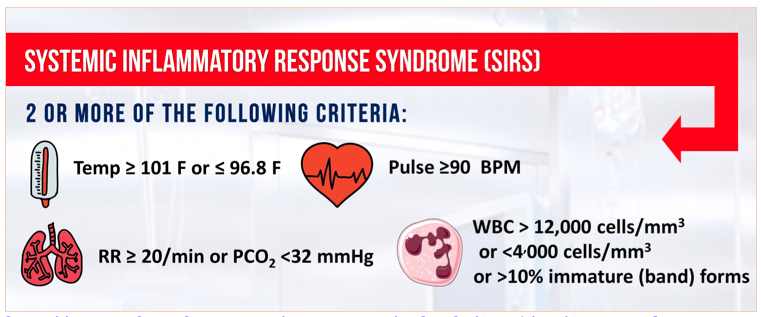
Sepsis, a maladaptive host-response to infection, is a leading cause of morbidity and mortality within the healthcare system. We have known about and discussed this disease for decades, but recently have begun to alter our criteria for its diagnosis. Since 1991, we have categorized sepsis as a derangement in physiologic or laboratory parameters caused by a host’s systemic inflammatory response to an infection [1]. Two of 4 criteria, listed below, must be met in addition to a suspected source of infection. Sepsis complicated by end-organ dysfunction was deemed severe-sepsis. And sepsis-induced hypotension refractory to adequate fluid resuscitation was deemed septic shock.

Source: http://www.admitologist.com/wp-content/uploads/2015/12/Sepsis-Info.jpg

Source: http://www.admitologist.com/wp-content/uploads/2015/12/Sepsis-Info.jpg

Using these definitions, multiple studies have resulted in consensus guidelines that reduce morbidity and mortality, including early adequate fluid resuscitation, obtaining blood cultures before antibiotic therapy, administration of broad-spectrum antibiotics within 1 hour of diagnosis, and use of norepinephrine as a first-line vasopressor to maintain MAP > 65 mmHg2.

Recently, due to presumed limitations in the original definition, a task force was convened and a new definition was proposed. They define sepsis as life-threatening organ dysfunction caused by a dysregulated host response to infection [3]. This organ dysfunction is calculated using the Sequential Organ Failure Assessment (SOFA) Score. While the original is quite cumbersome, the qSOFA score is a simplified version easily used at the bedside. While qSOFA has already shown promise in predicting patients with sepsis at risk for increased mortality [4], it remains to be seen whether it is useful at screening patients for sepsis early in the disease process.

Source: http://epmonthly.com/article/sepsis-gets-an-upgrade/

Source: http://epmonthly.com/article/sepsis-gets-an-upgrade/

Comparison of qSOFA score and SIRS criteria as screening mechanisms for emergency department sepsis. Haydar S, Spanier M, Weems P, Wood S, Strout T. Am J Emerg Med. 2017 Jul 6. pii: S0735-6757(17)30509-0.

This study was a retrospective chart review performed at a single academic tertiary care hospital. Its primary objective was to determine both the sensitivity and the diagnostic timeliness of the qSOFA score compared to SIRS criteria in a population of emergency department patients. While not explicitly stated, its secondary objective was to determine the test characteristics (including sensitivity, specificity, negative predictive value, positive predictive value, and the area under the receiver operatic characteristic curve) of qSOFA and SIRS to identify septic patients that would ultimately die in-hospital.

The sample of patients were drawn from a base population that was treated with antibiotics in the ED for a suspected infection, admitted, and ultimately expired or subsequently discharged with a Center for Medicare Services Diagnosis Related Grouping (DRG) for sepsis.

Data were extracted to fulfill the criteria for both qSOFA and SIRS, including respiratory rates, systolic blood pressures, heart rates, white blood cell counts, temperatures, altered mental status (AMS), and the times at which these parameters were documented. Pre-hospital data was excluded. Of note, both physician and nursing documentation were reviewed. Interestingly, laboratory values (i.e., WBC count) were considered to be present at the time of the blood draw, not at the time of result. Data was extracted by a single reviewer for all data points except for the timing of AMS. During a random sampling by two reviewers to determine reliability, the kappa value of 0.4 was deemed inappropriate, prompting a consensus review of all charts to determine initial time of AMS.

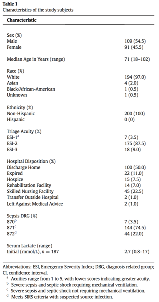
Of the 200 sampled patients, one was excluded due to transfer from an outside facility, leaving 199 for analysis (Table 1). Of note, median age was 71 years (range 18-102) and in-hospital mortality was 11.0% (n = 22). The majority of patients were white (97%, n = 194) and Non-Hispanic (100%, n = 200).

Table 1

Table 1

SIRS criteria outperformed the qSOFA score in sensitivity for diagnosing sepsis while in the ED, mean time to diagnosis, and median time to diagnosis (Table 2). The overall sensitivity for qSOFA was quite poor, and in particular, only 36.7% of patients met the AMS requirement. In determining in-hospital mortality, qSOFA had a much higher specificity and positive predictive value, but ultimately the overall performance as evidenced by the AUROC was relatively poor for both SIRS and qSOFA and they did not differ significantly (Figure 1).

Table 2

Table 2

Figure 1

Figure 1

qSOFA came after years of criticism towards the SIRS criteria. It has long been recognized that the SIRS criteria are not specific for infection, and that a variety of conditions including pain, trauma, and nonspecific inflammation can place patients in a SIRS-positive category. A recent study demonstrated that nearly half of hospital ward patients developed positive SIRS criteria at least once during their stay [5]. If not categorized appropriately, this could lead to inappropriate antibiotic utilization and fluid resuscitation.

However, the introduction of qSOFA the Sepsis-3 criteria has its own inherent limitations. Primarily, the focus has shifted towards hypotension and altered mental status as markers of end-organ dysfunction. While these features are intuitively associated with a higher disease burden (i.e., altered patients and those that are hypotensive are clearly sicker than those that are not), it shifts the focus from screening to prognostication. While high qSOFA scores have proven to correlate with in-hospital mortality [4], is this really the most important question for the emergency department physician?

More useful is a tool that screens positive for the largest proportion of potentially infected patients, leaving clinical judgment to further distill appropriate workup and treatment. This paper suggests that SIRS criteria are much better suited for this purpose. Compared to qSOFA, SIRS had a sensitivity for diagnosing sepsis that was over 36% higher, with a reduction in time to diagnosis by approximately one half.

The major strength of this paper is the wide net of inclusion. Essentially, all patients admitted and subsequently diagnosed with sepsis were included and randomly sampled. This group spanned a variety of ages, acuities, and types of infections. This was a strong attempt at making the data as generalizable as possible. However, the population at Tufts Medical Center in Maine, predominately White and Non-Hispanic may limit the external validity to more diverse practice environments.

Timing of diagnosis was another factor fraught with both pros and cons. While not explicitly stated, it seems intuitive that nursing documentation review in addition to physician notes would allow for expedited recognition of vital sign abnormalities and the onset of AMS. However, even with this inclusion, the retrospective determination of AMS onset without objective documentation practices is prone to error. One can imagine that late documentation in a busy emergency department setting may have contributed to the delay in time from arrival to documentation of qSOFA criteria. Furthermore, it seems odd that the laboratory results were considered to be present at the time of blood draw rather than at the time of result availability. Clinically, this is not how we would be able to diagnose sepsis in real-time, and it may have falsely hastened the time from ED arrival to documentation of SIRS criteria in this study.

Lastly, it is important to recognize that the Surviving Sepsis campaign has championed early recognition and treatment as the key principle to reducing morbidity and mortality. This has led many hospital systems to incorporate SIRS criteria into the electronic health record, to flag patients as early as possible for recognition. This is not as feasible with qSOFA, as the component of altered mentation is subject to individual interpretation.

In summary, sepsis is a critical diagnosis that must be made early to improve outcomes. The SEPSIS-3 campaign promotes the use of qSOFA criteria, which are clearly a prognostic marker for increased mortality. However, the SIRS criteria are more useful in screening for sepsis early in the disease process in emergency department patients.


Expert Commentary

As Dr. Ireland’s excellent review of this attempted validation study notes, what was originally heralded as a new paradigm in identifying the warning signs of sepsis (qSOFA) appears to in fact be (significantly) worse than the SIRS criteria we all know and (don’t) love.

As others have noted far more eloquently than I (see: pretty much anything that Josh Farkas of PulmCrit has written on qSOFA), it really shouldn’t be surprising to any of us that qSOFA is less sensitive than SIRS.  By including hypotension and altered mental status (just another word for end-organ dysfunction in the old paradigm), qSOFA is essentially screening for what was formerly known as ‘septic shock.’  Not a surprise that patients with 2 or more of these criteria don’t do well from a mortality standpoint.  This isn’t what I need from a screening test for sepsis in the ED though - I can’t think of the last time I walked into a patient’s room and found them altered, hypotensive and tachypneic and thought to myself, “Hmm, I wonder if they are sick or not?”

While SIRS isn’t perfect, it at least approaches the requisite sensitivity to make it a useful screening test.  It is important to remember that you as the clinician are tasked with providing the specificity - the warning signs of sepsis overlap with many other acute (and less acute) processes.  Patients screening SIRS positive may need aggressive management of conditions other than sepsis, and not every sepsis patient must receive the full sepsis bundle - evaluate each patient fully before initiating protocol-based care.

Ben_Schnapp-12.jpg
 

Benjamin Schnapp, MD

Assistant Residency Program Director, University of Wisconsin-Madison


How to Cite This Post

[Peer-Reviewed, Web Publication]   Ireland A, Iwaki K (2018, September 17). qSOFA SIRS.  [NUEM Blog. Expert Commentary by Schnapp B]. Retrieved from http://www.nuemblog.com/blog/qSOFA


Other Posts You May Enjoy


References:

  1. BoneRC, BalkRA, CerraFB, etal. American College of Chest Physicians/Society of Critical Care Medicine Consensus Conference: definitions for sepsis and organ failure and guidelines for the use of innovative therapies in sepsis. Crit Care Med. 1992;20(6):864-874.

  2. Dellinger RP, Levy MM, Rhodes A, et al. Surviving sepsis campaign: international guidelines for management of severe sepsis and septic shock: 2012. Crit Care Med 2013;41(2):580–637.

  3. Singer M, Deutschman CS, Seymour CW, et al. The third international consensus def- initions for sepsis and septic shock (sepsis-3). JAMA 2016;315(8):801–10.

  4. Freund Y, Lemachatti N, Krastinova E, et al. Prognostic accuracy of sepsis-3 criteria for in-hospital mortality among patients with suspected infection presenting to the emergency department. JAMA 2017;317(3):301–8.

  5. Churpek M.M., Zadravecz F.J., Winslow C., et al: Incidence and prognostic value of the systemic inflammatory response syndrome and organ dysfunctions in ward patients. Am J Respir Crit Care Med 2015; 192: pp. 958-964

  6. Comparison of qSOFA score and SIRS criteria as screening mechanisms for emergency department sepsis. Haydar S, Spanier M, Weems P, Wood S, Strout T. Am J Emerg Med. 2017 Jul 6. pii: S0735-6757(17)30509-0.

Posted on September 17, 2018 and filed under Infectious Disease.

Delirium as a symptom of UTI: physiology or pseudoaxiom?

Screen Shot 2018-07-08 at 3.00.34 PM.png

Written by: Ashley Amick, MD (NUEM alum '18) Edited by: Michael Macias, MD (NUEM alum '17) Expert commentary by: Alexander S Lo, MD, PhD


Asymptomatic bacteriuria (ASB) is a prevalent condition in the elderly population.  Bacterial colonization of the genitourinary (GU) tract increases with age and institutionalized status.  Though once thought to be pathogenic, randomized trials clearly demonstrate that treatment of ASB with antibiotics does not improve outcomes, except in pregnant patients and those undergoing GU procedures.  Emerging data even suggest there may be a protective effect of colonizing bacteria.  Conversely, there is increasing recognition of the dangers of inappropriate antibiotic use, both to the individual and the general population, and widespread agenda to limit unnecessary antimicrobial use. 

As the antibiotic stewardship movement marches forward, the treatment of ASB continues to be a central focus.  Recent guidelines emphasize that the presence of lower GU symptoms is the key distinction between UTI and ASB.  This strategy may be easily adopted in young otherwise healthy patients, but reaches a major stumbling block when considering the elderly population.  This is in part due to the fact that many clinicians believe that there is a causative relationship between UTI and delirium in the absence of other localizing symptoms or signs of systemic infection.  In other words, delirium is the symptom that substantiates a diagnosis of UTI in the presence of otherwise asymptomatic bacteriuria.  This concept, now generations old, is still taught in many medical school curricula.  The correlation between delirium and UTI is so well established in the minds of clinicians that many have never questioned whether this presumed association is rooted in data.

The concerning truth is that there is no reliable evidence to suggest that such a relationship between delirium and UTI exist.  A recent review of the literature found only five papers addressing this association primarily, all were observational and therefore lacked the ability to make conclusions about the degree of causation.  All studies were severely methodologically flawed, and none were case-control, cohort, or RTCs.  Additionally, there is no physiologic evidence or models to suggest that bacteriuria in the absence of systemic illness, results in cognitive dysfunction.  No known studies have ever shown that treatment of otherwise asymptomatic bacteriuria improves delirium outcomes.  Taking these data into account, the CDC and SHEA created guidelines specifically do not include delirium as a reason to treat potential UTIs in non-catheterized patients.  These represent a departure from earlier guidelines that included altered mental status as a symptom of UTI in the elderly.  The new SHEA recommendations have been tested in a large randomized trail and were found to be safe when compared to standard care.

Despite efforts to shift practice patterns in the direction of a more guideline-based management, ASB continues to be unnecessarily treated at high rates in the elderly.  One reason may be that anecdote is a powerful source of bias.  Many clinicians support their belief of a causative correlation between UTI and delirium by referencing cases where patient presented with confusion and were found to have a UTI.  The problem is, how was that “UTI” diagnosed?  The distinction is more than just semantics.  In the absence of GU symptoms and signs of systemic infection, then the clinician made the diagnosis solely on the basis of a UA and urine culture.  But as previously discussed, both a UA and culture will frequently be positive in both ABS and UTI, and cannot reliably distinguish between the two conditions. 

Many clinicians will cite the fact that the patients may improve following antibiotic administration, thereby confirming their suspicion of a presumed UTI-related delirium.  However, delirium frequently is short lived and self-resolving, therefore improvement is likely to be simply coincidental.  In addition, along with antibiotics administration patients also often receive intravascular volume, thereby improving hydration status, which is a frequent cause of delirium.  These factors confound the ability of the clinician to objectively interpret the causative relationship between the delirium and bacteriuria.  High quality randomized trials will be needed to further clarify these issues and assess is the high rate of concurrence of bacteriuria and delirium is due to causation or simply coincidence.


Expert Commentary

Over 50 million U.S. adults > 65 years of age (“older adults”), account for over 20 million Emergency Departments (ED) visits each year [1].  Many of these patients have unmet and complex underlying medical needs that are often understated by their chief complaints. The tempting application of traditional ‘one complaint; one algorithm’ approach taught to many emergency physicians, may often result in long-term, downstream, adverse outcomes.  One of those relevant to the accompanying blog, is the traditional “if grandma is delirious, look for and treat the UTI” doctrine.  A review of the literature proves that the evidence linking UTI’s to delirium in older adults is lacking [2]. Many older adults are bacteriuric; most do NOT have to be treated [3].  The delirium is not a reason to treat bacteriuria [4].  It is also just as likely that it is the other comorbid conditions causing the delirium, since 75% of older adults have two or more comorbid chronic conditions [5]. many of which have the potential to cause delirium at any time[6].   The patient may likely require admission for the delirium, but a more comprehensive investigation into its etiology is more helpful than treating the easy target of a contaminated urine sample

Alex_Lo.png

Alexander S Lo, MD, PhD

Assistant Professor of Emergency Medicine, Northwestern University 


Posts you may also enjoy


How to cite this post

[Peer-Reviewed, Web Publication]   Amick A, Macias M (2018, July 30). Delirium as a symptom of UTI: physiology or pseudoaxiom.  [NUEM Blog. Expert Commentary by Lo A]. Retrieved from http://www.nuemblog.com/blog/uti-part1


Resources

  1. Pines JM, Mullins PM, Cooper JK, Feng LB, Roth KE. National trends in emergency department use, care patterns, and quality of care of older adults in the United States. Journal of the American Geriatrics Society. 2013;61(1):12-17.

  2. Balogun SA, Philbrick JT. Delirium, a Symptom of UTI in the Elderly: Fact or Fable? A Systematic Review. Canadian geriatrics journal : CGJ. 2014;17(1):22-26.

  3. Finucane TE. "Urinary Tract Infection"-Requiem for a Heavyweight. Journal of the American Geriatrics Society. 2017;65(8):1650-1655.

  4. Ninan S. Don't assume urinary tract infection is the cause of delirium in older adults. Bmj. 2013;346:f3005.

  5. Working Group on Health Outcomes for Older Persons with Multiple Chronic C. Universal health outcome measures for older persons with multiple chronic conditions. Journal of the American Geriatrics Society. 2012;60(12):2333-2341.

  6. Kuluski K, Hoang SN, Schaink AK, et al. The care delivery experience of hospitalized patients with complex chronic disease. Health expectations : an international journal of public participation in health care and health policy. 2013;16(4):e111-123.

  7. McKenzie, Robin, et al. "Bacteriuria in individuals who become delirious." The American journal of medicine 127.4 (2014): 255-257.

  8. Balogun, Seki A., and John T. Philbrick. "Delirium, a symptom of UTI in the elderly: fact or fable? a systematic review." Canadian Geriatrics Journal 17.1 (2013): 22-26.

  9. Nace, David A., Paul J. Drinka, and Christopher J. Crnich. "Clinical uncertainties in the approach to long term care residents with possible urinary tract infection." Journal of the American Medical Directors Association 15.2 (2014): 133-139.

  10. Gau, Jen-Tzer, et al. "Interexpert agreement on diagnosis of bacteriuria and urinary tract infection in hospitalized older adults." J Am Osteopath Assoc 109.4 (2009): 220-226.

  11. Juthani-Mehta, Manisha, et al. "Interobserver variability in the assessment of clinical criteria for suspected urinary tract infection in nursing home residents." Infection Control & Hospital Epidemiology 29.05 (2008): 446-449.

  12. Schulz, Lucas, et al. "Top Ten Myths Regarding the Diagnosis and Treatment of Urinary Tract Infections." The Journal of emergency medicine (2016).


Posted on July 30, 2018 and filed under Infectious Disease.

Asplenia in The Emergency Department

From an infectious perspective, asplenia poses a serious risk factor in acquiring various types of infections. The spleen plays a critical role for the immune system in having a robust response to various encapsulated organisms... Read this week's post to learn more about managing these at risk patients. 

Necrotizing Fasciitis: Using EBM to Answer the Big Questions

Necrotizing soft tissue infections (NSTI) are exceedingly difficult to recognize in their early stages, and can resemble a cellulitis. A high index of suspicion must be maintained. This week we briefly summarize the evidence in diagnosis and management of this deadly condition.  

HIV Counseling in the ED: Commonly Asked Questions and How to Answer Them

Testing for HIV in the emergency department (ED) has become a vital topic and policy in hospitals across the country.  Early diagnosis of HIV is critical in decreasing transmission rates, in addition to providing better outcomes for patients, as early diagnosis often leads to earlier treatment. Today we discuss how to counsel patients with a new diagnosis of HIV in the ED. 

Posted on February 13, 2017 and filed under Infectious Disease.

Infestations

Skin infestations are frequently encountered in the ED, particularly among the homeless population, though data on the number of visits are lacking. While patients with infestations may seem like pests in the middle of a busy shift, these conditions can be a public health menace, and may be markers of serious underlying pathology. 

Posted on January 9, 2017 and filed under Infectious Disease.

Acute Retroviral Syndrome In The ED

Consider acute retroviral syndrome like endocarditis or meningitis or epidural abscess: another life threatening cause of a seemingly benign febrile illness that we must recognize. HIV infection, especially in its primary phase, represents a life threatening malady that could have serious implications on the patient and the public health at large if not diagnosed.