Written by: Jacob Stelter, MD (NUEM ‘19) Edited by: Kimberly Iwaki, MD (NUEM ‘18) Expert commentary by: D. Mark Courtney, MD, MCSI
Case
A 54 year old male with no significant past medical history presents to the ED around 11pm with the chief complaint of “swollen penis.” He appears notably distressed on initial assessment and complains of penile swelling and pain. Per the history, the patient states that he was having sexual intercourse with his wife, with her on top of him, when he felt a sudden “popping” sensation in his penis followed by swelling, detumescence and pain. Vitals are stable and exam is notable for an enlarged, swollen, ecchymotic, mildly angulated and deformed penis. Diagnosis?
Penile fracture. Yes, penises (or penes) can fracture. The penis can sustain multiple different forms of injury, including fracture, direct trauma, burns and strangulation. It is both dramatic and problematic and requires emergent evaluation. Three of these injuries will be discussed here – penile fracture, zipper injury and penile strangulation.
Penile Fractures
Anatomy and Pathophysiology
The penis consists of two corpora cavernosa on the dorsal aspect of the penis and one corpus spongiosum on the ventral portion of the penis, all wrapped in a tough fascial layer call the tunica albuginea (1). Penile fractures typically involve an injury to the tunica albuginea that becomes significantly thinner and more stretched during an erection (2). Usually, this involves the tunica albuginea of one of the corpus cavernosum, which can also become injured or lacerated (2, 3). The most common mechanism for penile fracture in the United States is during vigorous vaginal sexual intercourse when the erect penis is misdirected into the female pubic bone (2). This most often occurs with the female-on-top or “cowgirl” positions (2). In the Middle East, the most common practice that leads to penile fracture is “taghaandan” or “to click or snap when forcibly pushing the erect penis down to achieve detumescence” (4). In fact, one study conducted in Iran determined that 69% of penile fractures were related to this mechanism (4).

Figure 1: Normal Penile Anatomy (1)
Presentation
The clinical presentation of a penile fracture is typically a “popping” sensation or sound, followed by significant pain, swelling and detumescence of the penis, often with notable ecchymosis, swelling and deformity on exam (3). Clinically, this is referred to as the “eggplant deformity” (5). The “rolling sign” refers to the physical exam maneuver that identifies where the corporal fracture is located by rolling the penis and palpating a firm, immobile swelling that represents the hematoma overlying the fracture (5). Most often, this is a clinical diagnosis with little utility for imaging studies. However, if the diagnosis is unclear, ultrasound or MRI can be used to diagnose injuries to the underlying fascia or corpora cavernosa (6).
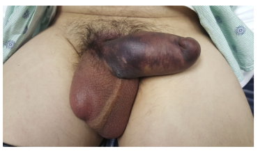
Figure 2: Penile fracture
Treatment
From an emergency department (ED) management perspective, if a penile fracture is suspected, an emergent urology consultation is indicated. Up to 38% of penile fractures can involve urethral injuries and, while they may present with hematuria and difficulty urinating, these signs are not reliable in ruling out a urethral tear (2). As a result, a retrograde urethrogram (RUG) or cystoscopy should be performed prior to Foley catheter placement (2). This is important, as acute urinary retention in the setting of a penile fracture is an emergency indication for Foley placement and an urologist may not be readily available to assist with this. Symptom control in the ED should include ice packs to reduce swelling, pain control and Foley placement as indicated (5). Current urological treatment guidelines recommend immediate surgical repair which includes hematoma evacuation, examination of the urethra, and repair of the corporal fracture and any tears in the surrounding fascia (5,2). If promptly repaired, within 36 hours, most patients have minimal residual effects after sustaining a penile fracture (3).
Zipper Injury
Zipper injuries involving the penis are another injury that can be encountered by the emergency physician. Most often, this occurs in children and is more common in uncircumcised males (1). Using a lubricant to attempt to separate the entrapped penis can be successful and is noninvasive (1). In fact, a recent study showed that the use of mineral oil to free a simulated entrapped penis was nearly 100% successful (7). If unsuccessful, using a wire cutter can help to cut the middle portion of the zipper that connects the two halves to help separate the zipper from the entrapped foreskin (1). Additionally, the zipper can be grasped above and below the entrapment to separate the teeth to free the penis (1). This can be painful, so using local or regional anesthesia is recommended (1).

Figure 3: Zipper median bar bisection (1)
Penile Strangulation
Penile strangulation injuries are seen occasionally in the ED. Most often, this results from self-application of rings to the base of the penis and often including the scrotum in an attempt to prolong erections or enhance sexual pleasure. Multiple case reports have been published, detailing various objects used as cock rings and methods to remove them. Being unable to remove the constricting ring is a medical emergency as it can cause a prolonged erection and can lead to ischemia and eventual necrosis of the penis (8). There are five published grades of injury resulting from penile strangulation (9):
Grade I: Penile edema with no urethral or skin injury
Grade II: Skin injury with constriction of the corpus spongiosum with penile edema and decreased sensation
Grade III: Injury to both the skin and urethra without urethral fistula
Grade IV: Urethral fistula formation due to division of the corpus spongiosum
Grade V: Penile necrosis or amputation of the distal segment
Multiple different tools can be used to remove these rings, including ring cutters found in most emergency departments, dental drills or pliers (8). Care must be taken to protect the skin of the penis underneath the ring and this can be done by using a tongue depressor or other protective guard. If it is difficult to get anything underneath the constricting band, blood can be drained from the corpora cavernosa in a manner similar to draining a priapism (8). If this is unsuccessful, an emergent urology consult is indicated to avoid permanent penile damage (8).
Take Home Points
Penile fractures are one of the most common injuries of the penis and are often sustained by vigorous sexual intercourse. ED management of penile fractures includes pain control, swelling reduction with ice, Foley placement once a urethral injury is ruled out and emergent urology consult for operative intervention.
Zipper injuries occur most frequently in young, uncircumcised males. Mineral oil is the best way to try to free a zipper-trapped penis. If unsuccessful, try bisection of the median bar of the zipper.
Penile strangulation occurs by the self-placement of constricting rings and can end up causing penile necrosis or amputation if the constricting object is not promptly removed.
Expert Commentary
One of the more difficult aspects of management of these injuries is reassurance and anxiolysis to the patient and perhaps their partner or parent in the case of a child with a zipper injury. There is often a morbid curiosity on the part of members of the treatment team including other physicians and nurses. For the sake of maintaining a calm patient who trusts in you as you approach the penis with a tool that looks like and may have come from the hospital mechanical maintenance room, try to avoid spectacle and treat the patient with dignity and respect and perhaps a bit of a benzodiazepine. This will make them feel more comfortable and make your job easier.
Additionally, don’t trust the history. There are many reasons patients may not be forthright about the events leading up to and causing a penile fracture. In my experience half of the patients I have taken care of have denied onset while having intercourse despite later revealing the truth. Point is….if it looks like a penile fracture, it almost certainly is, regardless of the history that may be unclear or inconsistent with your suspicion.
Finally, the seriousness of implications of untreated penile injuries at times needs to be impressed upon patients, family members, and treating physicians. Sometimes these patients arrive intoxicated and wanting to leave. This presents the usual difficulties in determining medical decision making capacity and preventing harm. Though the above Blog post notes most have a good outcome with repair, un-repaired penile fractures have a high incidence of subsequent deformity and erectile dysfunction.

D. Mark Courtney, MD, MSCI
Associate Professor
Department of Emergency Medicine
Northwestern University
How to Cite this Post
[Peer-Reviewed, Web Publication] Stelter J, Iwaki K. (2019, Dec 2). Penile Injuries. [NUEM Blog. Expert Commentary by Courtney DM]. Retrieved from http://www.nuemblog.com/blog/penile-injuries
References
Dubin J, Davis JE. Penile emergencies. Emerg Med Clin N Am 2011;29:485-499.
Chang AJ, Brandes SB. Advances in diagnosis and management of genital injuries. Urol Clin N Am 2013;40:427-438.
Cooper BL, Beres KP. Penile Fracture. J Emerg Med 2017;52(2):238-239.
Zargooshi, J. Penile fracture in Kermanshah, Iran: Report of 172 cases. J Urol 2000;164:364-366.
Jack GS, et al. Current treatment options for penile fractures. Rev in Urol 2004;6(3):114-120.
Bertolotto M, et al. Penile trauma. Chapter 125 in Abdominal Imaging. 2013. Pp. 1937-1946.
Oquist M, et al. Comparative analysis of five methods of emergency zipper release by experienced versus novice clinicians 2016;68(45):S116.
Chapman JD. A case of penile strangulation secondary to deliberate placement of a wedding band. J Clin Urol 2016;9(2):131-132.
Agarwal AA, et al. Penile strangulation due to plastic bottle neck: A surgical emergency. BMJ Case Rep 2014:1-2.