


Author: Sarah Sanders, MD (EM Resident Physician, PGY-2, NUEM) // Edited by: Jordan Kaylor, MD (EM Resident Physician, PGY-4, NUEM) // Expert Commentary: Elizabeth Dearing, MD
Citation: [Peer-Reviewed, Web Publication] Sanders S, Kaylor J (2016, July 12). Diagnosing Pneumothorax With Bedside Ultrasound [NUEM Blog. Expert Commentary by Dearing E]. Retrieved from http://www.nuemblog.com/blog/diagnosing-pneumothorax-ultrasound/
Case
A 28 year old male presents as a trauma activation following a motor vehicle collision. The patient was a restrained passenger in a car that was hit head on. He presents well appearing with ABCs intact and a midline trachea with no respiratory distress. However, he is saturating 93% on room air.
Can you see it? I wasn’t too confident. There is a very small left sided apical pneumothorax. In a rushed trauma scenario without a skilled radiologist, would it have been caught? Next time, consider grabbing an ultrasound.
Why Grab An Ultrasound?
According to the literature, ultrasound (US) is superior to chest x-ray (CXR) in diagnosing pneumothorax. Ding et al. published a meta-analysis of 20 articles and found US was 88% sensitive and 99% specific for pneumothorax versus CXR’s 52% sensitivity and 100% specificity. The increased sensitivity of US is echoed throughout the literature. Wilkerson and Stone evaluated emergency physicians using US to diagnose pneumothorax compared to AP CXR after blunt trauma and found similar results—US had a sensitivity of 86-98% compared with AP CXR sensitivity of only 28-75%.
Why Not Just CT Scan?
CT is the gold standard for diagnosis of pneumothorax. However, in an unstable trauma patient, getting a CT scan is not always possible, and it takes time. Fortunately, US can provide real-time information at the bedside in only seconds.
Ok, I Believe US Is Superior To CXR In Diagnosing A Pneumothorax. How Do I Use Ultrasound?


- Place the high-frequency linear probe in sagittal orientation on the chest wall.
- Remember, a pneumothorax is air, and air moves to the least dependent area of the chest.
- In an upright patient, start on the apical lateral lung.
- In a supine trauma patient, start on the anterior chest, in the 2nd or 3rd intercostal space, along the mid-clavicular line.
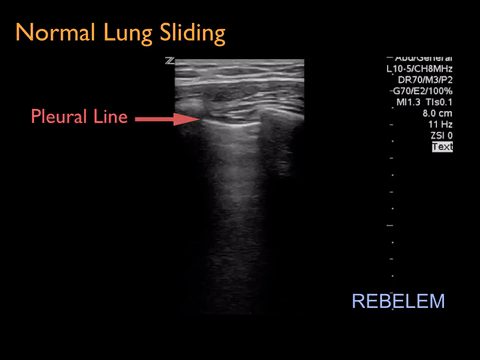
- Identify the landmarks of two ribs with posterior shadowing on the edges of the probe. The pleural line should be visualized between them. If you are unable to find the two ribs, move the probe caudally (towards the toes) until two ribs are visualized.
- Once you have found the intercostal space, scan medially and laterally to evaluate for lung sliding.
- Next, move to the next intercostal space and repeat.
It's Hard To Tell If The Lung Is Sliding.


Agreed. Try M-Mode.
M-mode, or motion mode, represents movement of structures over time. The user places the M-mode line over the particular area of interest and M-mode shows how the structures move towards and away from the probe over time.
What About False Positives?
You mean there can be absence of lung-sliding without a pneumothorax? That’s a valid concern. According to Lichtenstein et al, underlying lung disease (especially COPD and ARDS) makes diagnosis of pneumothorax with ultrasound more difficult and more likely to have false positives. Do keep in mind that small pneumothoraces can be managed without tube thoracostomy. Never forget that a piece of technology does not substitute for proper clinical judgement.
Take Home Points
- In a trauma, do not depend on the CXR to evaluate for a pneumothorax
- Turn the FAST exam into an EFAST exam to evaluate for pneumothoraces
- Start with the probe on the least dependent area of the chest to evaluate for air and look for lung sliding to rule out pneumothorax
- Use M-mode to evaluate for the seashore vs. barcode sign if you are unsure if lung sliding is present
- Identifying a lung point on ultrasound is pathognomonic for pneumothorax and can also be used to gauge the size of a pneumothorax
- Ultrasound should always be used in conjunction with good clinical judgement
Additional FOAMed Resources:
- Rebel EM: Ultrasound for Pneumothorax
- Emergency Ultrasound Teaching: Video Tutorial of EFAST
- NUEM Blog: Lung Ultrasound Basics
- EM Curious: US ABCs & Trauma Ultrasound
Expert Commentary
Dear Sarah,
Thank you for this great discussion regarding the utility of ultrasound in diagnosing pneumothorax. CT is certainly the gold standard for diagnosis however it exposes patients to radiation, takes more time to perform and can be an unsafe place for an unstable patient. Ultrasound is easy to perform, fast and does not expose patients to radiation. The traditional FAST exam has been performed in the United States since the 1990s in the evaluation of peritoneal and pericardial fluid in trauma patients. Over the past decade we have also seen an increase in the use of thoracic ultrasound and a subsequent increase in the use of the extended FAST or EFAST exam for trauma patients. The EFAST exam adds evaluation of the thoracic cavity for pneumothorax and hemothorax. Ultrasound, as you discussed, has been shown to be a better test than chest x-ray for pneumothorax in a supine trauma patient since air will layer along the anterior chest. Overall, ultrasound use in these patients can efficiently answer specific questions that can save a patient’s life. Here are few more tips to performing this exam:
+ Probe Selection
In patients who are being evaluated only for pneumothorax, the high frequency linear probe is the best choice. The higher the frequency, the better the resolution. However, we don’t use the highest frequency at all times because it comes at a cost – depth. When you increase the frequency (and therefore the resolution), you are unable to image deeper than several centimeters. In the setting of a trauma patient, you are undoubtedly already using the curvilinear probe (or phased-array) for the FAST exam. The curvilinear probe is my probe of choice for the lung exam. The curvilinear probe is a low frequency probe that will allow you to image structures that are much deeper and with decreased but adequate resolution. You should still have no trouble seeing the pleural line and lung sliding as long as you decrease your depth so that the pleural line is in the middle of the screen (usually a few centimeters). If you cannot see lung sliding at an appropriate depth setting, then M-mode may be helpful.
+ Technique
When you are using ultrasound to image the lung, like any other structure you have to look for landmarks to ensure you are in the proper orientation and location. For the lung exam, hold the probe in the sagittal plane with the probe marker toward the patient’s head. You should see ribs with rib shadow distally and the bright white horizontal pleural line just deep to ribs (referred to as the bat sign). In order to obtain the best image, you should be perpendicular to the pleural line (i.e. a-lines visualized). Start in the midclavicular line just below the clavicle and scan caudally until you visualize the diaphragm (tri-laminar structure that moves with respiration). On the left chest you may need to move more laterally to visualize lung sliding since the cardiac motion will interfere with your ability to assess for lung sliding. If you see absence of lung sliding, try sliding laterally to find the “lung point” which is pathognomonic for pneumothorax.
+ Imaging the lung should be done with a “dumb” machine
The normal lung is filled with air and is a poor medium for conduction of ultrasound waves. When the ultrasound wave hits aerated lung, it creates artifacts which are used to determine if any underlying lung pathology is present. Many of the machines that we use today have added features that help make the machine “smarter” and decrease the artifacts for imaging other structures such as kidneys or heart. To increase the artifacts and improve your lung ultrasound, turn these features (e.g. Tissue Harmonics, Multi Beam) off. This will usually make the image appear grainier but will also improve artifacts such as lung sliding.
+ False Positives
I think your point regarding false positives is an important one to make here. It is necessary for a clinician to understand the limitations of any test being performed. For thoracic ultrasound, absence of lung sliding can represent other underlying lung pathology in addition to pneumothorax. If you see absence of lung sliding, look for lung point to prove it is a pneumothorax. Additionally, subcutaneous emphysema will create a horizontal hyperechoic line (artifact from air) without lung sliding. However don’t forget your landmarks because on closer look this hyperechoic line will be above the level of the ribs (in the soft tissues).
Overall, ultrasound is just like any other test – it has limitations and you must interpret your findings with regards to the clinical context. Again, great discussion.

Elizabeth Dearing, MD
Assistant Professor; Department of Emergency Medicine; Division of Emergency Ultrasound; Vanderbilt University Medical Center
Other Posts You May Enjoy


References
- Husain LF et al. Sonographic diagnosis of pneumothorax. J Emerg Trauma Shock. 2012 Jan-Mar; 5(1): 76–81. http://www.ncbi.nlm.nih.gov/pmc/articles/PMC3299161/
- Wilkerson, R Gentry, and Michael B Stone. 2010. Sensitivity of bedside ultrasound and supine anteroposterior chest radiographs for the identification of pneumothorax after blunt trauma. Academic emergency medicine : official journal of the Society for Academic Emergency Medicine, no. 1. doi: 10.1111/j.1553-2712.2009.00628.x.http://www.ncbi.nlm.nih.gov/pubmed/20078434.
- Ding W, Shen Y, Yang J, He X, Zhang M. Diagnosing of Pneumothorax by Radiography and Ultrasound. Chest. 2011;140(4):859-866. doi:10.1378/chest.10-2946.
- Lichtenstein DA, Menu Y. A bedside ultrasound sign ruling out pneumothorax in the critically ill. Lung sliding. Chest. 1995. Nov: 108 (5): 1345-8.