

Author: Jessica Bode, MD (EM Resident Physician, PGY-2, NUEM) // Edited by: Andrew Ketterer, MD (EM Resident Physician, PGY-4, NUEM), Duc Pham, MD, & Jonathan Rich, MD // Expert Commentary: Jota Nakano, MD
Citation: [Peer-Reviewed, Web Publication] Bode J, Ketterer A, Pham D, Rich J (2016, July 5). LVAD Management In The ED [NUEM Blog. Expert Commentary by Nakano J]. Retrieved from http://www.nuemblog.com/blog/LVAD-management/
What are LVADs?

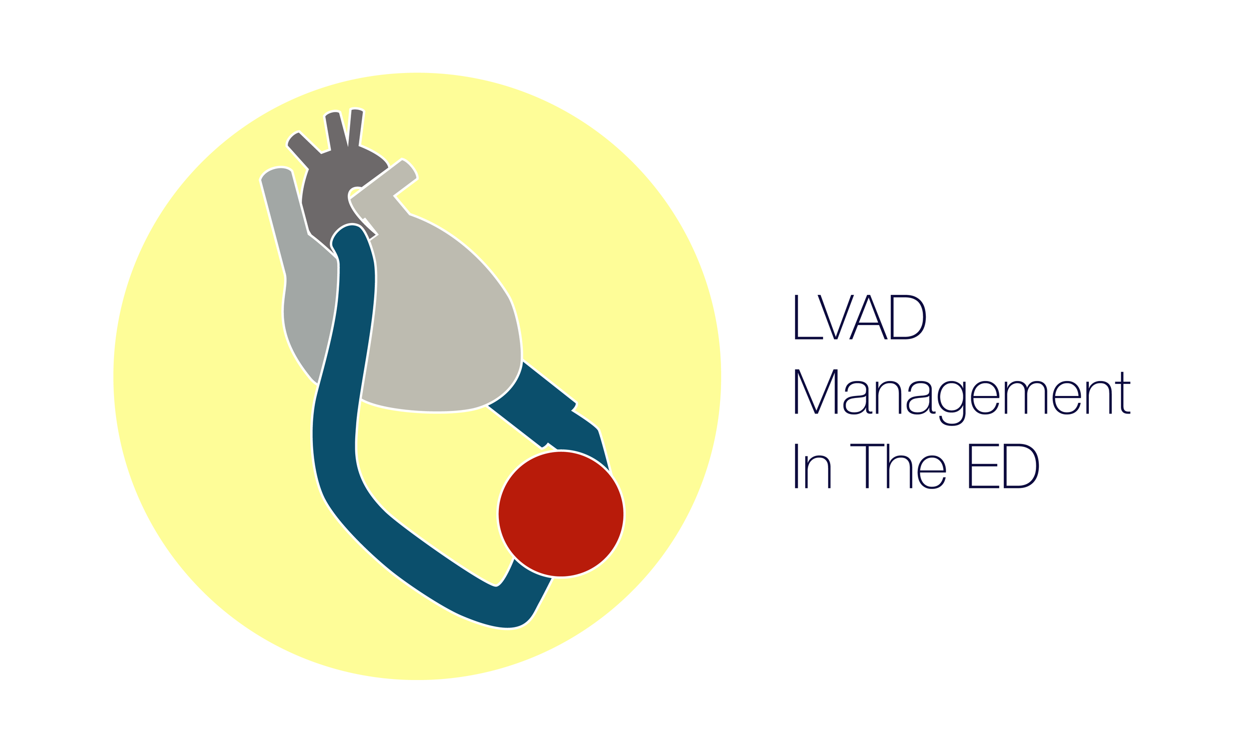
The Heartware HVAD Pump
Left Ventricular Assist Devices (LVADs) are surgically implanted pumps that essentially “take over” the job of the failing left ventricle, restoring normal blood flow to the body. These devices accomplish this by sucking in blood from the ventricle via a pump and propelling the blood into the aorta. The first VAD was implanted in 1984 and with an ever increasing number of patients with heart failure and fewer donor organs available, they have grown in popularity ever since. The landmark 2001 REMATCH paper demonstrated a significant survival increase [52% vs 25% at 1 year, p=0.002] with an improved quality of life. However, these earlier generation pulsatile VADs were insufficiently durable, usually lasting only 1-2 years. With advances in LVAD technology, currently over 90% of LVAD-supported patients will survive to 1 year post-implantation with the newer generation continuous flow LVADs and a minority of patients will continue to thrive on LVAD support for ten years or more.
Why are they important?
Currently around 2,300 Americans receive heart transplants per year, yet tens of thousands of patients are suffering from advanced heart failure and in need of a transplant. While the majority of centers perform between 10 and 19 heart transplants per year, the number of transplant centers has decreased from 243 in 1996 to 204 in 2007. The ongoing organ donor shortage has been the major limitation to the growth of this therapy, leaving many very sick patients without many viable options. This is where the LVAD comes into play. Additionally, there are many patients ineligible for heart transplantation but who would otherwise die without support of an LVAD.
Who gets VADs?
Anatomy of the LVAD
There are three major indications for LVAD implantation:
- Bridge to Transplantation: As a bridge device in end-stage heart failure transplant candidates, with the goal of stabilizing if not resolving their heart failure until they can receive a transplant.
- Destination Therapy: As destination therapy in end-stage heart failure patients who are refractory to medical treatment and are not transplant candidates, they would thus live the remainder of their life with an LVAD.
- Bridge to Recovery: For acute situations to allow for myocardial recovery after cardiogenic shock, myocardial infarction, postoperative cardiotomy, and ventricular arrhythmia refractory to medical management. This represents a minority of the LVAD implants.
How have VADs evolved?
First generation VADs usually relied on a pulsatile flow pump mechanism whereas newer models generate non-pulsatile, continuous flow. The advantages of these newer VADs include smaller units, fewer moving parts, and more efficient use of energy. The downside is that they generally require systemic anticoagulation and do not have a backup method, e.g. the hand pump, in the event of device failure.
What in the world do I do if I see one in the ED?
Chest X-ray demonstrating LVAD silhouette
- Remember, these are still patients just like you and me. And most general patient care principles will continue to apply with a few notable exceptions:
- ABCs and neuro status. Patients with continuous-flow LVADs may not have a palpable pulse or may have only a faint one. Obtaining a blood pressure is the best way to obtain assessment of circulation (more on this shortly).
- It is never wrong to call for help! Contact the hospital or local VAD coordinator on-call. The proximity and responsiveness of those teams may change the options available to you in the ED.
- Call for help! Seriously.
- As mentioned above, most modern VADs generate continuous rather than pulsatile flow, so you shouldn’t expect to feel a pressure pulse. Instead, check the blood pressure. Conventional cuffs may not work due to the non-pulsatile flow but you can work around this by using a manual cuff with a Doppler on top of the radial or brachial artery. Inflate and slowly release until you start hearing Doppler flow again, which can usually be relied upon to represent the patients mean arterial pressure (MAP). Recent studies have shown superiority of newer slow-cuff deflation devices but these are not widely available in EDs. However, if AT ALL IN DOUBT, an arterial line is still the gold standard, and though it is invasive, and somewhat time-consuming, it will provide the most reliable blood pressure assessment and should be done in the potentially unstable LVAD patient. . The MAP goals for LVAD patients are usually 70-90 mmHg though the coordinator should also have this information available.
- Evaluate the driveline, the part connecting the internal LVAD to the external control unit. Specifically examine for erythema, purulent drainage, or tenderness around the area indicating infection, just like you would with any other indwelling hardware.
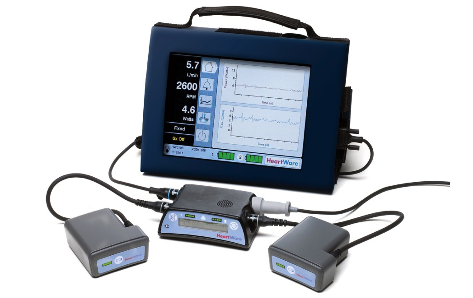
- Check the external control unit itself - usually patients and family members can help as they may be more familiar with the device than you are. Check the battery light indicator and assess for heat, which indicates malfunction.
- Get an EKG. A 2015 study of EKGs in patients pre- and post-LVAD showed that EKG changes associated with continuous-flow devices may include low limb-lead voltage, ventricular pacing, artifact (electrical), duration of the QRS > 120 milliseconds, ST-elevation in the lateral leads, and splintering of the QRS complex. Perhaps the most important information the ECG provides is the patient’s cardiac rhythm; it is not uncommon for an LVAD patients to be conscious yet in rapid ventricular tachycardia or, in extreme cases, even ventricular fibrillation.
- Consider CXR, echo, or CT depending on the circumstances. Note that LVADs ARE contraindications to MRIs.
- Labs: CBC, chemistry panel, LDH (to assess for hemolysis, an early indicator of possible LVAD thrombosis), lactate, troponin, blood cultures
Common complications of VADs
Most LVAD patients will present with normal, routine complaints such as URI, cough, abdominal pain, etc. that may not be related to the LVAD. That is, LVAD patients, will also have normal ED problems.
Bleeding and Thromboembolic Events
Bleeding is one of most common presenting chief complaints for LVAD patients, primarily due to a few potential compounding factors. First, they are at risk for an acquired von Willebrand’s deficiency owing to the large size of von Willebrand factor multimers which are broken down as they traverse through the mechanical device. Second, emerging evidence suggests that the relative absence of pulsatility may contribute to the development of intestinal AV malformations. Third, they are also usually anticoagulated with an INR goal of 2.0-3.0. Thus, if an LVAD patient presents with active hemorrhage and they are clinically unstable, do not hesitate to reverse the coagulopathy ( but this should be done in consultation with the VAD team unless the patient is truly unstable). In the same vein, bear in mind that the above mentioned need for anticoagulation and potential for bleeding dyscrasias puts VAD patients at higher risk of hemorrhagic stroke, with a reported incidence of up to 27% of patients with VADs.
Pump thrombus is another potential presenting complication, occurring in 1-2% of patients. It can usually be diagnosed by an elevation in the LDH, high powers and flow reading on the patients LVAD controller, and/or certain echocardiographic findings. The treatment includes the immediate reinstitution of anticoagulation if sub-therapeutic (i.e. IV heparin) and the immediate contacting of the VAD team to determine next steps. While some of these patients may be medically managed, many will require surgical LVAD exchange. An echocardiogram is also useful as an adjunctive tool in diagnosing RV failure, a complication that may occur both in the early days following implantation or as a late complication. Treatment options for VAD patients with RV failure follow those for the general population and include inotropes, volume unloading, and occasional pulmonary artery vasodilators.
Arrhythmias
Arrhythmias are common and seen in up to half of patients with LVADs, but treat these as you would in a patient without a VAD. Amiodarone is first line for ventricular arrhythmias and cardioversion/defibrillation are still viable options if an internal defibrillator is not in place. Many LVAD patients will tolerate VT for a period of time. If an LVAD patient is in VT but is otherwise hemodynamically stable and mentating normally, chest compressions and cardioversion is not immediately required. LVAD patients in VF or asystole can and should receive CPR, including chest compressions and cardioversion. Some LVAD patients may present with appropriate or inappropriate shocks from their ICD. The VAD team and electrophysiology (EP) should be immediately consulted.
Infection
Infection risk is of huge importance in LVAD patients. Although infection alone does not have negative prognostic value if the patient recovers and goes on to receive a transplant, it is the number one cause of death. In fact, sepsis accounts for more than twice the number of deaths caused by device failure. Presentations usually take the form of driveline infections, pump-pocket infections, or endocarditis. Broad spectrum antibiotics are crucial but make this decision in conjunction with the VAD specialist. The most common microorganisms isolated are Staphylococcus, Enterococcus and Pseudomonas, but when initiating appropriate therapy consider Candida coverage as well. It is important to obtain both driveline cultures and blood cultures followed by the early initiation of antibiotics.
Device Failure
Although uncommon, device failure is one of the most feared complications and can have catastrophic consequences. In the event of cardiac arrest, ABCs are paramount. Intubate, give IVFs, and assess the VAD itself as outlined above. Echo can be very helpful to assess RV and LV size and function, and you may be able to see a big VAD thrombus if there is one.
For years chest compressions were thought to be contraindicated in LVAD patients due to concern for dislodging the VAD and shearing the aorta, but a recent 2014 analysis looked at eight LVAD patients who received chest compressions for cardiac arrest. None of the eight patients had cannula dislodgement. Four of the eight patients had return of neurologic function, suggesting CPR may be viable, but the current thought is that this should only be done if there is a cardiac surgeon in house. Northwestern’s current policy is to initiate compressions, but follow your own institution’s guidelines.
Vasopressors, inotropes and placement of an intraaortic balloon pump are the mainstays of temporizing treatment for acute device malfunction.
Final Thoughts
In summary, VAD patients often present with normal medical problems just as in non VAD patients and should be treated accordingly. However, VAD patients may also present with highly complex conditions and require specialist input and intervention as soon as possible.
Here are a few quick pearls for Emergency Department management:
- Call for help immediately - this is the time to bring the specialists on board
- ABCs still apply. Get a blood pressure (and arterial line if you need one), and check an EKG.
- VADs are preload dependent, i.e. they can only pump blood if enough blood reaches them in the first place. Fluids are likely helpful but you can usually assume some degree of RV dysfunction so approach cautiously. When in doubt in the unstable patient, start an inotrope or pressor.
- LVADs are a contraindication to magnetic resonance imaging.
- In patients with continuous-flow pump devices, absence of a peripheral pulse is expected.
- Sometimes, even in LVAD patients, a hangnail is just a hangnail.
Expert Commentary
Hi Jessica:
Thank you for your excellent and thorough blog post on LVAD. More than 3,500 patients undergo LVAD implantation a year in the US since the survival benefit was demonstrated in the REMATCH trial. This number has already exceeded and is approaching nearly double the number of orthotropic heart transplants in the US. Also, destination therapy has gained more popularity in recent years. Thus, you can expect to see this cohort of patients more and more frequently in the ED settings. Of note, current devices implanted in the US - HeartMate II & III, and HeartWare - are all non-pulsatile.
Here are some comments on seeing a patient in the ED:
- Blood pressure: The newer devices like HeartMate III are also afterload dependent in addition to preload. Thus, it is important to keep the MAPs between 65 and 85 for this group of patients. Higher blood pressure is also known to be associated with greater stroke risk, especially hemorrhagic subtype.
- External control unit: You can see LVAD flows, a current speed setting (RPM), pulse index (HeartMate, not HeartWare), and power (watts). If the device is HeartWare, you can also check flow patterns with a HeartWare monitor.
- Labs: It is not particularly valuable to check troponin for the patients with LVAD because the disease process is more chronic (e.g., cardiomyopathy) rather than acute (e.g., myocarditis or acute MI). As you mentioned in the complication section, you may need to think about checking INR depending on the situation.
- Imaging studies: CXR and TTE are good modalities to know the fluid balance and LV/RV functions. Aortic valve is usually closed when an LVAD is working properly. However, you can suspect thrombus in an LVAD when you see that the aortic valve opens every beat on TTE (RAMP study). Elevated LDH (generally greater than 600) is another alarming sign of an LVAD thrombus (inside a pump), which cannot be seen with an echo in general because a pump is made of metal. CT scan with contrast can detect a thrombus in the outflow graft if it is large. CT chest/abdomen is also useful to evaluate infection (i.e. fluid collection around a driveline and/or a pump).
As for the common complications:
- Bleeding from a GI tract is a fairly common complication secondary to not only an acquired von Willebrand disease but arteriovenous malformation, which is associated with continuous non-pulsatile flow of the LVAD. Octreotide is another agent to treat GIB for this patient group.
- Stroke is one of the most devastating complications of LVAD. It is extremely difficult to treat given the necessity of anticoagulation and the risk of hemorrhagic conversion. Consider TTE to seek a possible source of embolism inside the heart and the pump.
- Patients with pump thrombus sometimes need additional anticoagulation (e.g., bivalirudin, eptifibatide) to heparin. Approximately half of the patients eventually need pump exchange. Please touch base with VAD team before you start any anticoagulation.
- Arrhythmias. Ventricular tachycardia is common due to the underlying disease. Many of the patients also have undergone ICD placement. Sometimes device interrogation per EP is necessary if an inadequate firing is detected. Ventricular arrhythmias can also occur secondary to a suck-down event, which an inflow of the pump interacts with an LV wall when the LV is under-filled. In this situation, the patient needs volume resuscitation as well as adjustment of the pump speed.
- Infection. Obtaining a blood culture is important before starting broad spectrum antibiotics. Again, CT can give us good information about driveline/pump pocket infection.
- Device failure. In benign cases, an exchange of the external controller resolves the problem. However, in case of complete malfunction of the device, the patients may need even emergency ECMO institution given the fact that these patients have only 10-15 % of ejection fraction and an intraaortic balloon pump may not be sufficient for the support.
Thanks again for sharing your thoughts on LVAD patients in the ED. Please remember that VAD coordinators (pager number 312-695-9611) and cardiothoracic surgery fellows/residents at Northwestern are more than happy to come to the ED and treat the LVAD patients with you 24/7.

Jota Nakano, MD, PhD
Instructor in Surgery-Cardiac Surgery; Bluhm Cardiovascular Institute of Northwestern University; Division of Cardiac Surgery
Other Posts That May Interest You


References
2004 Annual Report of the U.S. Organ Procurement and Transplantation Network and the Scientific Registry of Transplant Recipients: Transplant Data 1994-2003. Department of Health and Human Services, Health Resources and Services Administration, Healthcare Systems Bureau, Division of Transplantation, Rockville, MD; United Network for Organ Sharing, Richmond, VA; University Renal Research and Education Association, Ann Arbor, MI.
Rose EA, Gelijns AC, Moskowitz AJ, et al. Long-term use of a left ventricular assist device for end-stage heart failure. N Engl J Med 2001; 345:1435.
Partyka C, Taylor B. Review article: Ventricular assist devices in the emergency department. Emergency Medicine Australasia. April 2014;26(2):104-112 9p.
Lanier GM, et. al. Colombo PC. Validity and reliability of a novel slow cuff-deflation system for noninvasive blood pressure monitoring in patients with continuous-flow left ventricular assist device. Circ Heart Fail. 2013;6:1005–1012.
Martinez, SC, et. al. Characteristics of the Electrocardiogram in Patients with Continuous-Flow Left Ventricular Assist Devices. Annals of Noninvasive Electrocardiology, 2015; 20: 62–68.
Tsukui H, et. al. Cerebrovascular accidents in patients with a ventricular assist device. J Thorac Cardiovasc Surg. 2007; 134: 114– 123.
Argiriou M, Kolokotron S-M, Sakellaridis T, et al. Right heart failure post left ventricular assist device implantation. Journal of Thoracic Disease. 2014;6(Suppl 1):S52-S59. doi:10.3978/j.issn.2072-1439.2013.10.26.
Maniar S, Kondareddy S, Topkara VK. Left ventricular assist device-related infections: past, present and future. Expert review of medical devices. 2011;8(5):627-634. doi:10.1586/erd.11.36.
Rose EA, Gelijns AC, Moskowitz AJ, et al. Long-term use of a left ventricular assist device for end-stage heart failure. N Engl J Med 2001; 345:1435.
Shinar Z et al. Chest compressions may be safe in arresting patients with left ventricular assist devices (LVADs). Resuscitation. 2014 May;85(5):702-4. PMID: 24472494.