

Written by: Adam Eckburg, MD (NUEM ‘29) Edited by: Afolayan Oladeji, MD (NUEM ‘26)
Expert Commentary by: Seth Trueger, MD, MPH, FACEP (NUEM)
An ECG is handed to you. “Chest pain,” the operator states as the chief complaint. Your eyes quickly scan over the rate, rhythm, axis, intervals, and segments, all while you mentally filter through your many other competing tasks in a crowded Emergency Department. You are searching for the ST elevations and reciprocal depressions you have seen countless times before.
At the core of this task is a fundamental question: “Does this ECG suggest imminent risk to the patient sitting in front of me?”
Every EM provider knows the traditional STEMI criteria and the dangers associated with ECGs that meet them. However, recent evidence suggests that only ~60% of patients with acute myocardial infarction actually meet traditional STEMI criteria. (1) This has led to increasing efforts to recognize ECG patterns beyond classic STEMI criteria that still signal acute MI. These are known as STEMI equivalents.
In one study, cardiologists and emergency physicians correctly activated the cath lab only ~66% of the time when evaluating STEMI-equivalent ECGs. (2) Furthermore, evidence shows that patients with occlusive myocardial infarction who do not meet STEMI criteria may experience more than a tenfold delay in door-to-cath time despite sharing the same underlying pathophysiology. (1)
Developing mastery in recognizing these patterns is pivotal to strengthening the evidence-based heuristics we rely on as EM doctors. But as our experience with these patterns grows, we must also take the next step and ask: what does this actually mean for the patient?
Yes, an ECG may represent a “STEMI equivalent,” but that does not necessarily mean it carries risk equivalent to a classic STEMI.
Here, we will review the most common ECG patterns that signal acute myocardial infarction while also discussing which are more or less deadly - and, most importantly, what to do about them.
1. Isolated Posterior MI
Standard 12-lead ECGs do not directly visualize the posterior myocardium, making this a STEMI equivalent in most cases.
Findings include:
● Horizontal ST depression in leads V1 - V3 AND
● Tall R waves in V1 - V3 AND
● Upright T waves anteriorly AND
● ST elevation ≥0.5 mm in posterior leads V7–V9
This pattern comprises approximately 3.3–8% of all STEMIs. (3)
So, how deadly is it?
This represents transmural infarction of the posterior cardiac wall, so the answer is very deadly! This pattern will meet classic STEMI criteria once you obtain posterior precordial leads V7-V9. Call the cath lab right now!
A 2021 study found 90% of ACS patients with this pattern had an identified acute culprit lesion, almost universally in the left circumflex artery. (4)
Patients with isolated posterior MI experienced similar mortality to other STEMI patterns once longer door-to-balloon time was controlled for. (5)
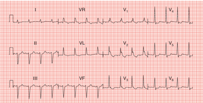
Isolated Posterior MI (6)
2. aVR ST Elevation with Diffuse ST Depression
This pattern lacks contiguous ST elevations required for STEMI criteria, but it often reflects severe global subendocardial ischemia.
Findings include:
● ST elevation in aVR (often ≥ 1 mm) AND
● Widespread ST depression across multiple leads
● Possible elevation in V1
So, how deadly is it?
That’s a controversial question right now. Ultimately, routine emergent STEMI activation is not warranted. (7)
For a time, this pattern was thought to represent acute left main occlusion. However, a cornerstone study conducted at the University of Arizona from 2014-2018 found that, among 79 patients with this pattern who underwent angiography, only 8 (around ~10%) had acute thrombotic coronary occlusion, and none of those occlusions were in the left main or left anterior descending arteries. (7)
Despite the low acute occlusion rate, this pattern still has exceptionally high mortality risk. In-hospital mortality for this pattern is as high as 31%, which is 5x that of STEMI without this pattern. (7)
aVR STE with multi-lead depression signals the presence of severe left main or triple vessel coronary disease, but there is currently no way to distinguish acute occlusion from chronic severe stenosis with superimposed demand ischemia. (8) Thus, high clinical suspicion for an acute occlusive event must be maintained, but emergent PCI is not warranted as a universal measure for this pattern given such high prevalence of conditions causing this pattern that are not amenable to catheter-directed solutions.
3. Hyperacute T Waves
These are often the earliest ECG manifestation of coronary occlusion and may precede overt ST elevation. They are associated with dynamic evolution over minutes!
Findings include:
● Tall, broad, symmetric T waves in contiguous leads
● Possible reciprocal ST depression
So, how deadly is it?
This pattern is incredibly high risk, as it is pathognomonic with acute coronary occlusion. It is also a favorable window for detection. The cath lab needs to be activated immediately!
Extensive transmural injury has not yet occurred, but delayed reperfusion leads to mortality comparable to STEMI. (9)
In a study of 147 anterior wall MIs, patients who presented with hyperacute T waves prior to development of ST elevation had a 0% in-hospital mortality rate. (9) You can save your patient's life by detecting this and acting quickly!
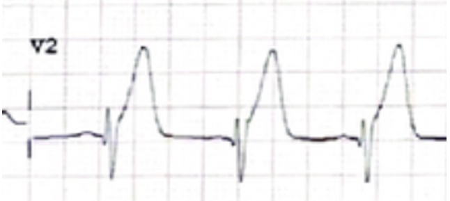
Hyperacute T Waves (10)
4. Left Bundle Branch Block or Right Ventricular Paced Rhythm (VPR) Meeting Modified Sgarbossa Criteria
Criteria include:
● Concordant ST elevation ≥1 mm in any lead with a positive QRS complex OR
● Concordant ST depression ≥1 mm in any of V1 - V3 OR
● Discordant ST elevation ≥1 mm and ≥25% of the preceding S-wave depth in any lead
So, how deadly is it?
If modified Sgarbossa criteria are met, call the cath lab immediately! Specificity for acute occlusion is as high as 99% for LBBB and 96% for VPR meeting criteria. (11)
Crude mortality is much higher for this STEMI equivalent. When adjusted for comorbidity of the patient population who typically presents with these patterns, however, mortality is similar to STEMI. (12)
Please note, new LBBB on its own is no longer a STEMI equivalent! That being said, do not dismiss a symptomatic patient with new or old LBBB not meeting modified Sgarbossa Criteria. Reach for POCUS or serial troponins in these patients if you have a high clinical suspicion. Don’t call the cath lab yet, though, as the false activation rate for these is near 40%. (13)
5. Wellens Syndrome
This pattern is classically associated with impending anterior wall MI.
Patients are often pain-free at the time of ECG acquisition.
Findings include:
● Deeply inverted or biphasic T waves in V2–V3
● Usually minimal ST elevation
In a study of 641 LAD occlusion patients, Wellens was identified in 6.7% of cases. (14)
So, how deadly is it?
Wellens on its own does NOT indicate acute occlusive infarction; rather, it is a signal of critical proximal LAD stenosis and high risk for OMI in the near future.
This pattern is best treated with urgent coronary angiography, typically within 24 hours. When treated with early invasive strategy, mortality is not increased compared to other NSTEMI presentations. (15,16)
In the meantime, start DAPT, anticoagulation, and a statin for your patient.
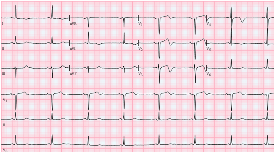
Wellens Syndrome (17)
Please see our prior NUEM blog post for further information on the different types of Wellens syndrome patterns: https://www.nuemblog.com/blog/stemi-to-omi
6. New Right Bundle Branch Block
This pattern must be associated with ischemic symptoms to be STEMI equivalent!
It is most commonly due to proximal LAD occlusion but also described in acute left main occlusion. (18)
Findings include: (19)
● Classic findings of RBBB, new compared to prior ECG
● Possible fascicular blocks, STEs, and/or STDs
● Possible concordant elevation in anterior leads with RSR’
● Possible discordant depression in anterior leads with RSR’
So, how deadly is it?
This is the deadliest but least recognized pattern discussed here. Recent literature supports emergent reperfusion consideration in these patients. (18) It is difficult to estimate exact incidence given frequent overlap with other concurrent ECG changes.
New or presumed new RBBB with ischemic symptoms was associated with 18.8% in-hospital mortality in one study, higher than any presentation of LBBB. More than half of these new RBBB patients were found to have complete obstruction. (20)
Concurrent ST-T wave changes are not required.
7. De Winter Sign
This is a rare but highly specific pattern associated with proximal LAD stenosis.
Findings include:
● Upsloping ST depression at J-point with tall symmetric T waves in precordial leads AND
● Absence of ST elevation in anterior leads
● Possible slight ST elevation in aVR
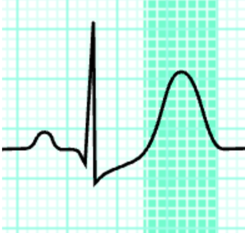
De Winter Sign (21)
In the same study of 641 LAD occlusion patients, De Winter was only identified twice (~0.3%). (14) Another study of 967 patients with anterior myocardial infarction identified De Winter pattern 30 times (~3.1%). (22)
So, how deadly is it?
Very deadly. This pattern almost always indicates acute occlusion and should be treated identical to an anterior STEMI with immediate angiography +/- PCI. Call the cath lab!
There is recent early evidence that De Winter pattern is more prevalent in those with preexisting multivessel coronary disease. As a result of cardiac preconditioning, De Winter Sign may be associated with lower in-hospital mortality (3% vs 18%, p<0.05) and less frequent malignant arrhythmic degeneration than those meeting classic anterior STEMI criteria. Still, this should not change our management in the acute setting. (22)
8. Aslanger's Pattern
This is one of the newest additions to STEMI-equivalent literature, first defined in 2020 by Aslanger et al. (23)
Look out for this pattern in patients with known multivessel coronary artery disease. It is classically described as suggesting an acute inferior RCA occlusion in a patient with other concurrent critical coronary artery stenosis. (23)
Findings include:
● ST elevation in lead III AND
● ST depression in at least one of V4 - V6 with a positive T wave AND
● ST elevation magnitude in V1 > V2
Without contiguous ST elevations in lead II or aVF, this pattern does not meet classic STEMI criteria and has been historically classified as NSTEMI.
So, how deadly is it?
It’s hard to tell given the limited data that exists. Preliminary findings suggests in-hospital mortality rates comparable to inferior STEMI (5% vs 4%, p >0.05). (23) Thus, for the time being, we should treat this pattern as we would an inferior STEMI. Call the cath lab immediately! Unlike other patterns, both emergent PCI and CABG are appropriate revascularization strategies. (24)
Classically denoted as an NSTEMI, this pattern was recently found to be associated with higher occlusive lesion rates on angiography and larger infarct size than typical NSTEMIs, prompting its new denotation as a STEMI equivalent. (23)
Although initially thought specific to RCA inferior occlusion, recent case reports have demonstrated this pattern in independent left-sided lesions as well, including LAD, LCx, and left main. (24,25,26,27)
For reference, here are the traditional STEMI patterns, classified by frequency and mortality (28,29,30):
Inferior location (classically involving RCA or LCx), frequency ~37-54% of all STEMIs with in-hospital mortality ranging from 3.6-5%
Anterior location (classically involving LAD), frequency ~33-41% of all STEMIs with in-hospital mortality ranging from 6.5-8.6%
Lateral location (classically involving Lcx), frequency ~3-13% of all STEMIs with in-hospital mortality similar to inferior STEMI
Multilocation, frequency ~17% of all STEMIs with the mortality higher than any of those above
References:
1. Kola M, Shuka N, Meyers HP, Zaimi Petrela E, Smith SW. OMI/NOMI: Time for a New Classification of Acute Myocardial Infarction. J Clin Med. 2024 Sep 2;13(17):5201. doi: 10.3390/jcm13175201. PMID: 39274412; PMCID: PMC11395726.
2. Shroyer S, Mehta S, Thukral N, Smiley K, Mercaldo N, Meyers HP, Smith SW. Accuracy of cath lab activation decisions for STEMI-equivalent and mimic ECGs: Physicians vs. AI (Queen of Hearts by PMcardio). Am J Emerg Med. 2025 Nov;97:193-199. doi: 10.1016/j.ajem.2025.07.061. Epub 2025 Jul 30. PMID: 40763602.
3. Rao, S, O’Donoghue, M, Ruel, M. et al. 2025 ACC/AHA/ACEP/NAEMSP/SCAI Guideline for the Management of Patients With Acute Coronary Syndromes: A Report of the American College of Cardiology/American Heart Association Joint Committee on Clinical Practice Guidelines. JACC. 2025 Jun, 85 (22) 2135–2237. https://doi.org/10.1016/j.jacc.2024.11.009
4. ECGAI. ECG-V1 Dataset. Roboflow Universe. Available at: https://universe.roboflow.com/ecgai/ecg-v1. Accessed May 7, 2026.
5. Meyers HP, Bracey A, Lee D, Lichtenheld A, Li WJ, Singer DD, Rollins Z, Kane JA, Dodd KW, Meyers KE, Shroff GR, Singer AJ, Smith SW. Ischemic ST-Segment Depression Maximal in V1-V4 (Versus V5-V6) of Any Amplitude Is Specific for Occlusion Myocardial Infarction (Versus Nonocclusive Ischemia). J Am Heart Assoc. 2021 Dec 7;10(23):e022866. doi: 10.1161/JAHA.121.022866. Epub 2021 Nov 15. PMID: 34775811; PMCID: PMC9075358.
6. Waldo SW, Brenner DA, Li S, Alexander K, Ganz P. Reperfusion times and in-hospital outcomes among patients with an isolated posterior myocardial infarction: insights from the National Cardiovascular Data Registry (NCDR). Am Heart J. 2014 Mar;167(3):350-4. doi: 10.1016/j.ahj.2013.11.011. Epub 2013 Dec 10. PMID: 24576519.
7. Harhash AA, Huang JJ, Reddy S, Natarajan B, Balakrishnan M, Shetty R, Hutchinson MD, Kern KB. aVR ST Segment Elevation: Acute STEMI or Not? Incidence of an Acute Coronary Occlusion. Am J Med. 2019 May;132(5):622-630. doi: 10.1016/j.amjmed.2018.12.021. Epub 2019 Jan 9. PMID: 30639554.
8. Kazemi E, Mansoursamaei A, Bijan M, Hosseinzadeh A, Sheibani H. The prognostic effect of ST-elevation in lead aVR on coronary artery disease, and outcome in acute coronary syndrome patients: a systematic review and meta-analysis. Eur J Med Res. 2022 Dec 21;27(1):302. doi: 10.1186/s40001-022-00931-5. PMID: 36539835; PMCID: PMC9769006.
9. Birnbaum Y, Sclarovsky S, Blum A, Mager A, Gabbay U. Prognostic significance of the initial electrocardiographic pattern in a first acute anterior wall myocardial infarction. Chest. 1993 Jun;103(6):1681-7. doi: 10.1378/chest.103.6.1681. PMID: 8141879.
10. Post-infectious acute myocardial infarction following COVID-19 infection in a young patient. ResearchGate Scientific Figure. Available at: https://www.researchgate.net/figure/A-Electrocardiogram-at-admission-showing-hyperacute-T-waves-from-V-2-V-4-leads-B_fig1_368968186. Accessed May 7, 2026.
11. Neeland IJ, Kontos MC, de Lemos JA. Evolving considerations in the management of patients with left bundle branch block and suspected myocardial infarction. J Am Coll Cardiol. 2012 Jul 10;60(2):96-105. doi: 10.1016/j.jacc.2012.02.054. PMID: 22766335; PMCID: PMC3402162.
12. Meyers HP, Limkakeng AT Jr, Jaffa EJ, Patel A, Theiling BJ, Rezaie SR, Stewart T, Zhuang C, Pera VK, Smith SW. Validation of the modified Sgarbossa criteria for acute coronary occlusion in the setting of left bundle branch block: A retrospective case-control study. Am Heart J. 2015 Dec;170(6):1255-64. doi: 10.1016/j.ahj.2015.09.005. Epub 2015 Sep 24. PMID: 26678648.
13. Erne P, Iglesias JF, Urban P, Eberli FR, Rickli H, Simon R, Fischer TA, Radovanovic D. Left bundle-branch block in patients with acute myocardial infarction: Presentation, treatment, and trends in outcome from 1997 to 2016 in routine clinical practice. Am Heart J. 2017 Feb;184:106-113. doi: 10.1016/j.ahj.2016.11.003. Epub 2016 Nov 10. PMID: 28224924.
14. Palladino N, Shah A, McGovern J, Burns K, Coughlin R, Joseph D, Cone DC. STEMI Equivalents and Their Incidence during EMS Transport. Prehosp Emerg Care. 2022 Jan-Feb;26(1):48-54. doi: 10.1080/10903127.2020.1863533. Epub 2021 Feb 5. PMID: 33320732.
15. Fujii T, Ikari Y. Clinical outcomes in acute coronary syndrome after presentation of unique electrocardiographic findings. J Electrocardiol. 2024 Jul-Aug;85:31-36. doi: 10.1016/j.jelectrocard.2024.05.100. Epub 2024 May 28. PMID: 38823213.
16. Zhou L, Gong X, Dong T, Cui HH, Chen H, Li H. Wellens' syndrome: incidence, characteristics, and long-term clinical outcomes. BMC Cardiovasc Disord. 2022 Apr 16;22(1):176. doi: 10.1186/s12872-022-02560-6. PMID: 35429969; PMCID: PMC9013025.
17. Jer5150. Wellens’ Syndrome.png. Wikimedia Commons. Published May 25, 2012. Available at: https://commons.wikimedia.org/wiki/File:Wellens%27_Syndrome.png. Accessed May 7, 2026
18. Roshan PK. Right bundle-branch block (RBBB) and acute coronary syndrome (ACS): A narrative review. Am J Emerg Med. 2026;99:325-334. doi:10.1016/j.ajem.2025.10.037. Available at: https://www.sciencedirect.com/science/article/pii/S0735675725006989. Accessed May 7, 2026.
19. McLaren J. ECG Cases 12: RBBB and Occlusion MI. Emergency Medicine Cases. Published August 11, 2020. Accessed May 7, 2026.
20. Widimsky P, Rohác F, Stásek J, Kala P, Rokyta R, Kuzmanov B, Jakl M, Poloczek M, Kanovsky J, Bernat I, Hlinomaz O, Belohlávek J, Král A, Mrázek V, Grigorov V, Djambazov S, Petr R, Knot J, Bílková D, Fischerová M, Vondrák K, Maly M, Lorencová A. Primary angioplasty in acute myocardial infarction with right bundle branch block: should new onset right bundle branch block be added to future guidelines as an indication for reperfusion therapy? Eur Heart J. 2012 Jan;33(1):86-95. doi: 10.1093/eurheartj/ehr291. Epub 2011 Sep 1. PMID: 21890488; PMCID: PMC3249219.
21. MrBean90. De Winter’s T wave (ECG).svg. Wikimedia Commons. Published January 23, 2012. Available at: https://commons.wikimedia.org/wiki/File:De_Winter%27s_T_wave_%28ECG%29.svg. Accessed May 7, 2026.
22. Alireza M, Nader A, Alireza F, Babak B, Bahare G. Traces of cardioprotection behind the uncertainty of the de winter pattern. J Electrocardiol. 2025 Sep-Oct;92:154056. doi: 10.1016/j.jelectrocard.2025.154056. Epub 2025 Jun 28. PMID: 40617188.
23. Aslanger E, Yıldırımtürk Ö, Şimşek B, Sungur A, Türer Cabbar A, Bozbeyoğlu E, Karabay CY, Smith SW, Değertekin M. A new electrocardiographic pattern indicating inferior myocardial infarction. J Electrocardiol. 2020 Jul-Aug;61:41-46. doi: 10.1016/j.jelectrocard.2020.04.008. Epub 2020 Apr 13. PMID: 32526537.
24. Zhong Z. Aslanger pattern: A case report. J Int Med Res. 2025 Mar;53(3):3000605251327380. doi: 10.1177/03000605251327380. Epub 2025 Mar 28. PMID: 40152677; PMCID: PMC11954596.
25. Agnese MAVD, Copetti AS, de Ferreira Filho SF, de Ferreira PR, Leiria TLL. Aslanger's pattern with acute lesion in the left coronary system: A case report. J Electrocardiol. 2024 Nov-Dec;87:153807. doi: 10.1016/j.jelectrocard.2024.153807. Epub 2024 Sep 15. PMID: 39305728.
26. Liu M, Liang L, Zhang T, Qin S, Zeng J, Zhong G. Aslanger's and de Winter's patterns associated with acute left coronary artery disease: A case report of two patients. J Electrocardiol. 2026 Apr 9;96:154256. doi: 10.1016/j.jelectrocard.2026.154256. Epub ahead of print. PMID: 41996725.
27. Victor A, P M N, K T, Sheriff N. Atypical Aslanger pattern in inferolateral myocardial infarction - A new variant to look out for? J Electrocardiol. 2024 Nov-Dec;87:153786. doi: 10.1016/j.jelectrocard.2024.153786. Epub 2024 Aug 30. PMID: 39244814.
28. Thygesen, K, Alpert, J, Jaffe, A. et al. Fourth Universal Definition of Myocardial Infarction (2018). JACC. 2018 Oct, 72 (18) 2231–2264. https://doi.org/10.1016/j.jacc.2018.08.1038
29. Tzimas G, Antiochos P, Monney P, Eeckhout E, Meier D, Fournier S, Harbaoui B, Muller O, Schläpfer J. Atypical Electrocardiographic Presentations in Need of Primary Percutaneous Coronary Intervention. Am J Cardiol. 2019 Oct 15;124(8):1305-1314. doi: 10.1016/j.amjcard.2019.07.027. Epub 2019 Jul 27. PMID: 31455501.
30. Wagner GS, Macfarlane P, Wellens H, Josephson M, Gorgels A, Mirvis DM, Pahlm O, Surawicz B, Kligfield P, Childers R, Gettes LS, Bailey JJ, Deal BJ, Gorgels A, Hancock EW, Kors JA, Mason JW, Okin P, Rautaharju PM, van Herpen G; American Heart Association Electrocardiography and Arrhythmias Committee, Council on Clinical Cardiology; American College of Cardiology Foundation; Heart Rhythm Society. AHA/ACCF/HRS recommendations for the standardization and interpretation of the electrocardiogram: part VI: acute ischemia/infarction: a scientific statement from the American Heart Association Electrocardiography and Arrhythmias Committee, Council on Clinical Cardiology; the American College of Cardiology Foundation; and the Heart Rhythm Society. Endorsed by the International Society for Computerized Electrocardiology. J Am Coll Cardiol. 2009 Mar 17;53(11):1003-11. doi: 10.1016/j.jacc.2008.12.016. PMID: 19281933.
Expert Commentary

Thank you Dr. Eckburg and Dr. Oladeji for this nice review. The idea of OMI instead or as an adjunct to STEMI has grown over the years; I have found thinking about it as “STEMI and STEMI equivalents” is a reasonable way to wrap my head around it. Rather than “only STEMIs benefit from immediate cath lab” or “STEMI is outdated and misses things!”, I see it as another example of “STEMI is a useful model and we can extend it a bit.” (Aside: this is also my general view of lactate in sepsis; vital signs are useful for identifying sepsis and as markers as resuscitation; lactate can help us find more patients with sepsis who likely benefit from further resuscitation.) Given how entrenched STEMI is in our systems, there is some interdisciplinary nuance in how I think about these. See also this great detailed review by Ricci and colleagues (including Stephen Smith, who has done a lot to spread the OMI word, and modify the Sgarbossa criteria) in Annals of EM (REF1).
1.) Posterior MI – I have a low threshold to get posterior- and right-sided ECGs in patients who either have anterior depressions or just a high level of concern. With STD in V1-V3, I immediately get a posterior ECG. With patients who look like they might be having an MI, I get a “5-pack”: 1- standard ECG; 2- repeat standard; 3&4- right & posterior; 5; another repeat standard ECG.
As there are more than 1 way to place posterior and right-sided leads, I immediately confirm with the tech which leads are which and mark it all on my ECG with an (industrial strength [REF2]) Sharpie:
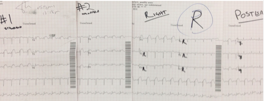
Especially when it’s a stressful situation like “is this patient having an (occult) STEMI equivalent, I want to make it as easy as possible to see which ECG is which.
2.) AVR – This one is a little trickier as it seems some of the earlier excitement about STE in AVR overstated how many of these were STEMI vs 3 vessel disease. Context is key, eg, does the patient look like they’re having a STEMI? Is Cardiology in-house? How easy is it to very quickly talk about the patient and ECG with a cardiologist?
3.) Hyperacute T-waves – I find this one harder to articuolate but easy to spot qualitatively. Big, wide (and not thinning out quickly as they go up), tall T-waves, especially bigger than the R wave, especially new tall T-waves in V1, are ominous.
4) LBBB/Smith-modified Sgarbossa – I also found the original Sgarbossa criteria confusing. You need ≥3 points; 2 of the criteria are each ≥3 on their own, and the 3rd is <3… so there are effectively 2 criteria. The Smith-modified Sgarbossa are a bit more intuitive: the first two are essentially the same – I simplify it to “CONCORDANCE BAD” and if there is concordance, look up the qualifying points. And now, VERY DISCORDANT STE is also bad.
5) Wellens Syndrome – I looked into this for my CCU talk as a PGY2 and there are 2 things that I love about Wellens: a) there’s not a ton to learn about it, so you can get up to speed very quickly, and, b) we spend a lot of time & effort trying to figure out which patients without STEMI or elevated troponins may still have UA/ACS, and patients with Wellens ECG do. They “should” be pain-free at the time of the ECG, and I do nto activate these but I do talk to a sufficiently senior cardiologist to suggest they should get a cath not now but soon, eg tomorrow (or later today if it’s feasible). And do not get stress tests for patients with Wellens ECGs, something like 1 in 4 will have a STEMI during the stress (REF3).
As for the rest, these are nice reminders that we keep learning and finding new ways to help patients when they need it. I remember when De Winter sign came out during my residency (REF4).
References
1. https://www.annemergmed.com/article/S0196-0644(24)01250-2/fulltext
2. https://pubmed.ncbi.nlm.nih.gov/20055641/
3. https://pubmed.ncbi.nlm.nih.gov/42661459/
4. https://www.nejm.org/doi/full/10.1056/NEJMc0804737
Seth Trueger, MD, MPH, FACEP
Associate Professor, Department of Emergency Medicine
Director, Digital Media, Center for Bedside Medicine
Northwestern University
Digital Media Editor, JAMA Network Open
How To Cite This Post:
[Peer-Reviewed, Web Publication] Eckburg, A. Oladeji, Afolayan. (2026, October 18). STEMI Equivalents [NUEM Blog. Expert Commentary by Trueger, S]. Retrieved from http://www.nuemblog.com/blog/stemi-equivalents





Blog regarding how to emergently manage acute radiological injuries.