

Overview of diagnosis and management of burns secondary to hydrofluoric acid.
Posts tagged #burn
Beyond the Burns: Toxic House Fire Gases

Written by: Adam Payne, MD (NUEM ‘24) Edited by: Emily Wessling, MD (NUEM ‘22)
Expert Commentary by: Justin Seltzer, MD (NUEM ‘21)

Expert Commentary
Congratulations to Drs. Payne and Wessling on an excellent post.
Management of toxic gas exposure from a house fire is essential knowledge for all emergency physicians. The two major toxic gases of interest are carbon monoxide and hydrogen cyanide; it is important to note that fires in other environments, such as factories or industrial sites, may result in alternative exposures based on the nature of the fire and materials present. The post goes into detail on the pathophysiology, signs, symptoms, and diagnosis of both carbon monoxide and cyanide poisonings, so this commentary will focus on the clinical approach.
I recommend the following simplified process to streamline diagnosis and treatment decision making:
1. Start high flow oxygen (15L NRB) immediately (or, if intubated, give 100% FiO2)
High flow oxygen reduces the half life of carbon monoxide from ~5 hours to ~90 minutes. Oxygen can be discontinued once carboxyhemoglobin normalizes (<2%)
2. Obtain a carboxyhemoglobin level, lactic acid level, and a venous blood gas (arterial is unnecessary unless oxygenation is also a concern) as soon as possible
Unfortunately, there is some disagreement as to what constitutes a “toxic” carboxyhemoglobin level. Weaver, et al. established 25% as an inflection point for the development of severe sequelae, which is now a commonly used (though not universally agreed upon) threshold value. Also, when evaluating a carboxyhemoglobin level, it is essential to consider the time period prior to the level being drawn to avoid false reassurance. For example, a level drawn after 90 minutes of high flow oxygen will be reduced by roughly 50% and interpreted accordingly. Further, do not rely on external co-oximeters alone to rule out carbon monoxide poisoning given limited sensitivity at the moment (though the technology will likely improve over time).
3. Treat with hydroxocobalamin empirically if symptomatic and/or if lactic acid elevated
Hydroxocobalamin is a low risk intervention with significant potential therapeutic benefit, so it should be given early if there is any clinical concern. The other major cyanide antidote, sodium thiosulfate, should only be used if hydroxocobalamin is not available as it has no efficacy advantage and an undesirable side effect profile. It is essential to avoid using nitrites, as inducing methemoglobinemia in the setting of coincidental carbon monoxide poisoning can dramatically worsen tissue hypoxia.
An elevated lactic acid is a surrogate for cyanide poisoning, specifically a level of 8-10 mmol/L or greater is sensitive and should prompt intervention. More modest lactic acid elevations are less likely to be related to cyanide poisoning and should not prompt intervention, especially in an asymptomatic patient, unless the level is persistently elevated despite adequate resuscitation. Cyanide levels, while diagnostic, are of no acute clinical utility since they are rarely available in a timely manner.
4. Consider hyperbaric oxygen therapy (HBOT) if readily available
HBOT for the treatment of carbon monoxide poisoning is controversial. While HBOT reduces the half life of carbon monoxide to roughly 20-30 minutes, HBOT is not used for this purpose alone. In fact, HBOT is primarily used to reduce associated cognitive, behavioral, and neurologic changes (collectively known as delayed neuropsychiatric sequelae). There is lower quality evidence for reduced myocardial infarction and mortality risk as well.
However, several factors limit HBOT use. Primarily, it is not universally available at most institutions, incurring the risk and cost of transport to a distant site. The benefit is thought to be highest when the treatment is performed early (ideally within six hours of exposure), which adds to the logistical burden. Additionally, many chambers are not operated on nights and weekends, and of those available at off hours, many are unable to accommodate intubated patients. Recognizing the controversial nature of HBOT, the 2016 ACEP position statement noted that HBOT or high flow, normobaric oxygen can be used to treat carbon monoxide poisoning; though it likely carries clinical benefit in certain situations, at this time HBOT is not the standard of care for severe carbon monoxide poisoning. Rather, it should be offered if it can be readily and reasonably arranged.
Keeping this in mind, the following are generally accepted indications for HBOT in the setting of carbon monoxide poisoning
Loss of consciousness associated with exposure
Altered mental status
Focal neurologic changes
Evidence of end organ ischemia (pH ≤ 7.1, EKG changes, elevated troponin, angina)
Pregnancy (with some resources citing a level ≥20%)
Carboxyhemoglobin level ≥25%
Importantly, these are not hard and fast rules and there is no firm guideline mandating when HBOT should or should not be used. As a result, it is prudent to involve a medical toxicologist early in the process.
In summary, a few key take home points:
Carbon monoxide and cyanide are strongly associated with house fires – assume exposure to both until proven otherwise
It is reasonable to treat any undifferentiated, symptomatic patient with high flow oxygen and hydroxocobalamin empirically; asymptomatic patients can wait safely for blood work on high flow oxygen alone
The decision making regarding use of HBOT, in particular, is complex – early consultation with a medical toxicologist is strongly encouraged

Justin Seltzer, MD
Toxicology Fellow
Department of Emergency Medicine
University of California, San Diego
How To Cite This Post:
[Peer-Reviewed, Web Publication] Payne, A. Wessling, E. (2022, May 2). Toxic House Fire Gases. [NUEM Blog. Expert Commentary by Seltzer, J]. Retrieved from http://www.nuemblog.com/blog/toxic-house-fire-gases
Other Posts You May Enjoy


Managing Minor Thermal Burns in the ED

Written by: Mitch Blenden, MD (NUEM ‘24) Edited by: Vytas Karalius, MD, MPH, MA (NUEM ‘22) Expert Commentary by: Matt Levine, MD

Expert Commentary
Dr. Blenden and Dr. Karalius provided an excellent handy, high-yield, quick reference of thermal burn considerations in the ED. There are some nuances of thermal burn care that I’d like to provide further commentary:
A pitfall is underestimating the severity of the burn when the patient presents within a few hours of the event. Burn appearance evolves over 24-48 hours. What initially appears as erythematous skin can be covered in bullae the next day. Consider a repeat examination in 24-48 hours, or at least discuss with the patient the possibility that this may occur and what to do if it does. Otherwise, if you initially diagnosed the patient with superficial burns and provided only instructions for superficial burns, which require little treatment or follow-up, the patient can be set up for a worse outcome when these burns subsequently declare themselves to be partial thickness.
For years, most non-facial burns were sent home with instructions to use silver sulfadiazine (AKA Silvadene) cream. This would require teaching of how to apply and remove it. The cream needs to be removed daily before applying a new coat (I always sent the patient home with tongue blades to scrape it off). The benefits of this are that it debrides some nonviable tissue when the cream is removed and provides a moist antimicrobial barrier. The down sides are that removal can be painful and some patients have difficulty performing this procedure, which requires teaching. Silver sulfadiazine can also cause skin staining. There is scant evidence recommending one topical antimicrobial over another. For these reasons, practice (including mine) has evolved in many places to simply prescribe whatever antibiotic ointment is on hand for ease of use and less painful and technically challenging application.
Another controversy is whether to debride blisters and bullae or leave them intact. This is another area without definitive evidence and practice is often guided by gestalt, local custom, or prior teachings. On one hand, intact bullae can be thought of as “sterile” coverings and may be less painful than dermal layers exposed to air and friction. On the other hand, when bullae rupture, the patient is left with dead skin which can be a nidus for infection. My practice has been to leave small blisters intact and debride large bullae if it seems like they will soon rupture and leave the patient with hanging skin fragments. If the patient has reliable follow up burn care then I may choose a less aggressive approach in debriding. Other clinicians are likely to give alternate approaches so ask your attendings what they do in these scenarios so you can develop a practice pattern that makes sense to you.

Matthew Levine, MD
Associate Professor of Emergency Medicine
Department of Emergency Medicine
Northwestern Memorial Hospital
How To Cite This Post:
[Peer-Reviewed, Web Publication] Blenden, M. Karalius, V. (2021, Oct 18). Managing Minor Thermal Burns in the ED. [NUEM Blog. Expert Commentary by Levine, M]. Retrieved from http://www.nuemblog.com/blog/managing-minor-thermal-burns
Other Posts You May Enjoy


Cold Injury Management in the ED
Written by: Sean Watts, MD (PGY-2) Edited by: David Kaltman, MD (PGY-4) Expert commentary by: Pinaki Mukherji, MD
Learning Objectives:
• Recognize the signs and symptoms of freezing injury
• Understand the pathophysiology of freezing cold injury and how it manifests in the delineated signs and symptoms
• Discuss the treatment practices for managing freezing injury in the emergency department
• Discuss barriers to effective treatments for these conditions, and current research to improve outcomes
Introduction
As interests in outdoor activities and the rates of homelessness have increased over the past twenty years, so has the level of patients presenting with cold injuries. Due to this increasing prevalence, it is important for emergency physicians to understand the signs, symptoms, and management of these injuries. This is especially true for emergency physicians working in areas where mountaineering, skiing, ice-climbing and other outdoor activities are popular, or in areas with significant cold exposure and large populations of undomiciled patients.
Cold injuries get divided into two categories: non-freezing injuries and freezing injuries. Non-freezing injuries include trench foot, pernio, panniculitis, and cold urticaria and are generally due to prolonged exposure to damp, non- freezing conditions. Freezing injuries include frostbite and its associated severity classifications. Of these categories frostbite remains the most severe and, and can present in a range of symptoms from clear blisters and cyanosis, to hard non-deforming necrotic skin.
Treatment of both non-freezing cold injuries and frostbite are similar, and co-presentation often occurs. However, if a patient presents with concomitant hypothermia, this should be treated first. The course of treatment for hypothermia should be guided by a patient's level of consciousness, shivering intensity, and cardiovascular stability in the field since accurate temperature readings cannot always be obtained. A rectal temperature should be obtained in the emergency department and active rewarming measures such as heat pads, or heated humidified oxygen should be utilized, and application of these rewarming devices should be applied to the areas of the body with the potential for greatest convective heat loss—back, axilla. While rewarming takes place, the practitioner should monitor vitals and provide CPR, AED, and intubation, as necessary.
Freezing Injury
The mechanism that accounts for frostbite is due to direct cell death due cold exposure and further cell death due to ischemia. As water freezes in tissues it expands—poking holes in the cellular membrane—leading to hyperosmotic cell death. This cell death is further exacerbated by what is known as the hunting reaction—an alternating freeze/ thaw cycle due to local alternating vasoconstriction and vasodilation. Emboli form in the vasculature due to endothelial damage, resulting in ischemia, leading to destruction of the microvasculature and localized cell death.
Frostbite can occur anywhere on the body but generally occurs on the distal extremities, face, nose, and ears. The injured area often appears pale and feels stiff and cold, and patients endorse stinging and numbness.
Now, frostbite gets divided into three zones: the zone of coagulation, the zone of hyperemia, and the zone of stasis. The zone of coagulation is distal and where the cellular damage is most severe. The zone of hyperemia is superficial, proximal, and has the least cellular damage. The zone of stasis is between the two and has the most potential for intervention to salvage tissue.
Frostbite also gets characterized into four different classifications schemes based on severity of injury and prognosis of recovery. First-degree frostbite, also referred to as frostnip, is characterized by partial skin freezing with erythema, edema, and has excellent outcomes. Second-degree injury is defined by full-thickness skin freezing, formation of substantial edema, erythema, and the formation of clear blisters. Second degree injury has the most potential for intervention with modest outcomes. Third-degree injury is defined by damage that extends into the subdermis with associated hemorrhagic blisters and necrosis of skin and necrosis of skin, appearing as a blue-gray discoloration. Fourth-degree injury is characterized by further extension into adipose tissue, muscle, and bone, with little edema that forms a dry black eschar. Fourth degree frostbite tends to have the “mummified” appearance of dry gangrene.

First Degree Frostbite

Second degree frostbite--note the clear filled blisters characteristic of this degree of frostbite
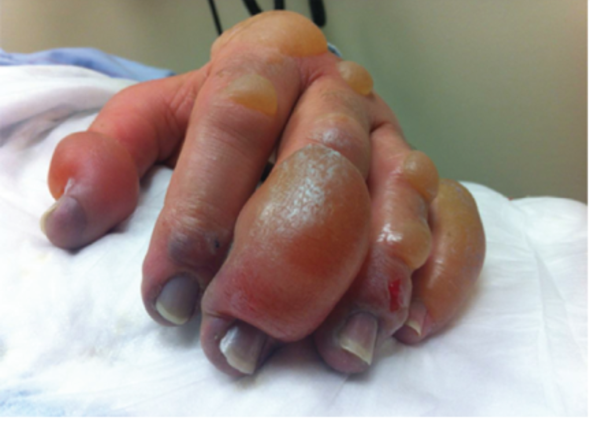
Third degree frostbite--note the areas of hemorrhagic blisters, characteristic of third degree frostbite

Fourth Degree Frostbite
In the field and the emergency department, treatment should be focused on preventing refreezing injury. It is imperative that active thawing measures not be initiated unless the thawed state can be maintained (remember the hunting reaction!). Preliminary measures to help thawing include hydration, administration of low molecular weight dextran (it has been shown to reduce blood viscosity and decrease thrombi), and NSAIDS that will reduce prostaglandin and thromboxane release. If it is possible to maintain a thawed state, active thawing can take place by submersion in a water bath maintained between 37 C and 39 C. The emergency physician will know the rewarming process is complete when the affected area becomes red or purple and is soft and pliable. If the affected area is an extremity, it should be elevated in order to prevent dependent edema from forming. When rewarming occurs patients will often note severe pain, and patients should be treated with parenteral opioid therapy. Other post-thaw therapy includes antithrombotic drugs—tPa has been used widely in addition to heparin, as well as vasodilating agents. When patients present within 24 hours, with multiple digits affected, or evidence of multiple limbs affected, intra- arterial tPA can be utilized along with intra arterial heparin. Iloprost has also been suggested for grade 2-4 frostbite when patients present <48 hours after injury. If blisters form, they should be treated with topical aloe vera cream every 6 hours, and tetanus immunization status should be assessed and given if needed.
One question that commonly arises in the emergency management of frostbite is does this injury need surgery? In general, early surgical intervention is not indicated for the management of frostbite. Studies have demonstrated that early surgery contributes to unnecessary tissue loss and poor cosmetic results. This stems from the inability to assess the depth of frostbite at its early stages and that tissue below blackened necrotic tissue is regenerating. Technetium (Tc)-99m scintigraphy often gets used after maximum rewarming therapy to predict long-term viability of affected tissue. Escharotomy is the only early surgical intervention indicated if the patient has range of motion or circulation abnormalities. Most patients with frostbite can be discharged from the emergency department with good follow up--barring a situation where the individual will simply be re exposed to cold temperatures or they require admission for pain management.
Future Directions
While non-freezing and freezing injuries continue to become common occurrences in austere environments and amongst undomiciled patient populations, developmental therapies to improve outcomes continue to be researched. For example, iloprost, as been gaining popularity, as it was recently used in Sweden with success. It is a prostacyclin analogue that mimic the effects of a sympathectomy and helps to prevent emboli from forming. In several studies it has proven more effective than tPA administration; however, it is not a currently approved FDA drug. More large scale clinical trials and cohort studies are needed, as many of these trials have low sample sizes.
Summary and Pearls
Suspect non-freezing or freezing injury in undomiciled patients or in patients with prolonged exposure in cold environments
Perform a thorough neurovascular exam of the afflicted digit/extremity, and attempt to grade if consistent with frostbite
Only begin rewarming if the warm state can be maintained
Frostbite, even when severe is not a surgical emergency
Consider iloprost or tPA for appropriate candidates
Expert Commentary
This is a nice overview of the spectrum of presentation of freezing injury. I would reinforce a few key points to give practical context in the treatment of these patients.
Systemic signs always take priority when resuscitating a cold injured patient. As such, the rewarming measures described will utilize dry heat, and target mental status and cardiovascular status. In contrast, in the rewarming of freezing injury, moist heat is always preferred.
As mentioned here, non-freezing and freezing injury coexist, and are not always easy to tell apart. While non-freezing injury may be outside of the scope of this blog post, severe cases of trench foot can appear similar to 2nd or 3rd degree frostbite injuries. Blister formation in non-freezing injury is rare, but can occur, and the sloughing of skin that occurs can be mistaken for ruptured blisters.
While the tissue damage of freezing injury can be severe, the deep necrosis that results is typically dry gangrene, whose natural course is auto-amputation. While a non-freezing injury like trench foot typically has better outcomes, severe cases can leave patients with circulatory compromise and non-intact skin, leading to wet gangrene and potentially sepsis.
Rapid rewarming of freezing injury (15 to 60 min.) is supported by animal models, with immersion being the preferred modality. The author above writes that rewarming should commence “if it is possible to maintain a thawed state.” For patients who have treatment initiated in the field, or en route to comprehensive care, this is a judgment not to be made lightly, as refreezing leads to much worse tissue destruction.
NSAIDs are indicated in the treatment of freezing injury, with an excellent safety to benefit ratio. The use of other agents aimed at improving consequences of thrombosis are less certain and should be reserved for more severe cases. Both tpa and iloprost have been associated with lower amputation rates in small studies and case series, along with PGE1 and isosorbide dinitrate as potential agents showing promise.
Low molecular weight dextran is recommended by Wilderness Medicine Society guidelines, with a minimal bleeding risk, but is often avoided if the patient is being considered for antithrombotic treatments such as tpa.
References
Sachs, C., Lehnhardt, M., Daigeler, A., Goertz, O. (2015). The Triaging and Treatment of Cold-Induced Injuries. Dtsch Arztebl Int., 112(44), 741-747. Doi:10.3238/arztebl.2015.0741
Petrone, P., Asensio, J., Marini, C. (2014). Management of accidental hypothermia and cold injury. Oct, 51(10):417-31. Doi: 10.1067/j.cpsurg.2014.07.004.
Imray, C., Grieve, A., Dhillon, S. (2009). Cold damage to the extremities: frostbite and non-freezing cold injuries. Postgrad Med J., Sep; 85(1007):481-8. Doi: 10.1136/pgmj.2008.068635

Pinaki Mukherji, MD
Assistant Professor
Department of Emergency Medicine
Department of Internal Medicine
Donald and Barbara Zucker School of Medicine at Hofstra/Northwell
References
Management of accidental hypothermia and cold injury. Petrone P, Asensio JA, Marini CP. Curr Probl Surg. 2014 Oct;51(10):417-31. doi: 10.1067/j.cpsurg.2014.07.004. Epub 2014 Jul 29.
Wilderness Medical Society practice guidelines for the out-of-hospital evaluation and treatment of accidental hypothermia: 2014 update.Zafren K1, Giesbrecht GG2, Danzl DF3, Brugger H4, Sagalyn EB5, Walpoth B6, Weiss EA7, Auerbach PS7, McIntosh SE8, Némethy M8,McDevitt M8, Dow J9, Schoene RB10, Rodway GW11, Hackett PH12, Bennett BL13, Grissom CK14.
Wilderness Environ Med. 2014 Dec;25(4 Suppl):S66-85. doi: 10.1016/j.wem.2014.10.010
Wilderness Medical Society Practice Guidelines for the acute treatment of Frostbite:2014 update. McIntosh SE1, Opacic M2, Freer L3, Grissom CK4, Auerbach PS5, Rodway GW6, Cochran A7, Giesbrecht GG8, McDevitt M9, Imray CH10, Johnson EL11, Dow J12, Hackett PH13. Wilderness Environ Med. 2014 Dec;25(4 Suppl):S43-54. doi: 10.1016/j.wem.2014.09.001.
Frostbite: a practical approach to hospital management. Handford C1, Buxton P2, Russell K3, Imray CE4, McIntosh SE5, Freer L6, Cochran A7, Imray CH8.Extrem Physiol Med. 2014 Apr 22;3:7. doi: 10.1186/2046-7648-3-7. eCollection 2014Gross, E., & Moore, J. (2012).
Using thrombolytics in frostbite injury. Journal of Emergencies, Trauma, and Shock, 5(3), 267. doi:10.4103/0974-2700.99709
Tintinalli, J. E., & Stapczynski, J. S. (2016). Tintinalli's emergency medicine: A comprehensive study guide. New York: McGraw-Hill.
Wrenn, K. (1991). Immersion foot. A problem of the homeless in the 1990s. Archives of Internal Medicine, 151(4), 785-788. doi:10.1001/archinte.151.4.785
Petrone, P., Kuncir, E., & Asensio, J. A. (2003). Surgical management and strategies in the treatment of hypothermia and cold injury. Emergency Medicine Clinics of North America, 21(4), 1165-1178. doi:10.1016/s0733-8627(03)00074-9
Gonzaga, T., Jenabzadeh, K., Anderson, C. P., Mohr, W. J., Endorf, F. W., & Ahrenholz, D. H. (2016). Use of Intra-arterial Thrombolytic Therapy for Acute Treatment of Frostbite in 62 Patients with Review of Thrombolytic Therapy in Frostbite. Journal of Burn Care & Research, 37(4). doi:10.1097/bcr.0000000000000245
How to Cite this Post
[Peer-Reviewed, Web Publication] Watts, S, Kaltman, D. (2020, Mar 16). Cold Management in the ED. [NUEM Blog. Expert Commentary by Mukherji, P]. Retrieved from https://www.nuemblog.com/blog/cold-injury
Featured


Understanding Inhalation Injury
Smoke inhalation injuries are one of the leading causes of morbidity and mortality in the United States. In emergency medicine, we must be aware of harbingers of impending airway issues that come along with this deadly injury. We discuss pathophysiology, diagnosis and management in this week's post.