Posts tagged #difficult airway

Accidental Tracheostomy Decannulation

Written by: Chezlyn Patton, MD (NUEM ‘27) Edited by: Keara Kilbane, MD (NUEM ‘25)
Expert Commentary by: Matt McCauley, MD (NUEM ‘21)


Introduction

Tracheostomy is a common procedure in the US with over 110,000 trachs placed annually (1). Complications occur at a rate of approximately 40-50%, however most complications are minor, with only 1% being catastrophic (1). Of these devastating complications, 90% occur within the first 10 days of placement. Overall approximately 15% of tracheostomies will be decannulated accidentally, and in a critical care setting, 50% of airway related deaths were associated with accidental tracheostomy decannulation (1, 2). 

One way to approach a decannulated tracheostomy tube could be with the acronym, IRMI; Investigate, Recannulate, Monitor, and Intubate (if you must). 

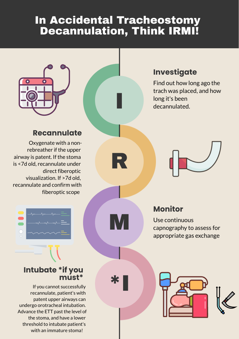
Investigate: How long ago was the tracheostomy tube placed? How long has it been out? What is the size of the trach? Is it cuffed or uncuffed? Why was the trach placed initially? 

  1. Cuffed vs. uncuffed: Is there a pilot balloon present? If yes, that indicates the trach is a CUFFED tracheostomy tube. 

  2. Size of tracheostomy tube: ALWAYS labeled on the neck flange. 

Figure 1: Size of trach tube identification on flange. Borrowed from https://tracheostomyeducation.com/tracheostomy-tubes/.

Figure 2: Tracheostomy tube parts labeled, borrowed from ©Linda L. Morris and M. Sherif Afifi, https://www.trachresource.com/table-of-contents/

Recannulate:  Oxygenate from above with non-rebreather or by blow by oxygen over the tracheostomy stoma if the patient is spontaneously breathing. If they are in respiratory distress or not spontaneously breathing, bag-valve-mask (BVM) oro-nasopharyngeal or over the stoma. You can use a pediatric mask to fit over the stoma or a LMA. Ensure to occlude the stoma if BVM from above or close the mouth if BVM from the stoma to prevent air leakage. Note, if the patient is ventilator dependent, you need a CUFFED tracheostomy tube. Obtain one tracheostomy tube of appropriate size, and a tube that’s a size down, as a stoma may begin to close the longer it’s out. For stomas less than 10 days old, grab a fiberoptic scope, as these will require recannulization under direct visualization. This is to minimize the risk of creating a false passage.

Figure 3: Depiction of creation of false passage through subcutaneous tissue when replacing tracheostomy tube. Borrowed from: Morris, L.L., Whitmer, A., & McIntosh, E. (2013). Tracheostomy care and complications in the intensive care unit. Critical care nurse, 33 5, 18-30 .

Otherwise, you can place blindly for the initial insertion. Ensure the obturator is placed inside the outer cannula tracheostomy tube prior to insertion, as it blunts the hard edge of the tracheostomy tube that can damage the membranous wall of trachea. If you meet any resistance, size down immediately, as the stoma has likely started to heal (even with a matured tracheostomy). Then remove the obturator and inflate the cuff to maintain placement (3,4).

Monitor: Once the trach tube is reinserted, it is important to monitor for appropriate placement and gas exchange. Continuous capnography is the gold standard for this. Additionally, the tube should be confirmed to be in the trachea through direct fiberoptic visualization of the trachea and carina. Be sure  to assess for complications such as creation of a false lumen, which could manifest as subcutaneous emphysema (5). 

Intubate if you must: If faced with a scenario where the tracheostomy tube cannot be passed through the stoma, and your patient is developing respiratory distress, you can intubate your patient orotracheally if they have a patent upper airway. The only exception to this is a patient who has had a laryngectomy, as those patients cannot be intubated orally and are obligate neck stoma breathers (6).

References

1.Bontempo, Laura J., and Sara L. Manning. "Tracheostomy emergencies." Emergency Medicine Clinics 37.1 (2019): 109-119.

2. Cheung, Nora Ham-Ting and Lena M. Napolitano. “Tracheostomy: Epidemiology, Indications, Timing, Technique, and Outcomes.” Respiratory Care 59 (2014): 895 - 919.

3. Rajendram, R., and N. McGuire. "Repositioning a displaced tracheostomy tube with an Aintree intubation catheter mounted on a fibre-optic bronchoscope." BJA: British Journal of Anaesthesia 97.4 (2006): 576-579.

4. Shah RK, Lander L, Berry JG, et al. Tracheotomy outcomes and complications: a national perspective. Laryngoscope 2012;122(1):25–9

5. Riley, Christine M.. “Continuous Capnography in Pediatric Intensive Care.” Critical care nursing clinics of North America 29 2 (2017): 251-258 .

6. McGrath B, Bates L, Atkinson D, et al, National Tracheostomy Safety Project. Multidisciplinary guidelines for the management of tracheostomy and laryngectomy airway emergencies. Anaesthesia 2012;67(9):1025–41.


Expert Commentary

Thank you for this concise summary of tracheostomy management. While most of the immediate complications of tracheostomy will occur in the ICU, these patients still frequent our emergency department with and without tracheostomy related emergencies. Despite this, patients with tracheostomy can be intimidating there is a general lack of knowledge about tracheostomy among healthcare professionals in general and emergency medicine trainees in specificity.1,2 

As you have outlined, understanding both the chronicity of the tracheostomy as well as the indication for the procedure are key history when managing a displaced tube. A patient who underwent tracheostomy for failure to liberate for the ventilator likely has a patent upper airway while one placed following an ENT surgery likely poses a significant challenge for orotracheal intubation! Obtaining this history (and handing off this key information when transitioning care) can be lifesaving. Significant care should be taken when replacing a tracheostomy through an immature stoma, the usual cited maturity date being 10 to 14 days old. These should always be replaced over a fiber-optic scope with visualization of the tracheal rings and carina prior to cuff inflation and ventilation by BVM or ventilator. Failure to do so can result in severe pneumomediastinum, pneumothorax, subcutaneous emphysema, and respiratory arrest if placement into a false tract is not recognized3 . Replacement of a tracheostomy tube into a mature tract can be done blindly with an obturator in place but care should also be taken if there are any signs of trauma or bleeding. When in doubt, play it safe and obtain fiber optic visualization!  

While trach replacement can be stressful under the wrong circumstances, patients with tracheostomy tubes can present with enumerable other emergencies. As with any high stress situation in resuscitation, it helps to fall back onto our ABCs, airway being of principle importance here. The immediate assessment of any patient with a tracheostomy tube in extremis should be focused on a singular question: can I ventilate the patient through this tube? In order for a patient to be effectively bagged or ventilated through a tracheostomy tube three things must be true. The tube must be patent and endotracheally placed, the tube must be cuffed, and the cuff must be inflated. Patency can be quickly assessed with passage of a flexible suction catheter. If this is unsuccessful, removal of the inner cannula (if present) and replacement with a fresh inner cannula can often resolve obstruction by secretions.  If obstruction is unable to resolve, you should oxygenate from above while preparing to replace the tracheostomy tube as you have elegantly outlined.  

The presence of a cuffed tube will be indicated by the presence of a pilot balloon, no reading of numbers or brand names needed! Finally, cuff inflation can be confirmed by palpation of the pilot balloon and assessing for any speech production or gurgling hear though the mouth. If the patient can phonate then the balloon is not properly inflated! If gentle inflation of the cuff does not resolve the air leak assume a ruptured cuff and replace the tracheostomy.  

Tracheostomy tube care and emergencies can be very intimidating but this procedure is a valuable tool for ICU and ventilator liberation. As emergency physicians, we need to be familiar with the nuances of these devices so we can safely manage the airway just as we would any sick patient. 

References

1. Whitcroft KL, Moss B, Mcrae A. ENT and airways in the emergency department: national survey of junior doctors’ knowledge and skills. J Laryngol Otol. 2016;130(2):183-189. doi:10.1017/S0022215115003102 

2. Darr A, Dhanji K, Doshi J. Tracheostomy and laryngectomy survey: do front-line emergency staff appreciate the difference? J Laryngol Otol. 2012;126(6):605-608; quiz 608. doi:10.1017/S0022215112000618 

3. Long B, Koyfman A. Resuscitating the tracheostomy patient in the ED. Am J Emerg Med. 2016;34(6):1148-1155. doi:10.1016/j.ajem.2016.03.049 

Matt McCauley, MD

Assistant Professor, Division of Critical Care  

UW BerbeeWalsh Department of Emergency Medicine  

Associate Medical Director  

UW Organ and Tissue Donation 


How To Cite This Post:

[Peer-Reviewed, Web Publication] Patton, C. Kilbane, K. (2024, Apr 15). Accidental Tracheostomy Decannulation. [NUEM Blog. Expert Commentary by McCauley, M]. Retrieved from http://www.nuemblog.com/blog/tracheostomy-decannulation


Other Posts You May Enjoy

Posted on April 15, 2024 and filed under Critical care, ENT.

C-Spine Intubation

Written by: Daniel Levine, MD (NUEM ‘24) Edited by: Zach Schmitz (NUEM ‘21)  Expert Commentary by: Matt Levine, MD

Written by: Daniel Levine, MD (NUEM ‘24) Edited by: Zach Schmitz (NUEM ‘21)
Expert Commentary by: Matt Levine, MD


The “Evidence” Behind Manual In-Line Stabilization During Intubation of Trauma Patients

Background

Even in the absence of frank head and neck trauma that may cause bleeding or distortions in usual anatomy, trauma patients present challenging airways because of cervical spine precautions. Standard-of-care technique according to EAST (Eastern Association for the Surgery of Trauma), West (Western Trauma Association), and ATLS (Advanced Trauma Life Support) guidelines for intubating acute trauma patients with known or potential cervical spine injury involves manual in-line stabilization (MILS). (1,2) This is a two-person technique whereby one provider performs laryngoscopy while another holds the patient’s neck in place.  The two most common techniques for this procedure are depicted below, one in which the stabilizer crouches down at the head of the bed (A), and the other where the stabilizer approaches from the side of the bed (B). (3)

(photo from Strange and Schafermeyer's Pediatric Emergency Medicine, 4th edition) (3)

(photo from Strange and Schafermeyer's Pediatric Emergency Medicine, 4th edition) (3)

Evidence

Like many practices in medicine, MILS has never been studied in randomized controlled trials, and the practice stems more from weak data and expert opinion. (4) The practice of spinal stabilization began during the 1970s after a retrospective review published in 1979 of 300 patients with acute cervical injuries who presented to Johns Hopkins hospital between 1950 and 1972. Although the main focus was on the effects of laminectomy and steroids, the review also found that 11 of the 300 patients developed neurologic deficits after reaching the hospital. Of the 11 patients, 7 developed these deficits “after neck immobilization was not provided”, with no clear comment as to whether immobilization was not provided during intubation or during some other process of the patient’s care. (5) These observations led to concerns that mobilization of the neck during intubation may worsen spinal cord injury, so manual in-line stabilization became standard of care in the 1980s.

Existing data for spinal stabilization comes from trials of cadaveric models, case series, and uninjured patients. Data from cadavers with post-mortem surgically created cervical spine injuries have shown mixed results on the effects of the amount of measured movement at the injured site with versus without MILS. For example, a 1993 study by Donaldson et al. found higher degrees of subluxation and angulation at C5-C6 during orotracheal intubation without MILS compared to with stabilization in five cadaveric specimens with injuries created in that area. (6) On the other hand, a 2001 Lennarson et al. study on cadavers found MILS significantly increased subluxation in C4-C5 during the same movements. (7) While it is somewhat counterintuitive that performing MILS might be associated with increased cervical motion, this may be explained by the laryngoscopist’s need to apply greater force with the laryngoscope in order to obtain an adequate view. This is what Santoni et al. (2009) found in a matched control study of 9 patients undergoing elective surgery. The patients in this study underwent two sequential laryngoscopies and oral intubations with a Macintosh 3 blade. Pressure transducers attached to the end of the blades detected higher maximum pressures at best glottic view with MILS compared to without. (8)

What is more clear in the literature on MILS than its effect on cervical motion is that it impairs glottic visualization and subsequent first pass intubation success. In the aforementioned Donaldson study on cadavers, MILS was shown to have a negative impact on Cormack-Lehane (CL) grade. (6) Similarly, in the aforementioned Santoni et al. study of 9 patients who underwent two sequential intubations with and without MILS, glottic visualization was worse in 6 patients with MILS, and intubation failure occurred in 2 of these 6 patients compared to no intubation failures among these patients when the intubation was performed without MILS. Thiboutot et al. (2008) performed a randomized controlled trial that further demonstrated this effect. In their study, 200 elective surgical patients were randomized to receive MILS or no MILS, and the primary endpoint was rate of failed intubation at 30 seconds with a Mac 3 blade. The rate of failed intubation was half in the MILS group (50%, 47/94), significantly higher compared to the control group (5.7%, 6/105). When they released manual in-line stabilization, they were able to intubate all patients. Secondary outcomes of rate of CL grade 3-4 as well as mean latency to successful intubation were also both significantly higher in the MILS group. (9) Additionally, these data were from patients undergoing elective surgery being intubated in the controlled OR setting by anesthesiologists. It is likely that the rate of failed intubation would be even higher in the chaotic emergency department environment with an acutely injured trauma patient. While 30 seconds is a somewhat arbitrary cutoff for a failed intubation, and it is quite possible many of the patients in the MILS group who “failed” may have been successfully intubated if a longer cut-off time were chosen, hypoxia caused by failed or delayed intubation is associated with poor outcome in central nervous system injury. (10)

Conclusion

In an ideal world, a large-scale randomized controlled trial of trauma patients studying the effects of MILS on mortality and important functional neurologic outcomes would help elucidate the utility of this commonly accepted practice. However realistically, completing such a study has significant obstacles. Cervical spine injuries are relatively rare (4% of trauma injured patients)4 and only a small fraction of those cases involve unstable injuries with potentially salvageable cord function. Thus, a study with sufficient power to detect any meaningful difference in outcomes would take many thousands of patients, many trauma centers, and many years to complete. Perhaps an even larger hurdle is the ethical and medicolegal hurdle of randomizing patients to not getting MILS and possibly putting them at risk of quadriplegia. (4) So what’s a clinician to do when faced with the common scenario of having to intubate a trauma patient? I personally like the approach that Dr. Reuben Strayer discusses in his video “Advanced Airway Management for the Emergency Physician” (link below). (11) To summarize his strategy:

Screen Shot 2021-10-17 at 10.22.17 AM.png

*The exception: in the rare situation where the patient has a highly suspected (e.g. obvious bony deformity, focal neurologic deficit) or known cervical spine injury, Dr. Strayer recommends lowering the threshold to perform a cricothyroidotomy. Additionally, he recommends considering an awake intubation approach in these patients. 


Another consideration is intubating using a hyper-angulated video GlideScope, which has been shown to have improved CL views and high rates of intubation success in c-spine immobilized patients. (12) That said, occasionally equipment availability or a bloody airway may preclude the use of video laryngoscopy in the trauma setting.

References

  1. Mayglothling J, Duane TM, Gibbs M, McCunn M, Legome E, Eastman AL, Whelan J, Shah KH; Eastern Association for the Surgery of Trauma. Emergency tracheal intubation immediately following traumatic injury: an Eastern Association for the Surgery of Trauma practice management guideline. J Trauma Acute Care Surg. 2012 Nov;73(5 Suppl 4).

  2. Brown CVR, Inaba K, Shatz DV, Moore EE, Ciesla D, Sava JA, Alam HB, Brasel K, Vercruysse G, Sperry JL, Rizzo AG, Martin M. Western Trauma Association critical decisions in trauma: airway management in adult trauma patients. Trauma Surg Acute Care Open. 2020 Oct 9;5(1)

  3. Leonard, J et al. "Strange and Schafermeyer's Pediatric Emergency Medicine, 4th edition." Chapter 24: Cervical Spine Injury. https://doctorlib.info/pediatric/schafermeyers-pediatric-emergency-medicine/24.html, accessed 5/7/21. 

  4. Manoach S, Paladino L. Manual in-line stabilization for acute airway management of suspected cervical spine injury: historical review and current questions. Ann Emerg Med. 2007 Sep;50(3):236-45. 

  5. Bohlman HH. Acute fractures and dislocations of the cervical spine. An analysis of three hundred hospitalized patients and review of the literature. J Bone Joint Surg Am. 1979;61:1119- 1142. 

  6. Donaldson WF 3rd, Towers JD, Doctor A, et al. A methodology to evaluate motion of the unstable spine during intubation techniques. Spine. 1993;18:2020-2023 
 

  7. Lennarson PJ, Smith DW, Sawin PD, Todd MM, Sato Y, Traynelis VC. Cervical spinal motion during intubation: efficacy of stabilization maneuvers in the setting of complete segmental instability. J Neurosurg. 2001 Apr;94(2 Suppl):265-70.

  8. Santoni BG, Hindman BJ, Puttlitz CM, Weeks JB, Johnson N, Maktabi MA, Todd MM. Manual in-line stabilization increases pressures applied by the laryngoscope blade during direct laryngoscopy and orotracheal intubation. Anesthesiology. 2009 Jan;110(1):24-31.

  9. Thiboutot, F et al. Effect of manual in-line stabilization of the C-spine on the rate of difficult orotracheal intubation by direct laryngoscopy; a randomized controlled trial. Can J Anaesth. 2009 Jun;56(6):412-8.  

  10. Chesnut RM, Marshall LF, Klauber MR, Blunt BA, Baldwin N, Eisenberg HM, Jane JA, Marmarou A, Foulkes MA. The role of secondary brain injury in determining outcome from severe head injury. J Trauma. 1993 Feb;34(2):216-22. 

  11. “Advanced Airway Management for the Emergency Physician”, uploaded by Reuben Strayer, https://vimeo.com/12440392

  12. Bathory I, Frascarolo P, Kern C, Schoettker P. Evaluation of the GlideScope for tracheal intubation in patients with cervical spine immobilisation by a semi-rigid collar. Anaesthesia. 2009 Dec;64(12):1337-41. 


Expert Commentary

So once again a review of a significant body of literature leaves a clinical question unanswered, leaving the practitioner to either follow dogma or make one’s own conclusions.  Like most of our medical decision making, this is a risk/benefit analysis.  So let’s go through the process.

Some background context to keep in mind:

Most cervical spine injury occurs from the initial traumatic event (primary neurologic injury).  Secondary neurologic injury is a cascade of events at the cellular level that worsen primary injury and is exacerbated by hypoxia and hypercarbia, which are frequent events in difficult/prolonged intubations.  These must be minimized when the brain or c spine are injured!  

The movements of the cervical spine that occur during ED care pale in magnitude to the cervical spine motion that caused the primary injury to occur.  These likely contribute less to neurologic outcome than secondary neurologic injury from other events during ED care like hypotension, hypoxia, and hypocarbia.

It’s too difficult to intubate with a collar on.  It must be carefully and temporarily removed.  As Dr. Levine taught us, MILS impairs glottic visualization and first pass intubation success.  Dr. Levine also taught us that we don’t know whether the injured cervical spine actually moves less or more with MILS during intubation attempts.

The synthesis:

These factors all lead me to agree with Dr. Strayer’s approach.  It is reasonable to minimize cervical spine motion as much as possible, but not at the expense of adequate glottic visualization. Maybe MILS helps minimize motion during intubation.  But abandon MILS when glottic visualization is suboptimal because MILS can be contributing to this, leading to hypoxia, hypercarbia, and secondary neurologic injury.  Practice MILS only until it is possibly prolonging airway success, because now it is more likely to be harming than helping.

Even more future questions remain.  Much of the prior literature is based on use of traditional orotracheal intubation techniques.  How much of that knowledge applies to the now widespread use of fiberoptic video intubations (i.e. Glidescope), which may have better first pass success rates and less neck motion?  Do we even need to perform MILS for these intubations?  Or can we reliably rapidly intubate with MILS and the Glidecope – so we can have our cake and eat it too?

Matthew Levine, MD

Associate Professor of Emergency Medicine

Department of Emergency Medicine

Northwestern Memorial Hospital


How To Cite This Post:

[Peer-Reviewed, Web Publication] Levine, D. Schmitz, Z. (2021, Oct 18). C-Spine. [NUEM Blog. Expert Commentary by Levine, M]. Retrieved from http://www.nuemblog.com/blog/cervical-spine-intubation


Other Posts You May Enjoy

Posted on October 18, 2021 and filed under Trauma.

Awake Intubation

Written by: Patricia Bigach MD, (NUEM ‘23) Edited by: Terese Whipple (NUEM ‘20) Expert Commentary by: Seth Trueger MD, MPH

Written by: Patricia Bigach MD, (NUEM ‘23) Edited by: Terese Whipple (NUEM ‘20) Expert Commentary by: Seth Trueger MD, MPH


Awake Intubation Final.png

Expert Commentary

Awake intubation can sound imposing but simply means the patient is still breathing on their own. It is mostly just a matter of using topical lidocaine instead of paralyzing, and sedating the patient a bit to tolerate it. It will almost always require some sedation – ketamine procedural sedation works very well as the patient’s protective reflexes will be intact (until we topicalize) as will their respiratory drive.

It does not take long! Just spray lido instead of pushing NMBA. This is the key concept. If time is really a factor, I atomize the larynx, push ketamine, and then reload and spray more lidocaine as I do laryngoscopy; everything else is just like every other ED intubation.

Glycopyrrolate is nice but if it’s not handy, not worth a delay.

I find nebulizing doesn’t add much, mostly just gets the mouth. I still nebulize if I can get it set up quickly while prepping everything else (and it can help tolerate the atomizer).

Small touches of propofol might help relax the ketamine-sedated patient as well, including spontaneous/dissociated movements and tightly closed mouths. Dexmedetomidine might not be fast enough for ED intubations.

I usually use hyperangulated VL (eg Glidescope S3) – we are usually doing this for predicted difficult intubation, and now not optimizing intubating conditions. Fiberoptic requires a fair amount of skill and time. One of the main things that demystified awake intubation for me is it is a medication choice; it doesn’t always mean awake-fiber optic.

In non-COVID times, I would keep the nasal cannula on at 5-15lpm to keep the patient as oxygenated as possible, which is even better than during RSI because they’re still breathing, now with extra oxygen.

The paradox of awake intubation is that we take the patients we predict to be the most difficult anatomically, and then don’t optimize intubating conditions (no NMBA). Part of the beauty of awake intubation is that we also gain a ton of information even if unsuccessful without losing much; if I get a partial view in non-NMBA circumstances I can make a judgment call about proceeding to paralysis (ie RSI) or calling for help, etc.

Sedation-only or ketamine-only intubation can sound like a good idea but neither makes sense to me. It takes a lot of sedation to knock out protective airway reflexes to allow laryngoscopy, i.e. enough to impair respiratory drive. Topicalization is not hard with atomizers. Similarly, ketamine keeps the airway reflexes intact, which is why it is so safe for procedural sedation, so hard to imagine that laryngoscopy won’t be an issue.

Seth Trueger, MD, MPH

Assistant Professor of Emergency Medicine

Department of Emergency Medicine

Northwestern University


How To Cite This Post:

[Peer-Reviewed, Web Publication] Bigach, P. Whipple, T. (2021, Aug 20). Awake Intubation. [NUEM Blog. Expert Commentary by Trueger, S]. Retrieved from http://www.nuemblog.com/blog/awake-intubation


Other Posts You May Enjoy

Posted on August 23, 2021 and filed under Airway.

Hanging Injuries

Written by: Vytas Karalius, MD, MPH (NUEM ‘22) Edited by: Nery Porras, MD (NUEM ‘21) Expert Commentary by: Kevin Emmerich, MD, MS

Written by: Vytas Karalius, MD, MPH (NUEM ‘22) Edited by: Nery Porras, MD (NUEM ‘21) Expert Commentary by: Kevin Emmerich, MD, MS


Today’s post was inspired by the near-hanging of young gentleman who ended up passing away due to complications related to his near-hanging. His parents decided to donate his organs to Gift of Hope, allowing the passing of his life to extend the lives of others. While we hope to never see cases like these, they are an inevitable part of our job as emergency medicine physicians. As with most rare and complex pathology, preparation and knowledge can help us with the management of these cases when things often get chaotic. Lastly, as emergency medicine physicians who see the sequelae of mental illness daily in their EDs, I encourage us all to advocate for better funding and access to mental health care in the United States.

Hanging Injury

Terms/Classification [1]

  • “Hanging” is used to describe a death from a form of strangulation that involves hanging from the neck.

  • “Near-hanging” is a term for patients who have survived an attempted hanging (or at least long enough to reach the hospital).

  • “Complete hanging” defines when a patient’s legs are fully suspended off the ground and the patient's bodyweight is fully suspended by the neck.

  • “Incomplete hanging” defines when some part of the patient’s body is still on the ground and the body's full weight is not suspended off the ground.

  • “Judicial hanging” classically refers to victims who fell at least the height of their body.

Epidemiology:

  • Hanging is the 2nd most common form of successful suicide in the US after firearms

  • Accounts for 23% of >34,500 suicides in 2007

  • In the jail system, hanging is the most common form of successful suicide

  • Increasing incidence in US

  • Risk Factors: male, aged 15-44 years, history of drug or alcohol abuse, history of psychiatric illness

Pathophysiology of Injury:

Spine/Spinal Cord:

  • When the drop is greater than or equal to the height of the victim, as in a judicial hanging, there will almost always be cervical spine injury.

  • The head hyperextends, leading to fracture of the upper cervical spine ("hangman's fracture” of C2) and transection of the spinal cord.

  • Cervical injuries are in non-judicial hangings are rare. [2] One retrospective case review of near-hangings over a 10-year period found the incidence of cervical spine fracture to be as low as 5%. [3]

Vascular:

  • The major pathologic mechanism of death in hanging/strangulation is neck vessel occlusion, not airway obstruction. [1,4]

  • Death ultimately results from cerebral hypoxia and global ischemia.

  • There are two mechanisms by which this happens:

    • Venous: The most implicated cause of death is actually venous obstruction. Jugular veins are superficial and easily compressible. Obstruction of venous outflow from the brain leads to stagnant hypoxia and loss of consciousness in as little as 15 seconds.

    • Arterial: The risk of damage to the major arterial blood flow to the brain (such as carotid artery dissection) is rare, but should suspected in patients. [4]

Cardiac:

  • Carotid body reflex-mediated cardiac dysrhythmias are reported, and likely a minor mechanism of death.

Pulmonary:

  • Airway compromise plays less of a role in the immediate death of complete hanging/strangulation. However, it is a major cause of delayed mortality in near-hanging victims. [1,4]

  • Significant pulmonary edema occurs through two mechanisms:

    • Neurogenic: centrally mediated, massive sympathetic discharge; often in association with serious brain injury and a poor prognostic implication.

    • Post-obstructive: strangulation causes marked negative intrapleural pressure, generated by forceful inspiratory effort against extra-thoracic obstruction; when the obstruction is removed, there is a rapid onset pulmonary edema leading to ARDS.

  • Aspiration pneumonia later sequela of near-hanging injury.

  • Airway edema from mechanical trauma to the airway, which can make intubation difficult.

  • Tracheal stenosis can develop later in the hospital course.

Other Injuries:

  • Hyoid bone fracture

  • Cricoid or thyroid cartilage injury [5]

Physical Examination:

  • "Ligature marks" or abrasions, lacerations, contusions, bruising, edema of the neck

  • Tardieu spots of the eyes

  • Severe pain on gentle palpation of the larynx (laryngeal fracture)

  • Respiratory signs: cough, stridor, dysphonia/muffled voice, aphonia

  • Varying levels of respiratory distress

  • Hypoxia

  • Mental status changes

Early Management/Stabilization:

  • ABCs as always

  • Early endotracheal intubation may become necessary with little warning.

  • Patients who are unconscious or have symptoms such as odynophagia, hoarseness, neurologic changes, or dyspnea require aggressive airway management.

  • If ETI unsuccessful, consider cricothyroidotomy; if unsuccessful, percutaneous trans-laryngeal ventilation may be used temporarily.

  • Judicious and cautious fluid resuscitation - avoid large fluid volume resuscitation and consider early pressors, as fluids increases the risk/severity of ARDS and cerebral edema.

  • Monitor for cardiac arrhythmias.

  • The altered/comatose patient should be assumed to have cerebral edema with elevated ICP.

Imaging/Further Testing:

  • Chest radiograph

  • CT brain

  • CT C-spine

  • CTA head/neck

  • Can consider soft-tissue neck x-ray, if CT not immediately available

Further Management:

  • In patients with signs of hanging/strangulation, there should be a low threshold to obtain diagnostic imaging/testing as discussed above.

  • Expect pulmonary complications early.

    • They are a major cause of delayed mortality in near-hanging victims, as stated above.

  • Early intubation and airway management are important.

  • Non-intubated patients with pulmonary edema may benefit from positive end-expiratory pressure ventilation.

  • Patients with symptoms of laryngeal or tracheal injury (e.g. dyspnea, dysphonia, aphonia, or odynophagia), should undergo laryngobronchoscopy with ENT. [4,6]

  • Tracheal stenosis has been reported during the hospital course. Address cerebral edema from anoxic brain injury, using strategies to reduce intracranial pressure or seizure prophylaxis. [4]

  • Address vascular complications seen on CTA and coordinate intervention with the appropriate specialty at your institution.

  • Therapeutic Hypothermia

    • There is some evidence for therapeutic hypothermia in those with cardiac arrest from hanging injury [7,8] and those who are comatose from hanging injury. [9-11] While the evidence is weak, in the absence of better evidence, it is reasonable to consider hypothermia treatment in all comatose near-hanging victims. [1,12,13]

  • When suicide is suspected, evaluate patients for other methods of self-harm (e.g. wrist lacerations, self-stabbing, ingestions). It is also important to consider drug and alcohol intoxication. [4]

Disposition:

  • Admit critically ill patients to the appropriate ICU-level care.

  • Admit patients with abnormal radiologic or endoscopic imaging to the appropriate service and level of care.

  • Even if the initial presentation is clinically benign, all near-hanging victims should be observed for 24 hours, given the high risk of delayed neurologic, airway and pulmonary complications. [14]

  • Observe asymptomatic patients with normal imaging.

  • Psychiatry/Crisis Team consult on all suspected intentional cases.

  • Emphasize strict return precautions as well as education about possible delayed respiratory and neurologic dysfunction when discharging patients.

  • Some patients may require transfer to a trauma center if the required services are not available at the initial receiving facility. [1]

Prognostication:

  • GCS 3/GCS 3T is a predictor of very poor outcome, [15-19] but there is mixed evidence on the GCS as a predictor of outcomes in GCS scores greater than 3, especially with regard to neurologic intactness. [3,19]

  • Recovery of patients with neurology symptoms is unpredictable. [4]

  • Patients presenting with cardiac arrest have a very poor prognosis, and might be the strongest predictor of poor prognosis. [4,8,16,18,20]

  • Other predictors of poor clinical outcome include:

    • Anoxic brain injury or cerebral edema on head CT [3,19]

    • Prolonged hanging time [18]

    • Cardiopulmonary arrest [8,11,19]

    • Cervical spine injury

    • Hypotension on arrival


Expert Commentary

We’ve all certainly been involved with a patient with reported hanging injury at some point in our time in the ED. They are usually unimpressive if a person does it as more of a gesture rather than a true suicide attempt. When they are unfortunately done “correctly,” they usually result in a trip to the morgue instead of the ED. When the swiss cheese holes align and a true hanging attempt results in a serious but not fatal presentation, things can get quite hairy. I’ve been a part of one such case, and will never forget it. Here are my two cents.

Airway

This should undoubtedly be treated as a predicted difficult airway, not only due to likely cervical spine trauma, but also possibly due to airway edema. Get your ducks in a row for this unless this patient is crashing in front of you. Get your consultants/help (if available), preoxygenate, airway adjuncts open and ready, backup airway supplies if your first plan fails. Most importantly, have a plan and discuss this with your team beforehand. Don’t be afraid to take an awake look with a hyperangulated video laryngoscope, especially if this patient presents with stridor. Ketamine can be your friend here. This should be an airway that you do not undertake without a scalpel, finger, and bougie ready just in case. I like to draw a line on the patient’s skin overlying the cricothyroid membrane beforehand.

Trauma

Self explanatory, but don’t be stingy here. Light this patient up from head to pelvis, including the neck angiogram. Document a repeat neuro exam every time you move this patient.

Overdose/psych

Don’t forget your Tylenol and salicylate levels, EKG in this suicide attempt. If you feel the need to add the useless urine drug screen, I suppose this is fine as well.

Kevin Emmerich, MD, MS

Emergency Medicine Physician

Methodist Hospital

Gary, Indiana


How To Cite This Post:

[Peer-Reviewed, Web Publication] Karalius, V. Porras, N. (2021, Aug 9). Hanging Injuries. [NUEM Blog. Expert Commentary by Emmerich, K]. Retrieved from http://www.nuemblog.com/blog/hanging-emergencies


Other Posts You May Enjoy

References

1. Walls RM, Hockberger RS, Gausche-Hill M. Rosen's emergency medicine : concepts and clinical practice. Ninth edition. ed. Philadelphia, PA: Elsevier; 2018.

2. Aufderheide TP, Aprahamian C, Mateer JR, et al. Emergency airway management in hanging victims. Ann Emerg Med. 1994;24(5):879-884.

3. Salim A, Martin M, Sangthong B, Brown C, Rhee P, Demetriades D. Near-hanging injuries: a 10-year experience. Injury. 2006;37(5):435-439.

4. Tintinalli JE, Stapczynski JS, Ma OJ, Yealy DM, Meckler GD, Cline DM. Tintinalli's emergency medicine: a comprehensive study guide. 9th. ed. New York: McGraw-Hill Education; 2019.

5. Tugaleva E, Gorassini DR, Shkrum MJ. Retrospective Analysis of Hanging Deaths in Ontario. J Forensic Sci. 2016;61(6):1498-1507.

6. Hackett AM, Kitsko DJ. Evaluation and management of pediatric near-hanging injury. Int J Pediatr Otorhinolaryngol. 2013;77(11):1899-1901.

7. Hsu CH, Haac B, McQuillan KA, Tisherman SA, Scalea TM, Stein DM. Outcome of suicidal hanging patients and the role of targeted temperature management in hanging-induced cardiac arrest. J Trauma Acute Care Surg. 2017;82(2):387-391.

8. Kim MJ, Yoon YS, Park JM, et al. Neurologic outcome of comatose survivors after hanging: a retrospective multicenter study. Am J Emerg Med. 2016;34(8):1467-1472.

9. Jehle D, Meyer M, Gemme S. Beneficial response to mild therapeutic hypothermia for comatose survivors of near-hanging. Am J Emerg Med. 2010;28(3):390.e391-393.

10. Lee BK, Jeung KW, Lee HY, Lim JH. Outcomes of therapeutic hypothermia in unconscious patients after near-hanging. Emerg Med J. 2012;29(9):748-752.

11. Hsu CH, Haac BE, Drake M, et al. EAST Multicenter Trial on targeted temperature management for hanging-induced cardiac arrest. J Trauma Acute Care Surg. 2018;85(1):37-47.

12. Borgquist O, Friberg H. Therapeutic hypothermia for comatose survivors after near-hanging-a retrospective analysis. Resuscitation. 2009;80(2):210-212.

13. Sadaka F, Wood MP, Cox M. Therapeutic hypothermia for a comatose survivor of near-hanging. Am J Emerg Med. 2012;30(1):251.e251-252.

14. McHugh TP, Stout M. Near-hanging injury. Ann Emerg Med. 1983;12(12):774-776.

15. Kao CL, Hsu IL. Predictors of functional outcome after hanging injury. Chin J Traumatol. 2018;21(2):84-87.

16. La Count S, Lovett ME, Zhao S, et al. Factors Associated With Poor Outcome in Pediatric Near-Hanging Injuries. J Emerg Med. 2019;57(1):21-28.

17. Martin MJ, Weng J, Demetriades D, Salim A. Patterns of injury and functional outcome after hanging: analysis of the National Trauma Data Bank. Am J Surg. 2005;190(6):836-840.

18. Matsuyama T, Okuchi K, Seki T, Murao Y. Prognostic factors in hanging injuries. Am J Emerg Med. 2004;22(3):207-210.

19. Nichols SD, McCarthy MC, Ekeh AP, Woods RJ, Walusimbi MS, Saxe JM. Outcome of cervical near-hanging injuries. J Trauma. 2009;66(1):174-178.

20. Gantois G, Parmentier-Decrucq E, Duburcq T, Favory R, Mathieu D, Poissy J. Prognosis at 6 and 12months after self-attempted hanging. Am J Emerg Med. 2017;35(11):1672-1676.

Posted on August 9, 2021 and filed under Airway, Critical care, Trauma.

Intubating the Pregnant Patient in the ED

Written by: Priyanka Sista, MD (NUEM ‘20) Edited by: Steve Chukwulebe, MD (NUEM ‘19) Expert Commentary by: Samir Patel, MD

Written by: Priyanka Sista, MD (NUEM ‘20) Edited by: Steve Chukwulebe, MD (NUEM ‘19) Expert Commentary by: Samir Patel, MD


Expert Commentary

Tip for #1 - While 3-5 minutes of 100% oxygen is ideal to achieve denitrogenation, in an emergency 8 vital capacity breaths (maximal inhalation and exhalation) with a high FiO2 source is sufficient in a cooperative patient.

Tip for #2 - Airway edema is even worse in preeclamptic patients, and Mallampati scores acutely worsen DURING labor. Don’t bother with direct laryngoscopy and go straight to the video laryngoscope if it’s available.

Tip for #3 - In this scenario, the ideal LMA or supraglottic airway is one that includes a port for passage of an OG tube. Your pregnant patient in the ER with increased aspiration risk is not likely to be NPO for 8 hours like they are for anesthesiologists before surgery.

Tip for #4 - The rapid sequence dose of rocuronium is 1.2 mg/kg. You can immediately reverse rocuronium with sugammadex 16 mg/kg if necessary. For cost purposes, succinylcholine is still the best choice unless medically contraindicated.

Tip for #5 - According to ACOG, if cardiac arrest occurs in a woman greater than 23 weeks gestation, and there is no return of spontaneous circulation after 4 minutes of correctly performed CPR, a perimortem c-section should be performed with the goal of delivering the fetus by the fifth minute.

Samir Patel.PNG

Samir K. Patel, MD

Assistant Professor

Northwestern University Feinberg School of Medicine

Department of Anesthesiology


How To Cite This Post:

[Peer-Reviewed, Web Publication] Sista, P. Chukwulebe, S. (2021, Jan 18). Intubating the pregnant patient in the ED. [NUEM Blog. Expert Commentary by Patel, S]. Retrieved from http://www.nuemblog.com/blog/intubating-the-pregnant-patient.


Other Posts You May Enjoy

Posted on January 18, 2021 and filed under ENT, Airway.

Intubation Positioning: Beyond Sniffing

Screen Shot 2019-01-30 at 1.01.19 PM.png

Written by: Katie Colton, MD (NUEM PGY-4) Edited by: Charles Caffrey, MD (NUEM Alum ‘18) Expert commentary by: Andrew Pirotte, MD


The patient

On a recent Thursday night in a single-attending ED, we received a call that a patient was several minutes out with respiratory distress.  He had been enjoying his routine post-dinner cocaine insufflation and became dyspneic, per bystanders. We could hear yelling in the back of the ambulance and EMS reported that they were only able to get an oxygen saturation, which was about 70%. 


The scene

Two minutes later, EMS rushed in with a patient who looked to be in his mid-50s who was clearly struggling to breathe; it required 6 people to transfer him to our cart.  He was altered, hypoxic, approximately 500 pounds, and needing an airway in the near future.  While there are many considerations for the difficult airway, what are particular positioning options that may increase the chances of success in this patient?


“Positioning is 90% of the battle”

Beyond the technical difficulties posed by the morbidly obese patient, there are physiologic differences that complicate their oxygenation and ventilation.  Due to the weight of the chest and larger abdomen they will have a decreased functional residual capacity and total lung capacity.  Supine position can complicate pre-oxygenation, endotracheal intubation (ETI), and cause hypotension in these patients.  

Many providers are still trained almost entirely in ETI with a supine patient, but there is growing evidence that a head-up position can improve pre-oxygenation and facilitate ETI.

Some authors advocate for an aggressive ramped position, using either a pre-formed foam ramp or a stack of pillows or blankets, like in the pictures below.  I would argue that unless you have a stack of pillows at the ready and a number of spare hands this technique may be difficult in the less-controlled setting.

(Simoni 2005)

(Simoni 2005)

sniff2.png

So then what?

Newer studies – and anecdotal experience - are showing good results with upright intubation through simple manipulation of the head of the bed.  One example of this is the back-up head-elevated position as seen below[2]. First, by brief placement of the patient into Trendelenburg, the patient is brought all the way to the top of the bed (1 in image), and then the back of the bed is ramped up to at least 30 degrees above the horizontal (2 in image), with the head placed into the “sniffing” position with a towel roll (3 in image).[2] In their retrospective analysis, Khandelwal et al. found a lower rate of intubation-related complications as compared to a supine cohort, 9.3% vs 22.6%.[2]

(Khandewal 2016)How to place the patient in the advised back-up, head-elevated position.

(Khandewal 2016)

How to place the patient in the advised back-up, head-elevated position.

A team from IU showed improved intubation success with head of bed elevation in both a simulated [4] and ED setting [3].  This approach allows the patient to be positioned during preoxygenation.  Redundant tissue falls away from the face and chest, improving the patients ability to breathe for themselves and the ease of BVM, if needed.  Khandelwal et al found lower risk of aspiration and esophageal tube placement. For every 5 degree increase in head of bed angulation above the horizontal, Turner et al found increased likelihood of first pass success with ETI.

(Turner 2017)

(Turner 2017)

In short, consider using the bed to your advantage in these difficult patients.  It takes time to overcome habits but there is good evidence for changing up your positioning plan.


Case Conclusion

The patient’s head of the bed was ramped up to 45 degrees. Utilizing rapid sequence intubation, the resident took one look with a size 4 Macintosh acquiring a Grade II view, and was able to place an 8-0 tube. A follow-up chest x-ray showed appropriate placement and frank pulmonary edema.  The patient was treated for pulmonary edema and admitted to the ICU.


Expert Commentary

As with all clinical excellence, the devil is in the details.  Skillful airway management requires attention to detail, notably patient positioning.  This case and review serve as a reminder that close attention to setup and positioning can help enhance successful airway management.

Positioning is critical to airway success.  Particularly in the setting of higher body-mass index patients, optimized positioning is a critical step to safe and successful airway management.  As suggested by the review, simply placing the patient in supine positioning is not optimal and should be avoided if possible.  Improved positioning can be achieved in several ways, but often the most straight-forward is by raising the head of the bed, or stacking towels and pillows.  This position is often referred to as the “sniffing” position. 

The sniffing position refers to bringing the sternal notch and the ear into the same plain (see blog post image).  This positioning not only improves ergonomics for the clinician, but provides enhanced laryngoscopy and endotracheal tube delivery success.  In addition, sniffing positioning compliments and enhances other airway optimization strategies. 

One significant benefit of the sniffing position is preventing collapse of soft tissue and occlusion of the airway.  The relief of redundant tissue with the sniffing position likely improves high-quality mask ventilation, as the tissue collapse into the posterior oropharynx is less prominent.  Physiological benefits of sniffing position also include decreased lung atelectasis and improved delivery of oxygen during airway preparation. Considering these points, utilization of the sniffing position (rather than supine positioning), vitally strengthens the airway management pathway.

Positioning remains crucial to optimized airway delivery. The sniffing position does not require expensive equipment or great skill; it is a straight-forward, useful, and impactful strategy to enhance airway management.  As emergency airway management continues to evolve, much focus has been on enhancing laryngoscopy.  In addition, there have been great strides in technology and monitoring equipment.  But even with the best equipment and technology, simple strategies such as optimizing positioning can lead to high-yield results. 

Special thanks to Dr. Jordan Kaylor and Dr. Matthew Pirotte

andrew_pirotte.png
 

Andrew Pirotte, MD

Department of Emergency Medicine, University of Kansas Hospital

Clinical Assistant Professor, University of Kansas Medical Center


How to Cite This Post

[Peer-Reviewed, Web Publication] Colton K, Caffrey C (2019, February 4). Intubation Positioning: Beyond Sniffing [NUEM Blog. Expert Commentary by Pirotte A]. Retrieved from http://www.nuemblog.com/blog/intubation-positioning.


Other Posts You May Enjoy


Resources 

  1. RF Simoni et al. Tracheal Intubation of Morbidly Obese Patients: A Useful Device. Brazilian Journal of Anesthesiology, 2005; 55: 2: 256-260

  2. N Khandelwal et al. Head-Elevated Patient Positioning Decreases Complications of Emergent Tracheal Intubation in the Ward and Intensive Care Unit. Anesth Analg. 2016 Apr;122(4):1101-7.

  3. Turner JS et al.  Feasibility of upright patient positioning and intubation success rates at two academic emergency departments. Am J Emerg Med. 2017 Feb 5. pii: S0735-6757(17)30100-6.

  4. Turner JS et al. Cross-over study of novice intubators performing endotracheal intubation in an upright versus supine position. Intern Emerg Med. 2016 Jun 14.


Posted on February 4, 2019 and filed under Airway.

Uvular Edema

Screen Shot 2018-08-08 at 10.58.39 PM.png

Written by:  Gabby Ahlzadeh, MD (NUEM PGY-4) Edited by:  Rachel Haney, MD (NUEM Alum '17) Expert commentary by: Gentry Wilkerson, MD (University of Maryland)


It’s a busy overnight and the nurses are speedily wheeling a gentleman from triage to the resuscitation bay. “It’s an allergic reaction, come quick!” You take a look at the patient: no respiratory distress, no lip swelling, no facial swelling, no hives, seems pretty comfortable. You ask a question and the patient has a hot potato muffled voice and seems like he has something stuck in the back of his throat. No wheezing, satting well on room air, no trismus, the patient opens wide and all you see is uvula: edematous, enlarged, looks like a very large grape. The patient is tolerating his secretions well otherwise. Is this an allergic reaction w/no other systemic symptoms and no clear precipitant? Is this uvulitis?  Is there a peritonsillar abscess? Does this person need to be intubated?

3 recent cases demonstrate the wide variety of ways that uvular edema can present:

Case 1

A male took a few shots of aged whiskey prior to sleeping then woke up a few hours later with a swollen uvula, no other allergic symptoms. He improved with IM epinephrine x3, NP scope showed normal appearing epiglottis and vocal cords; he was admitted to the ICU for close monitoring and had resolution of symptoms within 24 hours.   

Case 2

An older gentleman on an ACE-inhibitor  presented with 2 weeks of sore throat.  He had isolated uvular edema years ago after drinking vodka. He avoided vodka since then, but drank tequila the night prior to presentation and woke up with a grapelike uvula. We treated him for infectious and allergic causes and he was also admitted for respiratory monitoring.

Case 3

A man in town for business who had previously had uvular edema and was usually able to manage his symptoms at home with Benadryl and IM epi, but had forgotten his epi pen in a different suitcase. He improved after steroids, epinephrine, antihistamines. After a period of observation he wanted to leave. We discharged him with steroids and an epi pen and recommended he follow up with an allergist.

Differential for uvular edema:

  • Epiglottitis – fever, drooling, anxiety, airway obstruction, think of kids, get lateral neck films and be careful examining the kids- they need to be kept calm as agitation can worsen airway obstruction!

  • Retropharyngeal abscesses or cellulitis – midline or unilateral swelling of posterior oropharynx, stridor tachypnea, won’t extend the neck, fever, intense pain with swallowing, think CT scan

  • Peritonsillar abscess – tonsillar swelling, deviated uvula, fever, sore throat, trismus, hot potato voice (only 1 case of uvulitis w/PTA has been reported).

  • Angioedema – AKA Quincke’s edema, foreign body sensation, grape like in appearance, uvular hydrops, maybe eosinophilia on CBC, similar occurrence in the past

  • Viral exanthem – vesicular lesions

  • Severe pharyngitis – pharyngeal edema sore throat, palatal petechial, tonsillar enlargement, exudates

  • Mechanical Trauma – ulceration of the uvula, compression from LMA or ETT, recent ENT procedure

  • Drugs – inhaled cocaine, cannabis, herbal medicines like the juice of the squirting cucumber or Ecballium elaterium, used as a homeopathic remedy for sinusitis. Can be the exposure itself or as a result of thermal injury.

  • Hereditary angioneurotic edema (HANE) – autosomal dominant genetic form, lack of C1 esterase inhibitor protein, think about w history of repetitive episodes of uvular edema, family history, confirmed with blood samples showing low C1 esterase levels. If suspected, can use bradykinin receptor antagonist (Icatibant) or complement C1 inhibitor concentrate (Berinert, Cinryze).

 Literature:

  • Most cases are case reports and involve exposure to drugs, inhaled substances.

  • One Spanish study from 2010 found that of 58 patients who presented with uvular edema, 75.9% presented with isolated uvular edema; 55.1% were idiopathic with predisposing factors of being overweight, longer uvula, GERD, and having a tendency to snore. Recurrent episodes were more common in the idiopathic group as well

  • Snoring has been found to precipitate uvular angioedema in patients taking ACE inhibitors

  • While it seems there is no specific data about management of idiopathic cases, most are treated as caused by an allergic reaction

  • There is no clear cut recommendation about whether these individuals should be admitted for respiratory monitoring or whether isolated uvular edema can truly obstruct the airway

Management:

  • If infectious etiology (fever, pain), treat as such and the uvular edema is likely reactive. Think group A streptococci, Haemophilus influenzae, Streptococcus pneumoniae

  • Keep patient in upright position to minimize airway obstruction

  • Rule out epiglottitis ASAP with lateral neck films or NP scope if patient can tolerate

  • Allergy cocktail: antihistamine, epinephrine, steroids, H2 blocker; discharge with Epi pen

  • Uvula irritates posterior OP causing nausea, so Zofran can help

  • Topical epinephrine or inhaled nebulized epinephrine for vasoconstriction to decrease edema

  • Needle decompression of uvula has been done in the past with only anecdotal evidence

  • Rhinolaryngoscopy to rule out epiglottitis if patient is not improving over time; might be a good idea to have the ETT loaded onto the scope just in case there is cord edema or acute airway obstruction during the procedure.

  • If intubation is needed, the uvula will certainly be in the way so reach for the fiberoptic scope or just clamp the uvula and pull it to the side.

  • Consider observation for airway monitoring


Expert Commentary

This blog post is an interesting discussion about the patient presenting with isolated uvular swelling. The uvula is the fleshy structure that hangs from the soft palate in the posterior pharynx. It is composed of glandular and connective tissue with interspersed muscle fibers. Seromucous glands within the uvula produce much of the total volume of saliva.  Patients presenting with uvular complications will often have some combination of dysphonia, dysphagia, and dyspnea.

The underlying cause of uvular swelling can be due to trauma, infection, inflammation, and angioedema due to allergic reactions and non-allergic mechanisms. Performing a comprehensive history and physical will often help provide guidance about the cause of the problem. However, up to half of all cases of uvular swelling will have no identifiable cause. Trauma to the uvula can occur as a result of direct physical contact, thermal or cold exposure, and vibration (as with snoring). Uvular hematoma has been seen in cases of thrombolytic administration. Isolated infection of the uvula is very uncommon. It usually occurs in the setting of more widespread infection as with pharyngitis, tonsillitis, or epiglottitis. Pathogens responsible include Haemophilus and Streptococcus species as well as due to candidal infections.

Angioedema is a term that describes the physical exam finding of transient, nonpitting swelling of subcutaneous tissue or of the submucosal layer of the respiratory or gastrointestinal tracts. Isolated uvular angioedema has been called Quinke’s edema in recognition of Heinrich Quinke’s contribution to the understanding of angioedema. Most forms of angioedema result from increased levels of either histamine or bradykinin. Histaminergic angioedema is typically allergic or immunologic. Bradykinin-mediated forms of angioedema include hereditary angioedema, acquired angioedema, ACE-inhibitor induced angioedema. The term “angioneurotic edema” is archaic and refers to the earlier belief that angioedema was the result of neurologic or psychiatric disturbances. Differentiating between histaminergic and bradykinin-mediated forms of angioedema can be difficult due to the lack of available testing in the Emergency Department. Histaminergic forms may be associated antecedent exposure to a possible allergen and subsequent development of urticaria and pruritus whereas bradykinin-mediated forms are not.

Regardless of the cause of uvular swelling, the most important component of treatment is airway management. There is no definitive point at which it can be clearly determined that a definitive airway needs to be obtained. The decision must be made based on a combination of factors including rapidity of disease progression, anatomic considerations that may make intubation more difficult and equipment available to the clinician. Once the decision to intubate is made, it should be performed by the most experienced provider with anticipation of a difficult airway. Many experts suggest preparing a “double set-up” where the neck is prepped for a cricothyroidotomy in the event of a failed airway.

Performance of nasopharyngoscopy is somewhat controversial but I believe that it is of great importance to fully understand the extent of disease. The clinician should be aware that any physical manipulation of the airway may result in worsening of the swelling and therefore he or she should be prepared to immediately secure the airway.

Frequently, cases are treated with a shotgun approach where patients are treated with epinephrine, steroids and antihistamines. In cases of hereditary angioedema there are now a number of FDA-approved medications that act by replacing C1 esterase inhibitor (C1-INH), inhibiting kallikrein mediated breakdown of high molecular weight kininogen (HMWK) into bradykinin or inhibiting the bradykinin B2 receptor. Despite case reports and case series none of these have been shown to be effective in ACE inhibitor induced angioedema. Fresh frozen plasma contains both C1-INH and angiotensin converting enzyme (ACE, also known as kininase II), which may help to reduce the swelling associated with bradykinin-mediated forms of angioedema. FFP also contains HMWK and kallikrein, which may result in increased formation of bradykinin. Any concern for infection should prompt the clinician to provide appropriate antimicrobial or antifungal coverage.

Any patient that has swelling involving the airway will need close monitoring until the swelling resolves. Most will require admission to an intensive care unit where prompt airway management can occur in the event of clinical deterioration. In 1999, Ishoo et al performed a single-center, retrospective review of patients admitted over an eleven-year period with angioedema due to all causes.  They found the following factors were associated with an increased risk of need for definitive airway: voice change, hoarseness, stridor and dyspnea. Patients were categorized by the location of angioedema into 4 non-continuous stages. Application of this categorization has limitations as there have been numerous advances in management in the two decades since this was published.

wilkerson.png
 

Gentry WIlkerson, MD

Assistant Residency Program Director, University of Maryland Medical Center


How to Cite this Post

[Peer-Reviewed, Web Publication]   Ahlzadeh G, Haney R (2018, September 3). Uvular Edema.  [NUEM Blog. Expert Commentary by Wilkerson G]. Retrieved from http://www.nuemblog.com/blog/uvular-edema


Other Posts You May Enjoy


References:

  1. Alcoceba E, Gonzalez M, Gaig P, et al. Edema of the Uvula: Etiology, Risk Factors, Diagnosis and Treatment. J Investg Allergol Clin Immunol. 2010;20(1):80-3. http://www.jiaci.org/issues/vol20issue1/12.pdf

  2. Evans TC, Roberge RJ: Quincke's disease of the uvula. Am J Emerg Med 1987;5:211-216.

  3. Goldberg R, Lawton R, Newton E et al. Evaluation and management of acute uvular edema. Ann Emerg Med.1993;22:251-255

  4. Kuo DC, Barish RA. Isolated uvular angioedema associated with ace inhibitor use. J Emerg Med 1995;13:327–30

  5. Rasmussen E, Mey K, Bygum A. Isolated oedema of the uvula induced by intense snoring and ACE inhibitor. BMJ Case Reports, vol 2014; 2014.

  6. Roberts J. Acute angioedema of the Uvula. Emergency Medicine News. 2001;23(7):7-12.

  7. Welling A. Enlarged uvula (Quincke’s Oedema) – A side effect of inhale cocaine? – a case study and review of the literature. International Emergency Nursing. 2008;16(3):207-10.


Posted on September 3, 2018 and filed under ENT.