REBOA

Screen Shot 2019-01-02 at 5.08.24 PM.png

Written by: Andew Cunningham, MD (NUEM PGY-4) Edited by: Bill Burns, MD (NUEM Alum ‘17) Expert commentary by: Zaffer Qasim, MBBS, FRCEM, FRCPC(EM), EDIC


REBOA: Ready for Prime Time?

 For years, the resuscitative thoracotomy has been the sole weapon in the physician’s arsenal against a loss of a perfusing pressure in the crashing trauma patient. With the advent of new endovascular technologies, novel methods to control hemorrhage are being refined, among them Resuscitative Endovascular Balloon Occlusion of the Aorta or REBOA. With this newer method getting a lot of attention in the emergency and trauma communities, it’s time to take a look at what it is, how successful it is, and where we are going with it.


What Is It?

  • Resuscitative endovascular balloon occlusion of the aorta (REBOA) is a possible alternative to resuscitative thoracotomy in cases of non-compressible torso hemorrhage (NCTH) that present to the Emergency Department in extremis.1

  • REBOA works via the insertion of a catheter through the femoral artery to allow an endovascular balloon to be deployed within the aorta, allowing for bleeding control and augmentation of afterload in hemorrhagic shock.2

  • REBOA can be deployed in Zone III of the Aorta, as depicted in the image below, for pelvic hemorrhage, or in Zone I of the Aorta for abdominal hemorrhage.3

Borrowed from Reference 3

Borrowed from Reference 3

  • The primary indications for REBOA include:

    • PEA arrest secondary to abdominal or pelvic hemorrhage within 10 minutes of the onset of arrest

    • Severe hypovolemic shock secondary to abdominal or pelvic hemorrhage

    • Unstable hemodynamics refractory to volume resuscitation in patients with abdominal or pelvic hemorrhage2

  • The major contraindications include age older than 70, significant comorbidities, prolonged PEA arrest (lasting longer than 10 minutes),  or high suspicion for proximal aortic injury (REBOA may exacerbate bleeding from thoracic sources).2

Does It Work?

  •  A study at U of Arizona showed that 45% of patients who received thoracotomy may have benefited from REBOA based on autopsy results, but only 32% of the patients receiving a thoracotomy did not have a contraindication for it, and of those who did not have a contraindication, only roughly half would have potentially benefited. Compared to prior literature, this may suggest that REBOA is not as useful in patients in extremis.1

  • Although the literature does suggest that REBOA reduces the amount of overall hemorrhage, there is still no definitive evidence in humans of a decrease in mortality.4

  • There are still risks of complications in humans, including arterial injury and limb ischemia.3 In animal models, REBOA has also resulted in renal failure, liver failure, intestinal ischemia, and multiple other injuries which result from occlusion of the aorta.5

  • Given that REBOA still obstructs distal flow, just like cross-clamping the aorta in a resuscitative thoracotomy, it is still reserved as a last resort maneuver. The effects of aortic occlusion can be reviewed below6:

Borrowed from Reference 6

Borrowed from Reference 6

Where Is It Going?                      

  •  An alternative to REBOA may be Selective Aortic Arch Perfusion (SAAP); in this similar yet separate endovascular approach, a catheter that has two ports is utilized instead of the single-port REBOA catheter. This allows for both occlusion of the aorta and selective administration of blood, pressors, or other medications directly to the heart and brain. Where REBOA may be useful for exclusively shock, SAAP may have advantages in cardiac arrest secondary to trauma.7

  • A new, smaller REBOA catheter, the 7 French ER-REBOA, may cause fewer injuries and also allows for simultaneous blood pressure monitoring.8

  • Partial REBOA, or P-REBOA, allows for controlled blood flow to the body distal to the area of occlusion, in efforts to limit ischemia.5 

Is It Feasible for ER Docs to Perform?

  •  Yes! Some of the larger studies performed in Japan required placement by either a surgical or emergency medicine-trained attending.9

  • In England, there are case of REBOA being deployed in the pre-hospital setting to act as a bridging method for resuscitation during transport.7 As the relay between the hospital and Emergency Medical Services, it is an EM physician’s responsibility to be aware of this method and its utility in her area.

Take-Home Points

  • REBOA is a newer up-and-coming method of controlling hemorrhage secondary to abdominopelvic trauma that may act as an alternative to resuscitative thoracotomy.

  • Although more data still needs to be collected, REBOA has not yet shown to clearly improve mortality, and does come with certain risks and complications.

  • There are more novel methods of REBOA undergoing research and development, including SAAP, ER-REBOA, and P-REBOA, which may strengthen the utility of REBOA and reduce some of the complication risks.

  • REBOA is within an EM physician’s scope of practice, and may play a role in EMS in the future. As such, it is our duty to be aware of it and follow along with its developments.


Expert Commentary

Thanks for a great post on an evolving temporary hemorrhage control concept.  Hemorrhage, and torso hemorrhage in particular, remains the largest cause of death in trauma in the first 24 hours.  In the right patient, REBOA can be another effective procedure in the emergency physician’s toolbox.  Some additional points to consider:

1.     Access is key!  The rate limiting step is early common femoral artery (CFA) access.  It’s important to emphasize accessing the common as placing the sheath in one of the smaller branch vessels could increase the risk of iatrogenic injury.  I advocate using ultrasound to define the anatomy and routinely placing a CFA arterial line in your “big sick” patients to maintain skills.  As you state, this step is well within the wheelhouse of the Emergency Physician, and the foundation to build on to train in placing REBOA

2.     Patient selection is critical! The available data generally has the inclusion/exclusion criteria listed, but definitions on who is “unstable” vary. In my opinion, an arbitrary blood pressure cutoff of <90mmHg in someone with torso hemorrhage should not automatically trigger REBOA. I think these patients should get a CFA line, and then proceed to REBOA only if not responding to initial resuscitative measures or rapidly deteriorating to imminent arrest.

3.     Placement before arrest will likely lead to better outcomes.  The evolving data shows that the group that benefits most in terms of mortality are the nonresponders who will imminently arrest unless they have a lifesaving procedure.  In the arrested patient, as mentioned, determining the time of arrest is crucial. This can certainly be challenging with prehospital arrest.

4.     While the data does not show improved mortality compared to thoracotomy, there does seem to be a trend to improved neurologically intact survival – this is our ultimate goal and speaks to the ability to use REBOA proactively, before traumatic arrest happens

5.     It is absolutely critical that REBOA is used in a system that can rapidly deliver these patients to definitive care (OR/IR). The consequences of prolonged balloon occlusion as listed are dire.  Based on collective clinical experience and translational animal data, I would not recommend occluding beyond 45 minutes to 1 hour in Zone 1.

6.     REBOA in the US is currently used only at level 1 and 2 trauma centers. I think (as the British have shown), the biggest benefit is likely at smaller centers and ultimately prehospital.  Success here will be based on procedural considerations (like p-REBOA to prolong safe inflation times), appropriate training, and systems issues (expedited transfer to definitive care). 

zaffer.png
 

Zaffer A. Qasim, MBBS, FRCEM, FRCPC(EM), EDIC

Assistant Professor of Clinical Emergency Medicine

UPenn Medicine


How to Cite This Post

[Peer-Reviewed, Web Publication]  Cunningham A, Burns W (2019, January 7). REBOA [NUEM Blog. Expert Commentary by Qasim Z]. Retrieved from http://www.nuemblog.com/blog/REBOA


Other Posts You May Enjoy


References

  1. Joseph B, Ibraheem K, Haider AA, et al. Identifying potential utility of resuscitative endovascular balloon occlusion of the aorta: An autopsy study. J Trauma Acute Care Surg. 2016;81:S128-S132.

  2. Resuscitative Endovascular Balloon Occlusion of the Aorta (REBOA). Vol 2016. LIFTL.

  3. Napolitano LM. Resuscitative Endovascular Balloon Occlusion of the Aorta: Indications, Outcomes, and Training. Crit Care Clin. 2017;33:55-70.

  4. Morrison JJ, Galgon RE, Jansen JO, Cannon JW, Rasmussen TE, Eliason JL. A systematic review of the use of resuscitative endovascular balloon occlusion of the aorta in the management of hemorrhagic shock. J Trauma Acute Care Surg. 2016;80:324-334.

  5. Perkins ZB, Lendrum RA, Brohi K. Resuscitative endovascular balloon occlusion of the aorta: promise, practice, and progress? Curr Opin Crit Care. 2016;22:563-571.

  6. Russo RM, Neff LP, Johnson MA, Williams TK. Emerging Endovascular Therapies for Non-Compressible Torso Hemorrhage. Shock. 2016;46:12-19.

  7. Bebarta V. REBOA - Ready for Prime Time? ACEP EM Education.

  8. Weingart S. Podcast 170 - the ER REBOA Catheter with Joe DuBose. Vol 2016. EMCrit Blog.:Available at [http://emcrit.org/podcasts/er-reboa].

  9. Saito N, Matsumoto H, Yagi T, et al. Evaluation of the safety and feasibility of resuscitative endovascular balloon occlusion of the aorta. J Trauma Acute Care Surg. 2015;78:897-903; discussion 904.


Posted on January 7, 2019 and filed under Trauma.

Herpes Zoster

Screen Shot 2018-10-28 at 5.51.42 PM.png

Written by: Kevin Dyer, MD (NUEM PGY-3) Edited by: Simiao Li-Sauerwine, MD (NUEM Alum ‘18) Expert commentary by: David Zull, MD


The Patient

A 76-year-old man with a history of metastatic colon cancer (on Bevacizumab), HTN, and DM2 presents with a skin rash on his back wrapping around to his groin. He went on to say that 4 days ago he began having a dull, aching pain in this region and about 18 hours ago this rash appeared. He denied having a rash in any other location, and also denied oral sores, vision changes, confusion, neck pain, fevers, cough, or shortness of breath. His last infusion of bevacizumab was 9 days prior to ED presentation and he did not receive the shingles vaccine.

Upon examination he had a unilateral erythematous vesicular rash in the distribution of his T12 and L1 dermatomes that was classic for Herpes Zoster (HZ) along with erythema at the base that was concerning for an underlying cellulitis. His labs were remarkable for a white count of 2,200 and an absolute neutrophil count of 1,200.

Given his immunocompromised status, he was given Acyclovir 750mg (10mg/kg) IV and Ancef 2g IV in the ED and admitted for continued IV anti-viral therapy.


Discussion

To me, this case was much more interesting than the average case of HZ given the patient’s immunocompromised status and it served as an important reminder that there are several pitfalls and complications surrounding HZ that the ED physician must be aware of and constantly on the lookout for.


Post-Herpetic Neuralgia (PHN)

PHN is defined as pain that persists more than 30 days after the onset of rash. Both the incidence (8-70%) and duration are directly correlated with the patient’s age.[1-3] Symptoms of PHN include pain, numbness, dysesthesias, and allodynia. While PHN is not applicable to the patient with an acute attack of HZ, it is included here because it is the most common complication of HZ and should be included in the counseling given to patients who are discharged from the ED.


Herpes Zoster Ophthalmicus

Herpes Zoster Ophthalmicus (HZO) is an ocular and peri-ocular disease that is the result of virus reactivation in the trigeminal ganglion.

Patients with HZO may present with a vesicular eruption along the trigeminal dermatome, conjunctivitis, episcleritis, and lid droop. Clinicians should also pay particular attention the nose and be on the lookout for vesicular lesions at the base, side, or tip of the nose. Commonly known as Hutchison’s sign, lesions on the nose indicate involvement of the nasociliary branch and are strong predictors of ocular involvement, which occurs in 50-72% of HZO cases.[4,5]

Corneal involvement can lead to significant vision loss and should be evaluated for via fluoroscein staining in all patients with suspected HZO. Keratitis may present as punctate or dendriform lesions that are easily mistaken for Herpes Simplex Keratitis.[5]

[6]

[6]

Herpes Zoster Oticus

Often referred to as Ramsay Hunt Syndrome (RHS), Herpes Zoster Oticus manifests as a triad of ipsilateral facial paralysis, ear pain, and vesicles in both the auditory canal and auricle. RHS is considered a polyneuropathy affecting cranial nerves V, VII, VIII, IX, and X.7 Involvement of these nerves leads to tinnitus, hearing loss, hyperacusis, dysgeusia, and vertigo. It is worth noting that when compared to those with Bell’s Palsy, patient’s with RHS were found to have more severe palsy and a less favorable recovery.[8]

Panel A displaying a crusted lesion in the ear of a patient with RHS. Panel B highlights the CN VII palsy that can be seen in RHS.[9]

Panel A displaying a crusted lesion in the ear of a patient with RHS. Panel B highlights the CN VII palsy that can be seen in RHS.[9]

Disseminated Zoster

Disseminated cutaneous HZ is a complication that is often accompanied by visceral organ involvement.[10] Though it is more common in immunocompromised hosts, it can also be seen in immunocompetent patients, as well. As the name implies, patients with disseminated zoster will have midline-crossing vesicular skin lesions in multiple dermatomes. Visceral involvement can be life threatening and may present as fulminant pneumonia, hepatitis, or encephalitis.11

[12]

[12]

Skin Infection

Patients with HZ lesions are at an increased risk for skin superinfection regardless of immune status. The most likely causative agents are Staphylococcus and Streptococcus species. When encountering a patient with HZ, the clinician should be sure to look for signs of bacterial infection and treat with antibiotics in addition to antivirals.


Treatment

In immunocompetent patients, antiviral therapy should be started within 72 hours of rash occurrence or if new lesions continue to occur as this is a sign of ongoing virus replication.[13] For immunocompromised patients, antiviral therapy should be started regardless of when the lesions appeared.

The three antivirals currently recommended for use are acyclovir, valacyclovir, and famciclovir, with dosing regimens as follows:

                  Acyclovir – 800mg, 5 times per day for 7 days

                  Valacyclovir – 1000mg, 3 times per day for 7 days

                  Famciclovir – 500mg 3 times per day for 7 days


A meta-analysis of placebo-controlled trials have shown that patients who received acyclovir within 72 hours of rash onset are more likely to have resolution of neuritis and PHN.[14] A commonly used alternative option is valacyclovir which, when compared to acyclovir, has equal efficacy in the resolution of cutaneous lesions, and the added benefit of accelerated resolution of neuritis.[15] The TID dosing is also thought by many clinicians to lead to better patient compliance. The caveat to this, however, is valacyclovir is much more expensive than acyclovir and can be cost prohibitive depending on the patient’s insurance status.

Patients should be treated with intravenous acyclovir 10 mg/kg if they are immunocompromised,  have disseminated zoster, are found to have sight threatening disease on exam, or if they have symptomatic meningitis, encephalitis, or myelitis.[12]


Disposition

The overwhelming majority of patients with HZ can be treated as outpatients with an oral agent as discussed above. Admission is indicated for patients who meet the criteria for IV acyclovir as outlined in the previous section.


Take Home Points

When caring for a patient with herpes zoster, be on the lookout for herpes zoster ophthalmicus, herpes zoster oticus, signs of disseminated zoster, and underlying cellulitis.

Initiate antivirals if the onset of rash is within 72 hours of presentation or if the patient is immunocompromised.

Oral antivirals are acceptable for the majority of patients with herpes zoster, but those who are immunocompromised, have disseminated zoster, sight threatening involvement, or signs of CNS involvement should be treated with IV acyclovir.


Expert Commentary

It is estimated that up to one third of the population will develop Herpes Zoster virus infection (shingles) sometime during our lifetime, such that it is a very common ED presentation.    Emergency physicians should be keenly aware of the complications of HZV and be able to recognize it early.  Antiviral treatment within the first 72 hours of the skin eruption can limit progression of lesions, halt development of new lesions, minimize infectious risk to household contacts, decrease the risk of complications of HZV infection, and in particular ameliorate the severity of post herpetic neuralgia.

Ramsay Hunt syndrome is typically limited to a triad of Bell’s palsy (7th Nerve palsy), ear pain and vesicles in the auditory canal and external ear.  The skin lesions are often subtle and frequently limited to just a few papules such that the syndrome is often missed.  Other cranial nerves (5,8,9,10) can also be involved in Ramsay Hunt syndrome, most notably 8th nerve involvement with tinnitus, hearing loss or hyperacusis, and vertigo. 

Herpes Zoster ophthalmicus is usually limited to punctate keratitis and is not vision threatening.  Dendritic lesions can occur in HZV but they are referred to as pseudodendrites because they are composed of heaped up epithelial cells with no epithelial defects. These pseudodendrites do not take up fluorescein since the epithelium remains intact but fluorescein often collects at the edges thereby outlining the dendrites.  In contrast, the dendrites of HSV are comprised of denuded epithelium which do take up fluorescein.  The HSV dendrite can lead to corneal erosion, scarring, and even perforation.  HZV can also affect the corneal stroma and not the corneal epithelium leading to white rounded opacities with no overlying fluorescein uptake.  Pink eye can result from conjunctivitis or iritis but is often overlooked.   The most feared ocular complication of V-1 Zoster is acute retinal necrosis which leads to rapidly progressive vision loss often complicated further by retinal detachment.

Disseminated herpes zoster is defined as the presence of more than 20 lesions outside the involved dermatome. This unusual presentation is more common in immunosuppressed patients.  Lesions in an ipsilateral adjacent dermatome are usually not of concern.  Disseminated zoster can resemble acute varicella or smallpox, as well as acute vesiculating drug eruptions like erythema multiforme and toxic epidermal necrolysis.   Disseminated HZV is distinguished by the onset primarily in one dermatome before dissemination of lesions.  Systemic organ involvement can occur with dissemination of HZV including pneumonia, hepatitis, aseptic meningitis, encephalitis, motor neuropathy, myelitis, GBS, and CNS vasculitis with stroke.

Post herpetic neuralgia, a common complication of HZV infection, is best prevented by prompt antiviral therapy in the acute phase.  Although steroids may have some benefit in ameliorating the severity of pain in the acute setting, they have not been shown to decrease the development of PHN.  Pain management of PHD frequently requires use of TCA’s, gabapentin or pregabalin. 

A common complication of HZV dermatomal lesions is secondary bacterial infections with staph aureus, usually with an impetiginous superficial purulent discharge with or without local cellulitis.   Staph septicemia and toxic shock syndrome is a rare complication in dermatomal zoster but is well described in primary varicella infections in neonates and immunocompromised children.

The emergency physician should have a low threshold for initiating antiviral therapy acute shingle, typically Valcyclovir 1gm TID or Acyclovir 800mg five times per day, for 7 days in immunocompetent hosts with uncomplicated disease, or 14 days in compromised hosts and those experiencing complications of HZV.

David_Zull.png
 

David N. Zull, MD, FACEP, FACP

Professor of Emergency Medicine & Internal Medicine

Northwestern University


How to Cite This Post

[Peer-Reviewed, Web Publication]  Dyer K, Li-Sauerwine S (2018, December 17). Herpes Zoster [NUEM Blog. Expert Commentary by Zull D]. Retrieved from http://www.nuemblog.com/blog/herpes-zoster


Other Posts You May Enjoy


Resources

  1. Choo PW, Galil K, Donahue JG, Walker AM, Spiegelman D, Platt R. Risk factors for postherpetic neuralgia. Archives of internal medicine 1997;157:1217-24.

  2. Gnann JW, Jr., Whitley RJ. Clinical practice. Herpes zoster. The New England journal of medicine 2002;347:340-6.

  3. Ragozzino MW, Melton LJ, 3rd, Kurland LT, Chu CP, Perry HO. Population-based study of herpes zoster and its sequelae. Medicine 1982;61:310-6.

  4. Zaal MJ, Volker-Dieben HJ, D'Amaro J. Prognostic value of Hutchinson's sign in acute herpes zoster ophthalmicus. Graefe's archive for clinical and experimental ophthalmology = Albrecht von Graefes Archiv fur klinische und experimentelle Ophthalmologie 2003;241:187-91.

  5. Pavan-Langston D. Herpes zoster ophthalmicus. Neurology 1995;45:S50-1.

  6. Shaikh S, Ta CN. Evaluation and management of herpes zoster ophthalmicus. American family physician 2002;66:1723-30.

  7. Adour KK. Otological complications of herpes zoster. Annals of neurology 1994;35 Suppl:S62-4.

  8. Robillard RB, Hilsinger RL, Jr., Adour KK. Ramsay Hunt facial paralysis: clinical analyses of 185 patients. Otolaryngology--head and neck surgery : official journal of American Academy of Otolaryngology-Head and Neck Surgery 1986;95:292-7.

  9. Espay AJ, Bull RJ. Petrositis in Ramsay Hunt Syndrome with multiple cranial neuropathies. Arch Neurol 2005;62(11):1774-5.

  10. Locksley RM, Flournoy N, Sullivan KM, Meyers JD. Infection with varicella-zoster virus after marrow transplantation. The Journal of infectious diseases 1985;152:1172-81.

  11. Jantsch J, Schmidt B, Bardutzky J, Bogdan C, Eckardt KU, Raff U. Lethal varicella-zoster virus reactivation without skin lesions following renal transplantation. Nephrology, dialysis, transplantation : official publication of the European Dialysis and Transplant Association - European Renal Association 2011;26:365-8.

  12. Dworkin RH, Johnson RW, Breuer J, et al. Recommendations for the management of herpes zoster. Clinical infectious diseases : an official publication of the Infectious Diseases Society of America 2007;44 Suppl 1:S1-26.

  13. Cohen JI, Brunell PA, Straus SE, Krause PR. Recent advances in varicella-zoster virus infection. Annals of internal medicine 1999;130:922-32.

  14. Jackson JL, Gibbons R, Meyer G, Inouye L. The effect of treating herpes zoster with oral acyclovir in preventing postherpetic neuralgia. A meta-analysis. Archives of internal medicine 1997;157:909-12.

  15. Beutner KR, Friedman DJ, Forszpaniak C, Andersen PL, Wood MJ. Valaciclovir compared with acyclovir for improved therapy for herpes zoster in immunocompetent adults. Antimicrobial agents and chemotherapy 1995;39:1546-53.


Posted on December 17, 2018 and filed under Dermatology.

Topical Hemostatics

Screen Shot 2018-10-28 at 5.33.44 PM.png

Written by: Alex Ireland, MD (NUEM PGY-3) Edited by: Andrew Moore, MD (NUEM Alum ‘18) Expert commentary by: Joseph Posluszny, MD


Ireland (1).png

Expert Commentary

The above summary of the mechanism of actions, indications for and limitations of topical hemostatic agents is comprehensive and thorough.

As with most summaries of hemostatic agents, we focus on the mechanism of action or aspect of coagulation by which the agent works.  Clinically, it may be easier to take a different approach.

Sometimes, the most difficult aspect of applying a topical hemostatic agent is determining the appropriate agent given the clinical scenario- all work in some fashion, but which will work best?

To help approach this, an initial question to help frame what to do in the trauma bay or ED would be: does this wound require just a hemostatic agent as a covering/dressing to promote hemostasis or are both assisted hemostasis and pressure needed to control the bleeding?  For superficial, low volume, but persistently bleeding wounds, topical agents like Dermabond, thrombin (with gelfoam) and Surgicel are ideal.  For deep, complex wounds that require hemostatic agents and pressure, QuikClot Gauze, Ativene and Combat Guaze are more effective.

Hospitals and EMS systems purchase a variety of topical hemostatic agents.  It is imperative to become familiar with these agents and be prepared for their indications before you are presented with bleeding uncontrolled by conventional dressings or pressure.

Joseph-posluszny.png
 

Joseph Posluszny, MD

Assistant Professor of Surgery

Trauma and Critical Care, McGaw Medical Center of Northwestern University


How to Cite this Post

[Peer-Reviewed, Web Publication]  Ireland A, Moore A (2018, December 10). Topical Hemostatics  [NUEM Blog. Expert Commentary by Posluszny J]. Retrieved from http://www.nuemblog.com/blog/topical-hemostatics


Posted on December 10, 2018 and filed under Trauma.

Implicit Bias

Screen Shot 2018-10-28 at 5.45.53 PM.png

Written by: Vidya Eswaran, MD (NUEM PGY-3) Edited by: Meghan Quigley, MD (NUEM Alum ‘17) Expert commentary by: Bernard L. Lopez, MD, MS, CPE, FACEP, FAAEM


First, do no harm, it’s an oath we take when we are given the title of physician.  On the surface it sounds easy enough - nobody practices medicine intending to harm patients. Yet as we all know, mistakes are easy to make, especially in the emergency department. We work in a fast-paced, high acuity environment with limited information and time to make decisions. We depend on pattern recognition, heuristics and, often, generalizations to aid in our decision-making. Accordingly, we open ourselves to inadvertent errors, especially if our decision aids are based on stereotyping. Implicit biases are defined as unconscious beliefs that affect our understanding, actions and decisions. These biases can be both favorable and unfavorable, and are activated involuntarily - without the individual’s knowledge.[1] Implicit biases are ubiquitous - we all have them. Our biases are learned at a young age and are based on our cultural backgrounds and upbringing.[2] Biases do not make us racist, sexist, ageist, or any other -ist, however it is imperative that we understand that these biases exist and that they can affect our patient care.


Studies show that race contributes to differences in ESI scoring for triaging patients in ED waiting rooms,[3] to analgesic administration, and even to the therapeutic options offered to patients with coronary artery disease.[4] These disparities are presumably multifactorial, but implicit biases likely have a role to play. When using the Implicit Association Test, the current gold standard in implicit bias assessment, physicians routinely express an implicit pro-white/anti-black bias.[2] Just because we have these biases, however, does not mean we have to act upon them. In one study, Johnson et al. found that emergency physicians expressed more pro-white/anti-black bias when their EDs were overcrowded and they were personally caring for over 10 patients.[5] Such stressful environments may cause us to fall back onto our implicit biases. So what can we do to prevent biases from affecting our clinical care? Here are ten easy strategies that physicians and hospitals can implement to create a safer environment for our minority patients:


1.         Take an Implicit Association Test online (and take them often!). Our biases are not immutable, and data over time is more valuable than a single result. With the results of your IAT in hand, take a minute at the end of your shift to evaluate your patient interactions. Do you think your biases affected your patient care? What could you do in the future to prevent this from happening? Simple awareness that biases exist can make a significant impact.

2.         We utilize our stereotypes when we do not know our patients. Healthcare disparities have been shown to decrease when providers take a minute to learn a personal, not necessarily medically relevant, fact about their patients.[6] Where are they from? Do they have kids? What do they do for work? When we think about patients as individuals we are much less likely to generalize or stereotype.

3.         Do you live in the same community as your patients? If not, get to know where your patients come from. Ask a community organizer to arrange for a tour of neighborhoods where your patients live or invite patients to speak at your hospital about their experiences both inside and out of the healthcare system.

4.         Recognize situations when you are susceptible to unconscious biases, and take steps to prevent those situations from arising. This may require buy-in from hospital administrators to help ease the cognitive burden or additional tasks (reading triage EKGs, accepting interhospital transfers, etc) during busy shifts. This will likely help boost physician wellness as well!

5.         In some cases it is impossible to ease cognitive load from an administrative standpoint. It’s up to us then to develop coping strategies during these times. Being well fed and hydrated are important first steps. Make sure to go to the bathroom. Take two minutes out of your shift for breathing or meditation exercises. These steps can go a long way to improve your physical and mental wellbeing and prevent mistakes.

6.         Promote racial diversity at all levels of your hospital/departmental organization. Increased exposure to people of diverse backgrounds has been shown to decrease prejudices and racial anxiety.

7.         Encourage administrators to consider surveying patients on their ED experience and how biases may have impacted their patient-physician interaction.

8.         Support projects that encourage positive images of persons of color, LGBT and women. Distribute stories and pictures widely that portray stereotype-busting images. Biases can be mitigated just by seeing these positive images on a regular basis.

9.         Encourage and participate in research regarding biases and health disparities and test interventions to combat them. Without data, we are nothing.

10.   Consider professional training sessions on implicit bias and diversity and inclusion for your physician group. Encourage discussion - you’ll learn that you are not the only one with biases and can learn how your colleagues deal with similar struggles.


Expert Commentary

This is a great post on the topic of unconscious bias and how it affects us as physicians (and as human beings).  Unconscious biases are simply part of how we are wired as human beings.  We have them and cannot help but have them. They affect almost every human interaction and are especially important in healthcare as poor interpersonal communications may lead to mistreatment and misdiagnosis. However, while we cannot help but have them, we can work with them. 

Stereotypes, while often thought of in a negative way, are simply patterns of the mind developed by our conscious and unconscious experiences and are examples of our unconscious biases.  These unconscious patterns help us to function in a speedy and efficient manner. Pattern recognition can help but can also have negative consequences.  We use and rely on our pattern recognition (stereotypes) to move quickly in times of stress (such as a busy ED).  This makes us more "efficient" in completing tasks.  However, we may erroneously complete these tasks if we have biases that take us down the wrong pathways. 

Awareness that these unconscious cognitive biases exist and can have both positive and negative consequences in our dealings with people is the key take-home from this post. Recognizing that they come into play the busier and more stressful things get might help us to slow down a little to make better decisions.   The suggestion in the ten-item list of getting to know the members of the community, encouraging positive images of those with whom you may be unfamiliar, participating in research and promoting racial diversity all help to change our unconscious biases and potentially lessen its negative effects.

If you take the IAT, recognize that it is not a measure of racism, sexism, or any other -ism - it is a measure of association only.  If you can reduce the cognitive load (much easier said than done in a busy ED), take whatever steps one can (this typically requires systems changes) to lessen the urgency to make too quick of a decision based on unconscious biases.

Working with your unconscious bias is possible.  It is necessary for providing the best care possible to our patients.

lopez.png
 

Bernard L. Lopez, MD, MS, CPE, FACEP, FAAEM

Associate Provost for Diversity and Inclusion, Thomas Jefferson University


How to Cite This Post

[Peer-Reviewed, Web Publication]  Eswaran V, Quigley M (2018, December 3). Implicit Bias [NUEM Blog. Expert Commentary by Lopez B]. Retrieved from http://www.nuemblog.com/blog/implicit-bias


Other Posts You May Enjoy


Resources:

  1. https://www.jointcommission.org/assets/1/23/Quick_Safety_Issue_23_Apr_2016.pdf

  2. Chapman E, Kaatz A, and Carnes M. Physicians and Implicit Bias: How Doctors May Unwittingly Perpetuate Health Care Disparities. J Gen Intern Med. 2013 Nov, 28(11): 1504-1510.

  3. Vigil JM, Coulombe P, Alcock J, Kruger E, Stith SS, Parshall M, Chichowski SB. Patient Ethnicity Affects Triage Assessments and Patient Prioritization in U.S. Department of Veterans Affairs Emergency Departments. Medicine (Baltimore). 2016 Apr;95(14):e3191.

  4. Just Medicine

  5. Johnson T, Hickey RW, Switzer GE, Miller E, Winger DG, Nguyen M, and Hausmann LR. The Impact of Cognitive Stressors in the Emergency Department on Physician Implicit Racial Bias. Acad Emerg Med. 2016 Mar; 23(3): 297-305.

  6. Blair IV, Steiner JF, Havranek EP. Unconscious (Implicit) Bias and Health Disparities: Where do we go from here? Perm J. 2011 Spring; 15(2):71-78.

Posted on December 3, 2018 and filed under Ethics.

The UTI that isn’t: Why a common condition presents such a diagnostic challenge.

Written by: Ashley Amick, MD (NUEM Alum ‘18) Edited by: Michael Macias, MD (NUEM Alum ‘17) Expert commentary by: Alexander Lo, MD

Written by: Ashley Amick, MD (NUEM Alum ‘18) Edited by: Michael Macias, MD (NUEM Alum ‘17) Expert commentary by: Alexander Lo, MD


This is Part 2 of the blog post on the diagnosis of UTIs. Check out Part 1 here

Urinary tract infection (UTI) is the most common commonly diagnosed infection in the United States.  However, a high incidence of diagnoses does not render those diagnoses appropriate.  Increasing evidence suggests that this common condition poses a serious diagnostic challenge.  Erroneously identified UTIs frequently result in inappropriate treatment, as well as delays in management of the true underlying pathology.  In an era where ever more terrifying multi-drug resistant organisms continue to emerge, increasing emphasis is placed on evidence-based practice and antimicrobial stewardship.   In the acute care setting, where information is limited and time is scarce, guideline-based management can aid the Emergency Physician (EP) in improving both individual and community-level outcomes.

Despite increased awareness of UTI’s role in antimicrobial stewardship and cost-effective care, leading interest groups have failed to create a consensus definition of UTI.  (For an interesting experiment ask your colleagues what they consider diagnostic criteria for UTI, and prepare for wide variability).   Generally speaking, UTI is a diagnosis arrived at by two core features: 1) laboratory testing suggestive of infection, of which urine culture is considered gold standard; and 2) clinical symptomatology. 

Herein lies a major quandary for the Emergency Physician EP – culture data is not available in a timely fashion, and determining what defines a “symptom” of a UTI is, at best, elusive.  In the absence of culture data, the EP must rely upon a urinalysis (UA), with or without microscopy, as a surrogate.  Certain elements of the UA are thought to be particularly predictive of a true infection, including leukocyte esterase, nitrite, white blood cells, red blood cells, and bacteria.  However, when considered either alone or in combination, there is variable sensitivity and specificity of nearly all elements of a dipstick or UA.  Even when both leukocyte esterase and nitrite are present, the sensitivity and specificity is too poor to definitively diagnose or exclude a UTI.

Part of the poor predictive performance of UAs may be attributed to poor collection techniques and the presence of chronic bactiuria.  Obtaining a clean-catch sample in the emergency department setting can be a formidable challenge.  Studies suggest less than 10% of ED patients use proper midstream clean-catch techniques.  Concerningly, 50% of patients with a contaminated urine sample receive inappropriate intervention and antibiotics.  Proper education on sampling techniques as well as and in and out catheterization when appropriate, should be routinely employed. 

Despite adequate sample collection, UA interpretation is frequently confounded by the presence of asymptomatic bactiuria (ASB).  While definitions vary, the Infectious Disease Societies of America (IDSA) define ASB as isolation of a specified quantitative count of bacteria (105 cfu/ml from clean catch specimens) in a patient without symptoms or signs referable to urinary infection, such as frequency, urgency, dysuria, or suprapubic pain.  ASB is common in the geriatric population, and prevalence increase with age and in institutionalized patients.  ASB, like UTI, will frequently yield a UA positive for bacteria, LE, nitrate, and pyuria, therefore rending the UA of little use in differentiating between these two conditions. Given these considerations, the clinical symptoms become the most important factor in making the correct diagnosis.

When considering the diagnosis of UTI, beginning with an assessment of patient signs and symptoms seems not only rational, but intuitive. However, in the ever-increasing drive for efficiency, UAs are frequently drawn indiscriminately to expedite work-up.  In a recent study of patient treated for UTI in an ED population, 2/3 of patients diagnosed with a UTI had a UA collected as part of an order set, often before being evaluated by a clinician.  It was also found that antibiotics were administered inappropriately in 59% of those patients, due to lack of clinical signs or symptoms to substantiate a diagnosis of UTI.  Going about the diagnostic work-up in a backwards way invites not only anchoring bias when a UA is positive, but places pressure on the clinician to treat a UTI that isn’t.  Clinicians require discipline in looking beyond an abnormal UA, and work to objectively determine if the criteria for UTI are met based on symptomatology – or better yet – order UAs only when symptoms warrant further investigation.

Determining what constitutes a symptom – at least a symptom that should prompt a urinalysis – remains controversial.  According to the CDC and SHEA guidelines, symptoms consistent with a UTI include fever and lower genitourinary symptoms such as dysuria, urgency, frequency, suprapubic pain, and costovertebral angle discomfort.  Noteworthy is the omission of falls, altered mentation, and general malaise in the elderly in the absence of an indwelling catheter.  (See the related post: ‘delirium as a symptom of UTI, physiology or pseudoaxiom?’ for further discussion)

According to the most contemporary guidelines, these nonspecific symptoms without localizing symptoms or fever, are no longer sufficient to support the diagnosis of UTI.  This represents a shift in not only traditional clinical teaching, but a departure from prior guidelines.  This change results from a realization that both asymptomatic bactiuria and altered mentation are prevalent in the geriatric population, and there is a paucity of evidence supporting a causal link between these findings.  Despite these new recommendations, altered mentation, confusion, weakness, and falls are among the most frequent reasons for obtaining a UA in the geriatric population.  In a population where ASB is prevalent, and procuring a clean urine sample is challenging, geriatric patients are at high risk of morbidity from inappropriate antibiotic therapy and unnecessary testing.  Perhaps more concerning is that with a presumptive diagnosis of UTI, little thought may be devoted to other potential diagnoses – at least until the patient fails to improve.


Expert Commentary 

Over 50 million U.S. adults > 65 years of age (“older adults”), account for over 20 million Emergency Departments (ED) visits each year [1].  Many of these patients have unmet and complex underlying medical needs that are often understated by their chief complaints. The tempting application of traditional ‘one complaint; one algorithm’ approach taught to many emergency physicians, may often result in long-term, downstream, adverse outcomes.  One of those relevant to the accompanying blog, is the traditional “if grandma is delirious, look for and treat the UTI” doctrine.  A review of the literature proves that the evidence linking UTI’s to delirium in older adults is lacking [2]. Many older adults are bacteriuric; most do NOT have to be treated [3].  The delirium is not a reason to treat bacteriuria [4].  It is also just as likely that it is the other comorbid conditions causing the delirium, since 75% of older adults have two or more comorbid chronic conditions [5]. many of which have the potential to cause delirium at any time[6].   The patient may likely require admission for the delirium, but a more comprehensive investigation into its etiology is more helpful than treating the easy target of a contaminated urine sample

Alex_Lo (1).png
 

Alexander S Lo, MD, PhD

Assistant Professor of Emergency Medicine, Northwestern University 


How to Cite this Post

[Peer-Reviewed, Web Publication]  Amick A, Macias M (2018, November 26). The UTI that isn’t: Why a common condition presents such a diagnostic challenge [NUEM Blog. Expert Commentary by Lo A]. Retrieved from http://www.nuemblog.com/blog/uti-part2


Other Posts You May Enjoy


Resources

  1. Little, P., et al. "Dipsticks and diagnostic algorithms in urinary tract infection: development and validation, randomised trial, economic analysis, observational cohort and qualitative study." Health Technol Assess 13.19 (2009): 1-73.

  2. Van Nostrand, Joy D., Alan D. Junkins, and Roberta K. Bartholdi. "Poor predictive ability of urinalysis and microscopic examination to detect urinary tract infection." American journal of clinical pathology 113.5 (2000): 709-713.

  3. Schulz, Lucas, et al. "Top Ten Myths Regarding the Diagnosis and Treatment of Urinary Tract Infections." The Journal of emergency medicine (2016).

  4. Bent, Stephen, and Sanjay Saint. "The optimal use of diagnostic testing in women with acute uncomplicated cystitis." The American journal of medicine 113.1 (2002): 20-28.

  5. Klausing, Benjamin T., et al. "The influence of contaminated urine cultures in inpatient and emergency department settings." American Journal of Infection Control (2016).

  6. Gupta, Kalpana, et al. "International clinical practice guidelines for the treatment of acute uncomplicated cystitis and pyelonephritis in women: a 2010 update by the Infectious Diseases Society of America and the European Society for Microbiology and Infectious Diseases." Clinical infectious diseases 52.5 (2011): e103-e120.

  7. Nicolle, Lindsay E., et al. "Infectious Diseases Society of America guidelines for the diagnosis and treatment of asymptomatic bacteriuria in adults." Clinical Infectious Diseases (2005): 643-654.

  8. Detweiler, Keri, Daniel Mayers, and Sophie G. Fletcher. "Bacteruria and Urinary Tract Infections in the Elderly." Urologic Clinics of North America 42.4 (2015): 561-568.

  9. Kiyatkin, Dmitry, Edward Bessman, and Robin McKenzie. "Impact of antibiotic choices made in the emergency department on appropriateness of antibiotic treatment of urinary tract infections in hospitalized patients." Journal of hospital medicine (2015).

  10. Horan, Teresa C., Mary Andrus, and Margaret A. Dudeck. "CDC/NHSN surveillance definition of health care–associated infection and criteria for specific types of infections in the acute care setting." American journal of infection control 36.5 (2008): 309-332.

Posted on November 26, 2018 and filed under Infectious Disease.

Serotonin Syndrome

Screen Shot 2018-10-27 at 2.12.12 PM.png

Written by: Jason Chodakowski, MD (NUEM PGY-3) Edited by: Evan Davis, MD (NUEM Alum ‘18) Expert commentary by: Benjamin Schnapp, MD (NUEM Alum ‘16)


Background

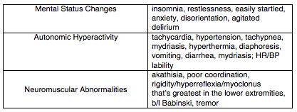
Serotonin syndrome is a condition characterized by increased serotonergic activity in the central nervous system. This can result from therapeutic use, inadvertent interactions, or intentional self-poisoning of any combination of drugs that have the net effect of increasing serotonergic neurotransmission. Serotonin syndrome most often causes mental status changes, autonomic and neuromuscular hyperactivity while severe cases may result in DIC, rhabdomyolysis, metabolic acidosis, renal failure, ARDS, and death.

Selective serotonin reuptake inhibitors (SSRIs) are the most commonly implicated group of medications, with other culprits including serotonin-norepinephrine reuptake inhibitors (SNRIs), monoamine oxidase inhibitors (MAOIs), and recreational drugs like cocaine, ecstacy, and amphetamines. The syndrome classically occurs after the initiation of a single drug, after increasing the dose of an existing medication, or simultaneous administration of two serotonergic agents. Serotonin syndrome involving a monoamine oxidase inhibitor may be especially severe and more likely to result in death. It’s important to keep in mind that antidepressants aren’t the only serotonergic agents, and that many commonly administered medications such as tramadol, meperidine, fentanyl, dextromethorphan, and linezolid among others have serotonergic activity.


Clinical Features

The majority of cases present within 24 hours, and most within 6 hours of a change or initiation of drug. The syndrome is characterized by the triad of mental status changes, autonomic hyperactivity, and neuromuscular abnormalities. The diagnosis, however, can be quite challenging as these three non-specific domains of symptomatology can present with very wide spectrum of severity.

Screen Shot 2018-11-02 at 6.15.23 PM.png


Diagnosis

There is no confirmatory test for serotonin syndrome, the diagnosis is entirely clinical. The best validated diagnostic criteria for serotonin syndrome is the Hunter Toxicity Criteria. To fulfill the Hunter Criteria, a patient must have taken a serotonergic agent and meet one of the following conditions:

  • Spontaneous clonus

  • Inducible clonus plus agitation or diaphoresis

  • Ocular clonus plus agitation or diaphoresis

  • Tremor plus hyperreflexia

  • Hypertonia plus temperature above 38ºC plus ocular clonus or inducible clonus


Differential Diagnosis:

Multiple other entities can present with a general sympathomimetic picture and should be considered in the differential diagnosis. These include:

  • Neuroleptic malignant syndrome (NMS)

  • Anticholinergic toxicity

  • Malignant hyperthermia

  • Intoxication from other sympathomimetic agents

  • Opioid withdrawal

  • Sepsis

  • Meningitis/encephalitis

  • Heat stroke

  • Delirium tremens

  • Thyroid storm

Neuromuscular findings, particularly myoclonus is an important distinguishing feature of serotonin syndrome from the above etiologies.  

Serotonin syndrome can typically be differentiated from other similar conditions by its characteristic neuromuscular findings of hyperreflexia and clonus. For example, conditions such as NMS, malignant hyperthermia, and sympathomimetic toxicity all typically lack these features.


Labs/Imaging

While serotonin syndrome is a clinical diagnosis labs and imaging can be useful for monitoring complications in severe cases and to help differentiate from other diagnoses in the differential above. The most severe complications of serotonin syndrome include DIC, rhabdomyolysis, metabolic acidosis, renal failure, and ARDS. Thus, you should consider checking the following to evaluate for these:

  • CBC

  • BMP

  • CK

  • LFTs

  • Lactic acid

  • DIC panel

  • UA

  • CXR

If the diagnosis is uncertain, you might also consider blood cultures, CT brain, lumbar puncture, urine toxicology screen and/or TSH to evaluate for other things on the differential.


Management

The mainstay of therapy for serotonin syndrome is supportive care. The main hallmarks of management include:

  • Discontinue the offending agent.

  • IV fluids to correct hypovolemia

  • Sedation with benzodiazepines. Options include Lorazepam 2-4mg and Diazepam 5-10mg, which can be repeated every 10 minutes.

  • Aggressive control of hyperthermia with standard cooling techniques. Since hyperthermia is often due to increased muscle activity in serotonin syndrome, consider early intubation and paralysis in severe hyperthermia.

  • Management of autonomic instability. Consider esmolol and nitroprusside for hypertension and tachycardia, while avoiding long-acting agent like propranolol. MAOIs can sometimes cause hypotension, treat this with pressors such as phenylephrine, epinephrine, and norepinephrine as necessary.

If benzodiazepines and supportive care fail to improve agitation and correct vital signs, you can consider cyproheptadine, an antidote of sorts with anti-histaminergic and anti-serotonergic activity. Cyproheptadine is only dosed orally, with a recommended initial dose of 12mg, followed by 2 mg every two hours until clinical response is seen. Symptoms from serotonin syndrome typically resolve within 24 hours.


Expert Commentary

Gathering an accurate medication history is the key to making this diagnosis.  As mentioned above, this is a clinical entity that generally manifests quite quickly after medication initiation.  Checking the medication list in the computer isn’t going to help you here - ask the patient what kinds of new medication they might have been prescribed or they might be taking.  Remember - selective serotonin reuptake inhibitors (SSRIs) are not the only class of medications that can affect serotonin and it may be the interacting agent that you’re looking for.  Besides the above, other common medications that can be problematic include triptans, ondansetron, valproic acid, carbamazepine, lithium, cyclobenzaprine, and even herbal supplements like St. John’s wort (patients may not think supplements are worth mentioning!). 

One underappreciated aspect of serotonin syndrome is the extent to which it occurs on a spectrum.  Studying for the boards and looking at the Hunter Criteria can leave one with the impression that clonus is necessary to make the diagnosis of serotonin syndrome.  This is not the case, and it has been suggested that many milder cases, presenting with only symptoms such as tachycardia, diarrhea, and restlessness, may be going missed.  Again, medication history is essential here.


Benjamin Schnapp, MD

Assistant Residency Program Director, University of Wisconsin-Madison Emergency Medicine


How To Cite This Post

[Peer-Reviewed, Web Publication]  Chodakowski J, Davis E (2018, November 19). Serotonin Syndrome  [NUEM Blog. Expert Commentary by Schnapp B]. Retrieved from http://www.nuemblog.com/blog/serotonin-syndrome


Other Posts You May Enjoy


 References

  1. Boyer, Edward W., and Michael Shannon. "The serotonin syndrome." New England Journal of Medicine 352.11 (2005): 1112-1120.

  2. LoVecchio, Frank, and Erik Mattison.. "Atypical and Serotonergic Antidepressants." Tintinalli’s Emergency Medicine: A Comprehensive Study Guide, 8e Eds. Judith E. Tintinalli, et al. New York, NY: McGraw-Hill, 2016,

  3. Mason, Peter J., Victor A. Morris, and Thomas J. Balcezak. "Serotonin Syndrome Presentation of 2 Cases and Review of the Literature." Medicine 79.4 (2000): 201-209.


Posted on November 19, 2018 and filed under Toxicology.

ECMO Initiation in the ED

Screen Shot 2018-08-07 at 3.02.25 AM.png

Written by: Kaitlin Ray, MD (NUEM PGY-3) Edited by: Evan Davis, MD (NUEM Alum ‘18) Expert commentary by: Colin McCloskey, MD (NUEM Alum ‘16)


ECMO Initiation in the Emergency Department

Introduction:

Extracorporeal membrane oxygenation (ECMO) provides prolonged cardiopulmonary support in severe acute respiratory or cardiac failure. As the science behind ECMO continues to grow and with promising data regarding its use in acute hypoxemic respiratory failure, cardiac arrest, and cardiogenic shock, ECMO use in the United States has increased over 400% in the last ten years. This has stimulated an interest in earlier applications of ECMO both in the emergency department (ED) and even in the prehospital setting [1]. Initiation of ECMO in the ED is a relatively new development, with 65% of programs <5 years old and the majority of programs with <3 cases per year. However, this number continues to grow [2].

There two main types of ECMO: venoarterial (VA) and venovenous (VV). While VV ECMO provides respiratory support, only VA ECMO provides both respiratory and hemodynamic support. ECMO drains blood from the native vascular system, then circulates it outside of the body via a mechanical pump where it passes through an oxygenator and heat exchanger. Hemoglobin then becomes saturated with oxygen, CO2 is removed, and blood then reinfuses into the circulation [4]. Venoarterial ECMO is more commonly utilized in the emergency department as eligible ED patients often have concurrent hemodynamic and respiratory collapse.

Emergency medicine physicians have an increasing responsibility to initiate ECMO and/or make the decision to transfer to an ECMO capable facility. Knowing this, it is critical that we are familiar with the types of ECMO available, understand the indications, contraindications, risks, benefits, and logistics of initiating this form of extracorporeal life support.


Indications/Contraindications:

The specific criteria and contraindications to ECMO vary from institution to institution, often making the decision and ability to initiate ECMO challenging. The Extracorporeal Life Support Organization (ELSO) provides specific guidelines for ECMO initiation. These indications include cardiogenic shock as defined as (1) hypotension and low cardiac output with inadequate tissue perfusion despite adequate intravascular volume and (2) persistent shock despite volume, inotropes, pressors, and possibly an intraaortic balloon pump. The ELSO also provides guidelines for ECMO in acute respiratory collapse, as well as contraindications for ECMO including: unrecoverable heart failure and not a candidate for transplant or LVAD, advanced age, chronic multi-system failure, compliance issues, terminal malignancy and prolonged CPR without adequate tissue perfusion. 

As emergency physicians, which patients should we consider as candidates for ECMO? Generally speaking, consider younger and healthier patients who experience an acute but reversible insult leading to rapid cardiopulmonary collapse.  Recall that ECMO should only be considered as a bridge to more definitive therapy. Examples of scenarios that fit this criterion include:

  • Massive pulmonary embolism

  • Myocardial infarction causing V-Tach/V-Fib or cardiogenic shock

  • Acute myocarditis or cardiomyopathy causing cardiogenic shock

  • Drowning

  • Hypothermia

  • Drug overdose causing cardiovascular collapse (such as beta-blocker or Ca-channel blocker)

  • Massive smoke inhalation, pulmonary contusion, or pulmonary hemorrhage causing refractory hypoxemia

As mentioned above, generally those with more chronic conditions such as end-stage heart failure, end-stage COPD, or those with chronic multi-organ failure, do not make good ECMO candidates. Other patient populations to consider in an ICU rather than ED setting include those with septic shock and/or ARDS. Note that patients with traumatic injury leading to hemorrhagic decompensation, although acute in nature, typically are not good candidates for ECMO as ECMO does not prevent further blood loss.

Additionally, we must consider what an ECMO-eligible patient clinically looks like. The majority of the patients who present with one of the above conditions will either be responsive to conventional therapies (intubation, fluids, inotropes), or they will be dead on arrival. However it is the rare, in-between patient that should be considered for ECMO. The condition of a good candidate would include things like: 

  • Persistent hypotension despite maximum conventional therapy

  • Persistent hypoxemia despite maximum ventilator therapy

  • Patients brought in in cardiac arrest but achieve periods of unsustained ROSC

  • Patients brought in with vitals but arrest in the ED

Unfortunately patients who arrest in the field, are brought to the ED already in cardiac arrest, or who do not achieve ROSC despite 30-45 minutes of well executed ACLS are unlikely to be appropriate ECMO candidates. The critical ECMO population is truly those who are flirting with life vs. death, especially the patients with intermittent periods of ROSC. Key exceptions to this include drowning and/or hypothermic patients. Generally these patients are better candidates for ECMO even if there has not been a recorded pulse, with the caveat that they should have been pulled out of the water or other environment quickly.


Risks/benefits:

ECMO is unique in that it provides full cardiopulmonary support without the physical trauma of chest compressions, thereby decreasing trauma, stress, and number of interruptions. Additionally, it provides a higher flow state than would otherwise be provided by manual compressions [2]. VV ECMO also minimizes barotrauma, volutrauma, and oxidative stress. However ECMO is not without risks and complications. The risk of bleeding is significant in the context of continuous anticoagulation and platelet dysfunction. Thromboembolism may lead to stroke or limb ischemia [4]. Infection may also occur secondary to indwelling lines/tubes [1].


Logistics:

ECMO is a costly intervention that requires a multi-disciplinary approach and an organizational commitment in order to proceed. Consideration must be given to the required equipment, blood bank capabilities, cannulation, and personnel availability. In order for ECMO to be successfully initiated from the ED, coordination between EMS, emergency medicine physicians, the cath lab, nursing staff, neurocritical care, cardiothoracic surgery, and the ICU is required [3]. When cannulating for VV ECMO, one may use a two cannula approach (femoral vein and internal jugular/SVC), or a single dual-lumen cannula (right atrium/IVC via the IJ). VA ECMO typically involves cannulation through the femoral artery and femoral vein [1]. If CPR is ongoing during cannulation attempts, programs may use a modified ACLS algorithm with a continuous epinephrine infusion at 0.7 mg/kg/min and minimization of pulse checks by utilizing continuous TEE monitoring [3]. Aggressive anticoagulation is required with continuous infusion of either unfractionated heparin or direct thrombin inhibitor, and efforts should be made to maintain platelet counts >50K and hemoglobin >12 mg/dL6,7.


Recap:

While initiation of ECMO from the emergency department is still a relatively new endeavor for many certified ECMO centers, the ED is in a unique position to bridge select patients in acute respiratory or cardiac failure to recovery using ECMO.  While institutional criteria for ECMO varies, the ELSO guidelines may be used as a reference to guide decision making in the absence of formal criteria. Generally speaking, pursue ECMO for younger, healthier patients with acute hemodynamic and/or respiratory collapse that is potentially reversible and unresponsive to conventional therapies. Typically patients with massive PE, MI, hypothermia, drowning, acute cardiomyopathy are the best candidates for ED ECMO. Contraindications generally include severe neurologic injury, end stage malignancy, advanced age, and irreversible multi-organ failure. Knowing that the emergency physicians are often the first to receive patients in acute cardiac and respiratory failure, it is critical that we are familiar with the types of ECMO available, understand the indications, contraindications, risks and benefits, and logistics of initiating this form of extracorporeal life support.


Expert Commentary 

This is a thoughtful and thorough overview of ECMO within the emergency department. I will limit my commentary to VA ECMO for cardiopulmonary failure (ECPR), given the enthusiasm for the topic in the FOAM world and my experience within a ED based ECMO program. Some broad themes I would like to highlight: Evidence, Patient selection and Systems of Care.

Evidence: There is a signal that ECPR is better than conventional CPR. A systematic review and metanalysis found that those with cardiac arrest who received VA ECMO had an association with increased neurologically intact survival, with a number needed to treat of 7 [1]. However, most of the data is retrospective and from single centers, making it subject to a number of confounders, as well as selection bias. Further, those who received ECPR were more likely to receive therapeutic hypothermia and percutaneous catheterization, both interventions known to improve outcome following cardiac arrest.  Another single center experience has been promising, with 9/18 patients surviving to hospital discharge with good neurological outcome [2]. This protocol involved EMS bypassing the ED and taking the patient to the cardiac catheterization lab where they were placed on ECMO and underwent catheterization. Those who had good outcome all had concomitant intervenable coronary artery disease. There are several centers that have similar experiences with published case series [3,4], but it depends thus far on quality patient selection and a viable system of care.

Patient Selection:  All the above trials had strict inclusion and exclusion criteria. Most established protocols include an age cutoff (65-75 depending on center), initial shockable rhythm, and a time from arrest to cannulation between 30-60 minutes. Pertinent exclusions include advanced comorbidities, initial asystole or prolonged downtime. This is done with the intent of cannulating patients with the best chance of surviving their ECMO run; namely young patients with likely coronary artery disease who need ECMO as a bridge to cardiac catheterization. VA ECMO’s other successful ED applications, namely pulmonary embolism [5], drug overdose [6], and acute myocarditis [7] all share the commonality that ECMO provides time for recovery or as a bridge to a viable intervention. A bridge to recovery must exist prior to any cannulation scenario; this cannot be understated.

Systems of care: Cannulation is just one step in a VA ECMO patients hospital course. When conceptualizing a successful ED ECMO program, the institutional commitment should be visualized: A patient requiring 5-7 days in the ICU, formal neuroprognostication and continuous goals of care discussions with family. You rightly include a logistics session in your review, but this system of care is paramount to a successful ECMO program. Prehospital EMS systems must be designed for quick recognition and transport of ECMO candidates.  Emergency physicians need to be trained, and maintain competency in ECMO cannulation; interventional cardiology must be willing to catheterize appropriate patients; ICU consultants must have a standardized protocol for post-arrest care and neurology/ICU must have an institutionally accepted neuroprognostication scheme. In parallel to this, the family discussions regarding prognosis and any transition of care should include social work, case management and palliative care professionals. Cannulation is as exciting a procedure an emergency physician can perform, but without a thoughtful, multidisciplinary system of care, these patients will do poorly.

In closing, VA ECMO in the emergency department is an exciting development to tertiary ED practice. More experience, and more data, will help define the niche of patients and the necessities of post-arrest care that provide these patients with the best outcome.

1. Ouweneel DM, Schotborgh JV, Limpens J, et al. Extracorporeal life support during cardiac arrest and cardiogenic shock: A systematic review and meta-analysis. Intensive Care Med. 2016;42(12):1922-1934.

2. Yannopoulos D, Bartos JA, Martin C, et al. Minnesota resuscitation consortium's advanced perfusion and reperfusion cardiac life support strategy for out-of-hospital refractory ventricular fibrillation. J Am Heart Assoc. 2016;5(6):10.1161/JAHA.116.003732.

3. Stub D, Bernard S, Pellegrino V, et al. Refractory cardiac arrest treated with mechanical CPR, hypothermia, ECMO and early reperfusion (the CHEER trial). Resuscitation. 2015;86:88-94.

4. Bellezzo JM, Shinar Z, Davis DP, et al. Emergency physician-initiated extracorporeal cardiopulmonary resuscitation. Resuscitation. 2012;83(8):966-970.

5. Yusuff H, Zochios V, Vuylsteke A. Extracorporeal membrane oxygenation in acute massive pulmonary embolism: A systematic review. Perfusion. 2015;30(8):611-616.

6. Wang G, Levitan R, Wiegand T, et al. Extracorporeal membrane oxygenation (ecmo) for severe toxicological exposures: Review of the toxicology investigators consortium (toxic). Journal of Medical Toxicology. 2016;12(1):95-99.

7. Nakamura T, Ishida K, Taniguchi Y, et al. Prognosis of patients with fulminant myocarditis managed by peripheral venoarterial extracorporeal membranous oxygenation support: A retrospective single-center study. Journal of intensive care. 2015;3(1):5.

mccloskey.png
 

Colin McCloskey, MD
NUEM Alum ‘16, Critical Care Anesthesiology fellow - University of Michigan Medical Center/University of Michigan Health System


How To Cite This Post

[Peer-Reviewed, Web Publication]  Ray K, Davis E (2018, November 12). ECMO Initiation in the ED  [NUEM Blog. Expert Commentary by McCloskey C]. Retrieved from http://www.nuemblog.com/blog/ECMO


Other Posts You May Enjoy


Resources

  1. Mosier, Jarrod M., et al. “Extracorporeal Membrane Oxygenation (ECMO) for Critically Ill Adults in the Emergency Department: History, Current Applications, and Future Directions.” Critical Care, BioMed Central, 17 Dec. 2015, ccforum.biomedcentral.com/articles/10.1186/s13054-015-1155-7.

  2. Tonna, Joseph E., et al. “Practice Characteristics of Emergency Department Extracorporeal Cardiopulmonary Resuscitation (ECPR) Programs in the United States: The Current State of the Art of Emergency Department Extracorporeal Membrane Oxygenation (ED ECMO).” Resuscitation, Elsevier Ireland Ltd, 10 Sept. 2016, experts.umich.edu/en/publications/practice-characteristics-of-emergency-department-extracorporeal-c.

  3. Tonna, Joseph E., et al. “Development and Implementation of a Comprehensive, Multidisciplinary Emergency Department Extracorporeal Membrane Oxygenation Program.” Annals of Emergency Medicine, Mosby Inc., 1 July 2017, cwru.pure.elsevier.com/en/publications/development-and-implementation-of-a-comprehensive-multidisciplina.

  4. Bartlett, Robert. Extracorporeal Membrane Oxygenation (ECMO) in Adults, 16 June 2017, www.uptodate.com/contents/extracorporeal-membrane-oxygenation-ecmo-in-adults.

  5. Extracorporeal Life Support Organization - ECMO and ECLS. “Guidelines for Adult Respiratory and Cardiac Failure.” Extracorporeal Life Support Organization - ECMO and ECLS > Resources > Guidelines, ELSO (Extracorporeal Life Support Organization), Dec. 2013, www.elso.org/Resources/Guidelines.aspx.

  6. Sklar MC, Sy E, Lequier L, et al. Anticoagulation Practices during Venovenous Extracorporeal Membrane Oxygenation for Respiratory Failure. A Systematic Review. Ann Am Thorac Soc 2016; 13:2242.

  7. Spinelli E, Bartlett RH. Anemia and Transfusion in Critical Care: Physiology and Management. J Intensive Care Med 2016; 31:295.


Posted on November 12, 2018 and filed under Cardiovascular.

Management of Myasthenia Crisis in the ED

Screen Shot 2018-10-27 at 1.48.45 PM.png


Written by: 
Kumar Ghandi, MD (NUEM PGY-3) Edited by: Arthur Moore, MD (NUEM Alum ‘18) Expert commentary by: Luke Rosiere, MD (NUEM Alum ‘12)


What do I need to know about myasthenia gravis?

Myasthenia gravis is the most common disorder of neuromuscular transmission. The disease can manifest as a combination of weakness in ocular, bulbar, and most importantly respiratory muscles. Myasthenia gravis is disease of the neuromuscular junction, in which autoantibodies are directed against nicotinic acetylcholine receptors (AChR) located on the postsynaptic end plate. These autoantibodies not only induce complement mediated destruction of receptors, but compete with acetylcholine for binding at the remaining receptors [1]. 

 
Picture1.png
 

In over 50% of cases myasthenic patients present with ocular symptoms including blurred vision, ptosis, diplopia. 15% of patients present with bulbar symptoms including ptosis, dysarthria, dysphagia, and fatigue when chewing [2]. Isolated respiratory failure is a rare sign. It is important to remember that myasthenic patients will often present specific muscle complaints and not a generalized muscle fatigue [2].

What is myasthenic crisis?

Acute myasthenic Crisis is defined as acquired myasthenia gravis severe enough to require intubation and mechanical ventilation [3]. Typically, between 10-20% of myasthenia gravis patients will experience an episode of myasthenic crisis, most commonly within the first two years [4].


Myasthenic Crisis vs. Cholinergic Crisis

Though cholinergic crisis is a quite rare it is important to differentiate cholinergic crisis from myasthenic crisis in the early evaluation of these patients. Cholinergic crisis is precipitated by excess use of cholinesterase inhibitors. This can manifest as both nicotinic and muscarinic toxicity. Nicotinic symptoms include weakness and fasciculations, while muscarinic symptoms include diaphoresis, tearing, increased oral secretions, diarrhea, and bradycardia [4].


What can trigger myasthenic crisis?

Common triggers for myasthenic crisis include infection, surgical intervention (thymectomy), pregnancy, childbirth, or tapering of immunosuppressive medications [5]. Medications are another large source of triggers for myasthenic crisis [1] [3] [6]. 50% of patients who receive treatment with high dose corticosteroids develop an early exacerbation and 10% will go on to require mechanical ventilation [6].

Screen Shot 2018-11-02 at 4.46.30 PM.png

What are the priorities of management of myasthenic crisis in the ED?

The tried and true emergency medicine axiom of Airway, Airway, Airway is critical for the management of myasthenic patients. Recognition of impending respiratory arrest, early intubation, and placement of a definitive airway has reduced mortality from 40% in the 1960’s to less than 5% today [7]. Evaluation of respiratory function and airway management for patients in myasthenic crisis presents multiple unique challenges in the ED.


How do I best assess respiratory function in the emergency department?  

Signs and Symptoms

Patients may describe dyspnea when laying flat. This is thought to be secondary loss of diaphragmatic excursion due to increased dependence of gravity during a myasthenic crisis [4]. Patients may also demonstrate severe dysphagia, with weak cough and inability to clear secretions. Other signs respiratory weakness includes, pausing mid-sentence to take a breath, use of accessory muscles, and tachypnea with shallow breathing [3]. A simple bedside evaluation includes having a patient take a deep breath and count out loud to twenty-five. If they are unable to reach five without pausing for a breath the patient may be bordering on respiratory failure [7].

Forced Vital Capacity

  • Assesses function of both inspiratory and expiratory muscle function.

  • Assess in both supine and sitting position as respiratory function tends to decline when supine.

  • Elective intubation should be considered for FVC <15 [1]. 

Maximal Inspiratory Pressure/Negative Inspiratory Force

  • Patient maximally inhales against a closed valve and force at the mouth is recorded.

  • Inspiration is a negative generating force, thus the more negative the value the stronger the inspiratory effort.

  • NIF of 0 to -30 cm H20 indicates profound respiratory muscle weakness.

  • Elective intubation should be considered for those with a NIF in the range of 0 to -30 [3].

Use of these tools are best utilized both in combination and serially with repeat assessment every two hours to assess for progressive respiratory failure. This data allows for objective assessment of respiratory function and assessment for the necessity of a well-controlled elective intubation.


What do I need to consider before intubating?  

Neuromuscular Blockers in Myasthenia Gravis:

Due to the pathophysiology of the disease, myasthenic patients due to mechanism of disease are typically resistant to neuromuscular blockade with depolarizing neuromuscular blocking agents such as succinylcholine. Due to the decreased number of receptors myasthenic patients typically require 2.6 times the dose [8]. In addition, use of cholinesterase inhibitors typically used to treat myasthenia gravis can prolong the effect of succinylcholine.

Though succinylcholine is still considered a safe agent, higher doses and inhibition of metabolism can lead to an unpredictable and lengthy paralysis. Typically, a non-depolarizing agent such as rocuronium is the preferred paralytic drug of choice. Myasthenic patients can be sensitive to non-depolarizing agents, starting at 0.1 to 0.2 mg/kg can often provide sufficient neuromuscular blockade [8].

Ventilator Settings:

Initial setting on the ventilator include:

  • AC/VC mode

  • Tidal volumes of 8-10 cc/kg of ideal body weight

  • PEEP 8-15cm H20 to prevent atelectasis and minimize work of breathing [4].

Can I use NIPPV to prevent intubation in myasthenic crisis?

Noninvasive ventilation has previously been shown to prevent intubation in patients in myasthenic crisis. Up to 20% of patients with myasthenic crisis could potentially be managed with non-invasive ventilation. Given the propensity for difficulty swallowing, these patients must be able to protect their airway and manage their secretions in order for NPPV to be a safe alternative. Early NIV has been shown to be associated with reduction in days of ventilator support, length of stay in the ICU, and rate of reintubation [9].


Disposition and Post Emergency Department Care

It is critical to alert Neurology early in the course of a possible myasthenic crisis. Often, myasthenic patients are well known to the department of Neurology, as these patients often frequent the hospital for mild exacerbations of myasthenic symptoms. Myasthenic crisis patients often require close observation in an ICU given their high risk for respiratory failure or need for ventilator management. Treatment options once leaving the ED often include, plasma exchange (PLEX) or intravenous immunoglobulin (IVIG). The table below serves as an excellent comparison for PLEX vs IVIG, knowing the modality of choice from the Emergency Department can help determine the vascular access needed to perform each modality [4]. 

Screen Shot 2018-11-02 at 4.46.48 PM.png

Expert Commentary

Myasthenic crisis is such a great topic.  I can't think of another case that employs such a breadth of expertise.  

While it is most exciting to talk about how to manage a patient in crisis, it can't be understated how critical we are in preventing such a crisis.  Look closely at that list of precipitating medications.  At least half of those are on my Top 20 most common prescription list.  Myasthenia is like prolonged QT.  If you don't think twice about your therapies, you can put this patient in a heap of trouble and plant them in the ICU for 2 weeks.  So, first thing I can say is, if you ever see myasthenia gravis on a PMH, whether they're there for a UTI or a hang-nail, think twice and three times about every medication you write for (including IV contrast).

When it comes to the recognition and management of a crisis, the above description is great.  

As you would suspect by the word "crisis," this won't be subtle.   

I'll add the following bullets:

1)  Great patient to monitor with continous capnography.  It will be your quickest indicator of deteriorating ventilation.

2)  Keep myasthenia in your differential for any undifferentiated hypercarbic respiratory failure.  Before you paralyze and intubate, try and get a brief history and physical to assess for intermittent weakness, bulbar nerve palsies, etc.  Otherwise, this potential diagnosis may be missed for days until they can get this patient off the vent

3)  Non-invasive ventilation sounds great, but probably won't help that much.  These crises last for days, and you can't keep a mask on for that long.  These patients often have dysphagia, so will aspirate.  Many patients have excess secretions that are better managed through an endotracheal tube.  If they need positive pressure, you're best off securing the airway before it's full of vomit.

4)  Succinylcholine 2 mg/kg or rocuronium 0.6 mg/kg.  Anesthesia literature often recommends puny doses (if any at all) of NMBAs.  They're expecting to extubate that patient in 3 hours and fear prolonged paralysis.  We are expecting them to be on the vent for days.  You need to ensure you have adequate intubating conditions, and paralytics will help.  Don't forget to keep your patient well-sedated if you think they're still paralyzed.

5)  We're not expected to decide how to cure this crisis.  Call neurology and they'll tell you their preference.  You'll probably start steroids and they'll decide if they want IVIg or plasma exchange.  None are going to work in 5 minutes anyway, so they can deliberate a little.

Rosiere.png
 

Luke Rosiere, MD

Northwestern University Emergency Medicine Class of 2012; Attending Physician at Northwestern Medicine Central DuPage Hospital


How To Cite This Post

[Peer-Reviewed, Web Publication]  Ghandi K, Moore A (2018, November 5). Management of Myasthenia Crisis in the ED.  [NUEM Blog. Expert Commentary by Rosiere L]. Retrieved from http://www.nuemblog.com/blog/myasthenia


Other Posts You May Enjoy


Resources

  1. Adams, James, and Erik D. Barton. Emergency medicine: clinical essentials. Philadelphia: Elsevier Saunders, 2013. Print.

  2. Clinical Manifestations of Myasthenia Gravis . (n.d.). Retrieved April 19, 2017, from https://www.uptodate.com/contents/clinical-manifestations-of-myasthenia-gravis

  3. Myasthenic Crisis . (n.d.). Retrieved April 21, 2017, from http://www.uptodate.com/contents/myasthenic-crisis

  4. Wendell LC, Levine JM. Myasthenic crisis. Neurohospitalist 2011; 1:16.

  5. Berrouschot J, Baumann I, Kalischewski P, et al. Therapy of myasthenic crisis. Crit Care Med 1997; 25:1228.

  6. EM:RAP. (2015, September). Retrieved April 20, 2017, from https://www.emrap.org/episode/september/drugsnottousein

  7. EM:RAP. (2006, January). Retrieved April 21, 2017, from https://www.emrap.org/episode/january2006/myasthenia

  8. Anesthesia for the patient with myasthenia gravis. (n.d.). Retrieved April 19, 2017, from http://www.uptodate.com/contents/anesthesia-for-the-patient-with-myasthenia-gravis

  9. Seneviratne J, Mandrekar J, Wijdicks EF, Rabinstein AA. Noninvasive ventilation in myasthenic crisis. Arch Neurol. 2008;65:54–58 


Posted on November 5, 2018 and filed under Neurology.

Bad Blood

Written by: Ade Akhetuamhen, MD (NUEM PGY-2) Edited by: Spenser Lang, MD (NUEM Alum ‘18) Expert commentary by: Matthew Levine, MD

Written by: Ade Akhetuamhen, MD (NUEM PGY-2) Edited by: Spenser Lang, MD (NUEM Alum ‘18) Expert commentary by: Matthew Levine, MD


bad-bood_25047755.png

Expert Commentary

Dr Akhetuamhen has provided a nice quick reference for topical hemostatic agents (THAs).  These agents have become more relevant in recent years, particularly in prehospital care, as the prehospital emphasis has shifted from resuscitating hemorrhage more towards hemorrhage control.  Much of our knowledge of these dressings come from battlefield studies of major hemorrhage.  Their use has been formally endorsed by the American College of Surgeons Committee on Trauma in 2014, particularly for junctional site hemorrhaging.  Dr Akhetuamhen has listed the properties of the ideal THA.  No current product fulfills all of these criteria.

Much of what we know about these agents comes from military studies.  There are limitations to these studies. There are fewer human studies, and these tend to be retrospective, observational, and based on questionnaires.  The possibility of reporting bias exists in these studies and study design made it impossible to control for the type of wound.  There are far more animal studies.  Animal studies allow for the ability to control for wound type, but are more difficult to simulate real life wounds from missiles or shrapnel.

Hemcon and Quickclot products were the earliest products studied by the military and became the early THA gold standards after they were determined to be more effective than standard gauze.  An earlier concern for Quickclot was exothermic reactions from the activated products that caused burns to patients.  As Quickclot transitioned its active ingredient from zeolite to kaolin, this concern diminished.  Quickclot is available in a roll called Combat Gauze that is favored by the military and available in our trauma bay.

Finally, there are some important practical tips for using these products.  THAs are not a substitute for proper wound packing and direct pressure.  Most topical hemostatic agent failures in studies were from user failure!  THAs must come into contact with the bleeding vessel to work.  Simply applying the THA over the bleeding areas does not mean it is contacting the bleeding vessel.  The product may need to be trimmed, packed, shaped or molded in order to achieve this.  Otherwise it is simply collecting blood.  After it is properly applied, pile gauze on top of it and give firm direct pressure for several minutes before checking for effectiveness. 

See what THA(s) you have available in your trauma bay, it is nice to know ahead of time before presented with a hemorrhaging patient what you have and in what form (a roll, sponge, wafer, etc).  Find out how the product is removed.  It may not be relevant to the patient’s ED stay but at some point the dressing needs to come off.   Some are left to fall off on their own.  The chitosan products are removed by soaking them.  When soaked, the chitosan turns slimy and can be slid off atraumatically.

Matt_Levine-33.png
 

Matthew Levine, MD

Assistant Professor of Emergency Medicine

Northwestern Medicine


How to Cite this Blog

[Peer-Reviewed, Web Publication]   Akhetuamhen A, Lang S (2018, October 29). Bad Blood.  [NUEM Blog. Expert Commentary by Levine M]. Retrieved from http://www.nuemblog.com/blog/bad-blood


Other Blogs You May Enjoy

Posted on October 29, 2018 and filed under Hematology.

Anatomic Approach to Ocular Complaints

Screen Shot 2018-09-27 at 11.51.29 AM.png

Written by: Philip Jackson, MD (NUEM PGY-3) Edited by: Jesus Trevino, MD (NUEM PGY-4) Expert commentary by: Rehan Hussain, MD


Ocular Complaints By Anatomy new format (1).png

Expert Commentary

Thank you for this excellent diagram, which demonstrates a thorough and systematic approach to the most common ocular complaints one would encounter in the ED.  In assessing a painful red eye, there is so much value in determining whether the discomfort is relieved with topical anesthetics, as you can reliably confine the pathology to the cornea or conjunctiva in those cases. However, a red eye associated with deep achy pain that is not relieved with tetracaine could be caused by scleritis, which is highly associated with autoimmune disease.  If cells and flare are visible in the anterior chamber, uveitis is more likely.  Any history of a recent eye procedure should prompt you to consider endophthalmitis, which can be visually devastating, and warrants urgent ophthalmology referral. 

In diagnosing conjunctivitis, you did a great job of highlighting the importance of a good history in determining the etiology . I would add that asking about sexual history can be valuable, as gonorrhea can present with copious purulent discharge, and chlamydia can present with chronic follicular conjunctivitis. Both would warrant systemic antibiotics and treatment of partners, in addition to eyedrops. 

Acute angle closure glaucoma is a diagnosis that should be considered in almost all cases of unilateral headache. It can mimic migraine, since both may present with nausea, vomiting, and visual disturbances. Checking the eye pressure is the most important step to making the diagnosis. 

Finally, acute onset of floaters and/or flashes always warrants an ophthalmology consult to rule out retinal detachment. Ultrasound is a useful tool to differentiate retinal detachment from posterior vitreous detachment (PVD) and vitreous hemorrhage. PVD and vitreous hemorrhages tend to move freely when the eye moves, whereas retinal detachment is anchored to the nerve but still flaps with eye movement.   

Once again, this diagram serves as a very helpful flowchart to trouble-shoot eye complaints in the ED.

hussain.png

Rehan M. Hussain, MD
Vitreoretinal Surgery Fellow

Bascom Palmer Eye Institute

University of Miami Health System


How To Cite This Post

[Peer-Reviewed, Web Publication]   Jackson P, Trevino J (2018, October 22). Anatomic approach to ocular complaints.  [NUEM Blog. Expert Commentary by Hussain R]. Retrieved from http://www.nuemblog.com/blog/ocular-complaints


Other Posts You May Enjoy

VBG vs ABG in the ED

Screen Shot 2018-09-27 at 11.40.11 AM.png

Written by: Emmanuel Ogele, MD (Cook County Stroger PGY-1) Edited by: Spenser Lang, MD (NUEM Alum '18) Expert commentary by: James Walter, MD


ABG’s vs VBG’s in the Emergency Department

  • Arterial blood gases (ABG’s) – blood sample taken directly from an artery used to gauge the metabolic environment, oxygenation, and ventilation status. Values such as pH, PCO2, PaO2, HCO3, and Base Excess obtained via ABG are considered the gold standard.

  • Venous Blood gases (VBG’s) – blood sample taken from either peripheral or central veins –can serve as an alternative to an ABG when evaluating patients with metabolic and respiratory disturbances.

    • Historically, values obtained via VBG have been criticized for a perceived lack of accuracy in all domains.

    • However, VBGs carry less risk of vascular injury, nerve damage, and cause much less pain to the patient along with lower risk for accidental needle-sticks as compared to ABGs


  • So the question remains – are values (such as pH, PCO2, and HCO3) truly disparate enough between ABG’s and VBG’s to actually change clinical practice?

    • Increasing data shows that for most clinical indications, data from VBG correlates well, and are just as useful as that from ABG.[1-4]

      • Zeserson et. al. conducted a prospective cohort study of 156 critically ill patients in the ED and ICU setting to evaluate the correlation between pH and pCO2 when derived from ABG vs VBG with added pulse oximetry for estimating PaO2 and concluded that arterial and venous pH and PCO2 had good correlation.

      • Byrne et al conducted a meta – analysis of 1768 subjects from 18 individual studies and found that peripheral VBG correlates well with ABG with respect to pH but found an unacceptably wide 95% prediction interval when looking at the pCO2.

      • A review article by Kelly AM summarized data comparing ABG and peripheral VBG variables in ED all-comers also concluded that venous pH had sufficient agreement however concluded with a word of caution: there is no data to support that this correlation is maintained in shock states.

    • Several studies have looked at the correlation between values obtained with VBG and compared them to ABG. These are summarized in Table 1.

Screen Shot 2018-10-10 at 6.26.27 PM.png

Table 1. Correlation of VBG to ABG values
** Widest limit of agreement from any single study included in the meta-analysis


  • For most parameters, there is good correlation. However, there are a few important scenarios that may be exceptions. Not surprisingly, the major exception is PO2; venous PO2 readings do not correlate well with arterial PO2. A workaround to this limitation is to estimate arterial oxygenation using SpO2.

  • The VBG analysis plus SpO2 provided accurate information on acid–base, ventilation, and oxygenation status for patients in undifferentiated patients ED and ICU.[2]

  • VBGs are acceptable to use in working up common conditions like COPD and DKA.[5,6] New data could potentially broaden the list of indications for VBG instead of ABG  

    • Ma OJ et al. conducted a prospective trial looking at the utility of ABG in patients presenting to the ED with suspected DKA and found that ABG analysis changed management of DKA 1% of the time and concluded that VBGs are a viable substitute.

Conditions that may affect the reliability of VBG

  • Hypercapnia.

    • When comparing VBG and ABGs, the average difference in CO2 reading was 5.7 mmHg. [1]

    • However, the limits of agreement (-17.4 to +23.9) in this study are too wide to allow reliable quantification of PCO2.

    • In sum, if you need a precise PCO2 number for clinical decision making, a traditional ABG is preferable.

    • One such scenario where a true PCO2 can be useful is evaluating for acute hypercapneic respiratory failure; however, a VBG still has some utility.

      • In the prospective study by AM Kelly 7 a PCO2 value above 45mmHg had a 100% sensitivity for true hypercapnia. This makes a VBG PCO2 value useful in screening for hypercapnia. 5

  • Shock Pearls

    • VBGs show increased discordance from ABGs in hypotensive patients.[8]

    • pH and PCO2 values may be wildly disparate in patients with severe circulatory failure.[9]

    • In sum, venous blood gases may be increasingly inconsistent with arterial blood gases in patients with increasing degrees of shock. No definitive data exists yet to tell us if VBGs are sufficient to replace ABGs in shock states.

  • Mixed Acid Base Disorders

    • There is insufficient evidence to confirm reliability of VBGs in these cases


In summary, VBGs can be used as a reliable alternative to ABGs in many clinical cases. The patients’ benefits of a VBG vs ABG are obvious – decreased pain, complications, and time. Clinical judgment must be used in deciding when to the substitute a VBG for a more traditional ABG. The evidence is mixed, and even non-existent in some clinical scenarios. In the future, noninvasive methods of evaluation, such as transcutaneous PCO2 monitoring and ETCO2, could allow for accurate for non-invasive and monitoring of the metabolic milieu.


Expert Commentary

ABGs vs VBGs in the Emergency Department: Expert Commentary 

Thank you for the opportunity to share some thoughts on this topic. The ABG vs VBG debate has been the source of a lot of discussion and at times disagreement between EM and IM. I am hopeful that we are starting to reach consensus on their respective advantages, disadvantages, and indications. When deciding on which test to obtain, here are a few questions to ask yourself:


1. What is my clinical question?

Diagnostic tests should be performed to answer a specific clinical question. Defining this question will help ensure you order the correct test, or perhaps appropriately order no test at all. For blood gas sampling this question might be: “Does my patient with a COPD exacerbation have significant hypercapnia?”; “Is my patient appropriately compensating for his metabolic acidosis?”; “Is my hyperglycemic patient acidotic?” If you can’t articulate a specific question, or if the answer to that question is unlikely to change your management (i.e., a question of “is my patient acidotic?” for a 70-year-old with urosepsis whose blood pressure has responded to 1L of fluid and looks well), then you can probably save your patient an unnecessary blood draw and avoid blood gas sampling altogether. This is certainly an issue for us in the ICU. Patients with arterial lines will have standing Q6hr ABG orders for 2 days before anyone asks if those blood draws are actually changing our management. Don’t order an ABG or VBG just because a patient has sepsis, or they have COPD, or you are “screening for badness.” Using a POC or rapid VBG with a metabolic profile to rapidly obtain lab values for patients presenting to the ER is reasonable. Outside of this situation, try to make sure you are asking a specific question and that answering that question is likely to change what you do.


2. Am I screening for hypercapnia?

If your clinical question is, “is my patient hypercapnic?” then a VBG is a great test. As noted above, a PvCO2 < 40 mmHg excludes hypercapnia. This can be an extremely helpful in the rapid workup of altered mental status and many other common presenting conditions. 


3. How accurate do I need my PCO2 value to be?

If the answer to this question is “not that accurate” then a VBG is probably fine. Having a rough estimate of PCO2 levels is usually adequate for the management of mild-moderate DKA, COPD exacerbations, and many other conditions managed in the ED. While a PvCO2 value of 18 mmHg or 75 mmHg may not exactly correlate with what you find on a PaCO2, they are abnormal enough to give you a good general sense of things.

If you are interested in performing a more refined blood gas analysis such as determining the chronicity of a respiratory acidosis, measuring shunt fraction, or accurately quantifying a hypercapnic patient’s true PCO2 then you probably need an ABG. As noted above, the correlation between PaCO2 and PvCO2 is often poor. 


4. Am I assessing oxygenation?

At times, obtaining a reliable SpO2 can challenging especially in patients with PAD, scleroderma, or shock. If you need an accurate assessment of oxygenation then you need an ABG. PvO2 values do not correlate well at all with PaO2.


5. Is my patient in shock?

As noted above, VBGs are much less accurate in shock. Unfortunately, this is where we are often most interested in frequent blood gas analysis. In the ED, I think ABGs are most useful (and underused) in critically ill acidotic patients who may or may not have appropriate respiratory compensation. This determination is hard to make on clinical grounds alone (i.e. the signs of early respiratory muscle fatigue can be subtle) and identifying fatigue may well change your management (pushing you to earlier NIV or mechanical ventilation). I would hesitate to solely rely on VBGs in this setting especially for patients in overt shock.


A few other points:

  • I do think the risks of an ABG as stated above and in other reviews (for example, https://lifeinthefastlane.com/ccc/vbg-versus-abg/) are overstated. A competent clinician should be able to obtain an ABG from a radial artery in a matter of seconds. If there are any concerns regarding anatomy or first stick accuracy, the use of a vascular ultrasound probe can remove any guess work from finding the best arterial access site. ABGs do require an extra needle stick for patients so clinicians should be discerning about their use. However, if one is indicated they shouldn’t be avoided for fear of causing a pseudoaneurysm or major bleeding. Compared to innumerable other invasive procedures and diagnostic tests performed in the ED, ABGs are pretty benign. For some reason, they are still frequently described like thoracotomies.

  • Remember the following rough corrections

    • Venous pH is 0.03 lower than arterial pH (venous pH 7.27 = arterial pH 7.3)

    • Venous PCO2 is 6 mmHg higher than arterial PCO2, but with wide variability; in general, difficult to predict arterial PCO2 from venous PCO2 (although a PCO2 can still be useful, as noted above).

James_Walter-44.png
 

James Walter, MD

Pulmonary and Critical Care, Northwestern Medicine

Medical Director of the Northwestern Lung Rescue Program


How to Cite This Post

[Peer-Reviewed, Web Publication]   Ogele E, Lang S (2018, October 15). VBG vs ABG in the ED.  [NUEM Blog. Expert Commentary by Walter J]. Retrieved from http://www.nuemblog.com/blog/vbg-abg


Other Posts You May Enjoy


 Resources 

  1. Kelly AM. Review article: Can venous blood gas analysis replace arterial in emergency medical care? Emerg Med Australas. 2010 Dec;22(6):493-8. doi: 10.1111/j.1742 6723.2010.01344.x. Review. PubMed PMID: 21143397

  2. Zeserson E, Goodgame B, Hess JD, Schultz K, Hoon C, Lamb K, Maheshwari V,Johnson S, Papas M, Reed J, Breyer M. Correlation of Venous Blood Gas and Pulse Oximetry With Arterial Blood Gas in the Undifferentiated Critically Ill Patient. J Intensive Care Med. 2016 Jun 9.

  3. Byrne, A. L., Bennett, M., Chatterji, R., Symons, R., Pace, N. L. and Thomas, P. S. (2014), Peripheral venous and arterial blood gas analysis in adults: are they comparable? A systematic review and meta-analysis. Respirology, 19: 168–175. doi:10.1111/resp.12225

  4. Kelly AM, McAlpine R, Kyle E. Venous pH can safely replace arterial pH in the initial evaluation of patients in the emergency department. Emerg Med J. 2001 

  5. McCanny, Venous vs arterial blood gases in the assessment of patients presenting with an exacerbation of chronic obstructive pulmonary disease.

  6. Ma OJ, Rush MD, Godfrey MM, Gaddis G. Arterial blood gas results rarely influence emergency physician management of patients with suspected diabetic ketoacidosis. Acad Emerg Med. 2003 Aug;10(8):836-41. PMID 12896883

  7. Anne-Maree Kelly, Elizabeth Kyle, Ross McAlpine, Venous pCO2 and pH can be used to screen for significant hypercarbia in emergency patients with acute respiratory disease, In The Journal of Emergency Medicine, Volume 22, Issue 1, 2002, Pages 15-19, ISSN 0736-4679, https://doi.org/10.1016/S0736-4679(01)00431-0.

  8. Shirani F, Salehi R, Naini AE, Azizkhani R, Gholamrezaei A. The effects of hypotension on differences between the results of simultaneous venous and arterial blood gas analysis. Journal of Research in Medical Sciences : The Official Journal of Isfahan University of Medical Sciences. 2011;16(2):188-194

  9. Adrogué HJ, Rashad MN, Gorin AB, Yacoub J, Madias NE. Assessing acid-base status in circulatory failure. Differences between arterial and central venous blood. N Engl J Med. 1989 May 18;320(20):1312-6.

Posted on October 15, 2018 and filed under Pulmonary.

Supratherapeutic INR

Screen Shot 2018-09-27 at 11.32.54 AM.png

Written by: Luke Neill, MD (NUEM PGY-3) Edited by: Logan Weygandt, MD (NUEM Alum '17) Expert commentary by: Abbie Lyden, PharmD BCPS


Expert Commentary

This is an excellent post on the management of supratherapeutic INR in patients taking vitamin K antagonist therapy – and as you described, there is not a one-size-fits-all approach. Warfarin is notorious for being one of the most difficult medications to manage based on narrow therapeutic index, variable dose response, clinically significant diet- and drug- drug interactions, delayed onset and offset of action and the need for frequent monitoring. Fortunately, warfarin does have an antidote in vitamin K. Yet, the choice of when to administer this antidote (and weaknesses of said antidote), along with other therapies including prothrombin complex concentrates (PCCs) and fresh frozen plasma, are not straightforward and depend upon a number of factors. In addition to your excellent teaching points, I have outlined some additional considerations below.

Life-threatening bleeding

In the setting of life-threatening bleed, guidelines dictate our therapeutic approach, which involves holding warfarin and administering 4-factor PCC and intravenous vitamin K (10mg slow infusion over 20-60 minutes)1.

Minor bleeding

The management of life-threatening bleeding is clear and requires aggressive therapy. But how do we manage the patient who has a moderately elevated INR but only a minor bleed? In the setting of minor bleeding (such as intermittent epistaxis), the goal is restore the INR to target range, without leading to subtherapeutic anticoagulation, thus introducing the risk of thrombosis. There is general consensus regarding holding a dose of warfarin in these scenarios but the choice to administer vitamin K has been debated. The choice of approach should depend upon the perceived risk of bleeding, extent of bleed, site of bleed, INR level (and trend in INR), comorbidities (including indication for anticoagulation) and risk of thromboembolism. The downsides to administering vitamin K are worth mentioning as they are sometimes superseded by our focus on providing active treatment (unquestionably necessary in the case of life-threatening bleed).  Excessive vitamin K dosing may result in warfarin resistance for 1-2 weeks which may require extensive bridging therapy once anticoagulation is restarted. For patients with high thromboembolic risk, poor adherence to medications, higher INR goals or comorbidities, this can become complicated and is certainly not without risk.

Elevated INR without bleeding

The 2012 ACCP guidelines recommend administration of oral vitamin K (2.5-5mg) to patients without an active bleed who have an INR>10 (1). Other experts and the 2008 ACCP guidelines use a more conservative cutoff of 9 (2). For those patients with INRs between 4.5 and 10 without evidence of bleeding, the 2012 ACCP guidelines suggest against the routine use of vitamin K. In this post, you make a great point regarding the management of INRs 5-9 without bleeding. That is, the administration of vitamin K may or may not be administered, depending on the risk of bleeding. Low dose vitamin K administration should be more strongly considered in patients with high risk bleed (elderly, prior bleed) and lower risk of thromboembolism. A retrospective review of 633 patients with elevated INR >6 identified risk factors for slow spontaneous lowering of supratherapeutic INR, including older age, higher index INR, lower warfarin maintenance dose, decompensated heart failure and active cancer (3). Knowledge of these risk factors may help guide decision-making when considering whether or not to administer vitamin K.
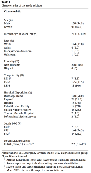
Urgent Surgery/Procedure

Patients meriting further discussion are those taking vitamin K antagonists who require urgent (same day) surgeries or invasive procedures. These patients are managed in a similar fashion to those with life threatening bleeding – that is to say, vitamin K (10mg IV) and 4-factor PCC. Of note, for those patients who can wait 24 hours, low dose vitamin K (1-2.5mg PO) is generally adequate for INR reversal. In these cases, 4-factor PCC and intravenous vitamin K can be avoided.

Specific Treatments

With regards to the specific treatments to reverse supratherapeutic INR, some points to keep in mind:

  1. Vitamin K (phytonadione): typically administered intravenous or orally.

  • For life-threatening bleed, intravenous vitamin K is preferred due to the faster onset of action (which is still delayed, ~3-8 hours) but faster than oral (onset ~24 hours)

  • Subcutaneous administration is generally avoided if possible due to erratic absorption

  • Intramuscular administration is generally avoided due to the risk of hematoma in an anticoagulated or overanticoagulated patient

2. Prothrombin complex concentrate (PCC):

  • 4-factor PCCs (factors II, VII, IX, X): preferred first line therapy for life-threatening bleed

  • Activated PCC (aPCC): factor VII is mostly present in the activated form, which is potentially more prothrombotic than unactivated PCC

  • Some PCC products contain heparin and should NOT be administered in a patient with a history of heparin-induced thrombocytopenia

In short, patients with life-threatening bleed taking vitamin K antagonist therapy require urgent evaluation and treatment with PCC and IV vitamin K. Treatment with PCC is paramount as INR can be corrected within 30 minutes, as opposed to several hours after IV vitamin K administration (onset dependent upon liver synthesis of new coagulation factors). For those patients with supratherapeutic INR without bleeding and patients with minimal bleed, a more gentle approach is indicated, which involves omission of warfarin dose +/- low doses of oral vitamin K to ensure correction of INR and prevention of bleeding but not subtherapeutic anticoagulation. Lastly, it is important to identify if there are additional explanations for the supratherapeutic INR by verifying if the patient was taking the appropriate dose of warfarin or whether they have recent dietary changes or new medications which may interact. If therapy is to be resumed, these questions are particularly helpful when deciding how and when to restart anticoagulation. 

  1. Holbrook A, Schulman S, Witt DM, et al. Evidence-based management of anticoagulant therapy: Antithrombotic Therapy and Prevention of Thrombosis, 9th ed: American College of Chest Physicians Evidence-Based Clinical Practice Guidelines. Chest 2012;141(2 Suppl):e152S.

  2. Ansell J, Hirsch J, Hylek E, et al. Pharmacology and management of the vitamin K antagonists: American College of Chest Physicians Evidence-Based Clinical Practice Guidelines (8th Edition). Chest 2008;133(6 Suppl):160S.

  3. Hylek EM, Regan S, Go AS, Hughes RA, Singer DE, Skates SJ. Clinical predictors of prolonged delay in return of the international normalized ratio to within the therapeutic range after excessive anticoagulation with warfarin. Ann Intern Med. 2001;135(6):393.

Abbie.png
 

Abbie Lyden, PharmD BCPS

Clinical Pharmacist, Emergency Medicine | Associate Professor, Pharmacy Practice

Residency Program Director, PGY2 EM Pharmacy

Northwestern Memorial Hospital


How To Cite This Post

[Peer-Reviewed, Web Publication]   Neill L, Weygandt L (2018, October 8). Supratherapeutic INR.  [NUEM Blog. Expert Commentary by Lyden A]. Retrieved from http://www.nuemblog.com/blog/supratherapeutic-inr


Other Posts You May Enjoy

Posted on October 8, 2018 and filed under Hematology.

End Tidal CO2 in Cardiac Arrest

Screen Shot 2018-09-27 at 11.16.07 AM.png

Written by: Alex Herndon, MD (NUEM PGY-2) Edited by: Andrew Moore, MD (NUEM Alum '18) Expert commentary by: Seth Trueger, MD, MPH

Introduction:

ER, Grey’s Anatomy, House, Chicago Med, The Good Doctor - across the nation millions tune in to their favorite medical dramas hoping to get a glimpse at what it’s like to be in the business of saving lives. As a newly minted emergency medicine intern, my eye caught on to the most recent addition, The Resident, intrigued by how the show would portray the medical profession. While watching and cringing at dramatized incidences of medical nonsense, one scene particularly stood out.

It was day one of residency and the new intern was leading the resuscitation of a patient who suddenly arrested. Fast-forward, the intern achieves return of spontaneous circulation, however he is immediately chastised by his senior resident who states, “her end-tidal CO2 was less than 15 for the entire code…”

As the credits rolled I was left agreeing with critic Dr. Esther Choo in that “this show feels like a most unfortunate and untimely addition to the medical drama genre”, given it is the least accurate medical show I’ve watched to date, except for this fleeting reference to End Tidal CO2 (ETCO2).


A Review of ETCO2 and its Applications:

Traditionally, ETCO2 has been used in order to assess proper endotracheal tube placement. Approximately 25 years ago anesthesiologists began using ETCO2 capnography because it was revealed that approximately 93% of anesthesia errors could have been prevented with additional capnography monitoring. In particular, the sensitivity of color change of colorimetric devices can be faulty at low concentrations of CO2, a particular concern when there is presumed decreased CO2 released from the lungs due to decreased cardiac output during an arrest, or if the ET tube is placed within the esophagus. The addition of capnography not only reinforces that the ET tube is properly placed, but its use has been extrapolated to indirectly assess cardiac output. (1)


The additional data ETCO2 supplies can be used in two key ways:


1. To assess quality of chest compressions

In cardiac arrest, ETCO2 waveform, while performing CPR, can serve as an indirect measurement of blood flow generated by chest compressions. The height of the ETCO2 waveform during CPR has been used as an indirect measure of adequate chest compressions, helping those involved in resuscitation monitor the effectiveness of their compressions in real time. In the awake adult, normal cardiac index lies between 2.5-4 L/min/m2, with an ETCO2 of 35-45 mmHg. On average during CPR, if adequate chest compressions are being delivered a cardiac index of 1.6-1.9 L/min/m2 can be generated, which correlates with ETCO2 pressures of 20mmHg.(1) ACLS guidelines define high quality chest compressions as achieving ETCO2 pressures of at least 10-20 mmHg. As rotating medical professionals deliver chest compressions, ETCO2 can be used to determine if they need to be deeper, if there is performer fatigue, or if there are other factors that might be inhibiting the ability to maintain ideal cardiac output outside of ineffective chest compressions. All in all, it provides a more accurate assessment of chest compression adequacy than visual estimation of compression depth.


2. To help predict return of spontaneous circulation (ROSC)

Numerous studies have shown that abrupt increases in ETCO2 pressures exceeding 10 mmHg that remain higher than preceding values suggest an increase in cardiac output and is indicative of ROSC, hence the incorporation of such measures in ACLS guidelines.(1) Patients with values less than 10 mmHg are more likely to die during CPR, and those with values greater than 10 during CPR were more likely to get ROSC. (1) However when comparing a mere 10 mmHg ETCO2 pressure to the minimal normal ETCO2 pressure of 35 mmHg, it can be difficult to argue 10 mmHg is enough. Multiple studies have aimed to drive this number up, in particular showing ETCO2 pressures higher than 16mHg were significantly associated with survival from CPR in the emergency department. However, the use of an absolute ETCO2 value was limited by the cause of cardiac arrest. Average ETCO2 pressures that achieved ROSC widely varied depending on whether cardiac arrest was purely cardiac versus pulmonary in etiology. (1) A 2015 meta-analysis of ETCO2 values associated with ROSC showed, on average, patients with ROSC after CPR had an average ETCO2 level of 25 mmHg, significantly higher than the current recommended 10 mmHg threshold.(2) Other than the aforementioned minimal ETCO2 threshold, it is important to follow ETCO2 trends, looking for the sudden increase in ETCO2 and maintenance of elevated levels associated with ROSC.


Furthermore, other studies have attempted to show how ETCO2 can be a tool in deciding to terminate CPR when ROSC isn’t achieved. One study from 1997 reported that ETCO2 less than 10 mmHg at the 20 minute mark is predictive of non-survivability in outside-hospital cardiac arrest patients thus should lead to terminating resuscitation efforts.(3) Current studies of use of ETCO2 in outside hospital arrest trends have shown 3-5 minutes of ETCO2 <10 mmHg are associated with a bad prognosis and has been used to terminate in-field resuscitation efforts.(4)


Next Steps:

While more data is needed in order to potentially reset the ETCO2 threshold used to assess adequate CPR and ultimately long-term survival post-arrest, others are looking at alternate applications of ETCO2. In 2016 Wang et al studied whether or not ETCO2 values could be used as a predictor of survival when looking at in-hospital versus outside-hospital cardiac arrests. They found that an initial ETCO2 level was predictive of not only sustained ROSC, but also survival to discharge.(2) Others have looked at the use of ETCO2 to determine potential effectiveness of defibrillation. It was found that ETCO2 less than 7 mmHg never resulted in effective shocks, whereas shocks delivered with ETCO2 greater than 45 mmHg were always successful. Ultimately the authors attributed the success to performing high quality chest compressions prior to defibrillation.(5)


Limitations of ETCO2 Capnography and Conclusions:

Similar to how ETCO2 can vary given the cause of cardiac arrest, ETCO2 can be influenced by other factors, thus altering how physicians interpret capnography. In particular, no studies have assessed the effect or epinephrine or sodium bicarbonate on ETCO2. Further complicating matters, the majority of cardiac arrest patients end up intubated, thus ventilator settings or over bagging can influence expired CO2 levels creating yet another confounding factor. These subtleties regarding ETCO2 need further exploration.   

Next time you are waiting for that outside-hospital cardiac arrest to roll through the Emergency Department entrance, arm yourself with ETCO2 capnography not only to aid your resuscitative efforts, but also help with your decision-making along the way. Its usefulness extends beyond simply achieving ROSC and has the potential to prognosticate whether patients will not only survive but thrive.


Expert Commentary:

Thanks for this nice overview of the data behind quantitative waveform ETCO2 in arrest. While it’s not the only tool in our armamentarium, it certainly can be helpful in assessing whether compressions are being effectively done, which can help the compressor modify their technique or location, add an element of motivation for the compressor, and help identify when the compressor is tiring out so we can switch someone else in before the official 2 minutes is up. Similarly, if there’s a big jump upward in the ETCO2 (eg, from 12 to 35), it’s reasonable to deviate from the usual 2 minutes and jump to a pulse check.

Using low ETCO2s is helpful in identifying futile codes; the general rule is that if the ETCO2 is consistently <10 after 20 minutes of well-done ACLS, the patient is very unlikely to come back. I find it important to point out that this is not a sensitive test, however, and many arrested patients will continue to have ETCO2 over 10 during long codes. I try to not get too focused on the ETCO2 as the only marker of when to terminate resuscitative efforts. Rather, it can help make a hard decision easier in some cases, but like most things, it’s not a magic bullet.

One other caution: don’t be mislead by a flat ETCO2 waveform during an arrest. If you don’t see any waves, then the airway is not in and either replace it or confirm intubation by other means (eg VL or gentle bougie insertion to holdup).

Here is a short screencast I put together back in 2013 on the 3 major uses of ETCOS: ETT confirmation, arrest, and monitoring in procedural sedation:

https://player.vimeo.com/video/57510987

seth trueger.png
 

Seth Trueger, MD, MPH

Assistant Professor of Emergency Medicine, Northwestern University


How To Cite This Post

[Peer-Reviewed, Web Publication]   Herndon A, Moore A (2018, October 1). End Tidal CO2 in Cardiac Arrest.  [NUEM Blog. Expert Commentary by Trueger NS]. Retrieved from http://www.nuemblog.com/blog/ETCO2


Other Posts You May Enjoy


Resources

  1. Kodali, B. Urman, R. Capnography during cardiopulmonary resuscitation: Current evidence and future directions. J Emerg Trauma Shock. 2014 Oct-Dec: 7(4): 332-340. Doi: 10.4103/0974-2700.142778

  2. Wang, AY. Initial end-tidal CO2 partial pressure predicts outcomes of in-hospital cardiac arrest. Am J Emerg Med. 2016 Dec;34(12):2367-2371. doi: 10.1016/j.ajem.2016.08.052.

  3. Morshedi, B. The role of ETCO2 in termination of resuscitation. J Emerg Med Services. 2017 Dec. <http://www.jems.com/articles/print/volume-42/issue-12/features/the-role-of-etco2-in-termination-of-resuscitation.html>

  4. Venkatesh, H. Keating, E. Can the value of end tidal CO2 prognosticate ROSC in patients coding into emergency department with an out-of-hospital cardiac arrest. Emerg Med J. 2017 Mar; 34(3): 187-189. doi: 10.1136/emermed-2017-206590.1

  5. Savastano, S et al. End-tidal carbon dioxide and defibrillation success in out-of-hospital cardiac arrest. Resuscitation. 2017 Dec;121:71-75. doi: 10.1016/j.resuscitation.2017.09.010.


Posted on October 1, 2018 and filed under Pulmonary.

Phytophotodermatitis

Written by: Luke Neill, MD (NUEM PGY-3) Edited by: Mitali Parmar, MD (NUEM Alum '18) Expert commentary by: Charles Pearce, MD

Written by: Luke Neill, MD (NUEM PGY-3) Edited by: Mitali Parmar, MD (NUEM Alum '18) Expert commentary by: Charles Pearce, MD


Case

A 7 yo female with no significant past medical history presents with a two-day history of worsening asymmetric rash on the left neck, upper middle chest, right thigh, and the dorsal aspect of both hands. The rash is described as a painful, burning, 3/10 pain and non-pruritic. She denies any history of allergies, especially any allergies to outdoor plants or foods, and denies using any new types of lotions, sunscreen, or other new chemical products. She denies history of trauma. She denies fevers, chills, or recent infections. Denies arthralgias, myalgias, fatigue, or weakness. Denies SOB or chest pain. Denies peeling of her skin or blistering.  She takes no medications.

She is currently attending an outdoor day-time summer camp and her mother first noticed the rash after picking her up at the end of camp two days ago. The mother, due to concern for possible physical abuse would like to know your opinion on its cause. You ask to speak to patient in private and she adamantly denies any physical abuse. You check her vitals.

Vitals/Exam: 

HR: 65   BP:120/70 Temperature: 96.4 RR:16 O2:100

General: patient well appearing, watching TV

Skin: Multiple small areas of blotchy erythema over left neck, upper middle chest, right thigh, and dorsal aspect of hands in different patterns with no symmetry. Some appear to represent a hand print. The rash on the chest also appears to streak vertically.

Head: normocephalic, atraumatic

HEENT: oral mucosa moist, PERRL, EOM intact, TMs clear bilaterally

Neck: supple, trachea midline

Cardiovascular: regular rate/ rhythm, no murmurs, rubs, or gallops

Chest: non-tender

Pulmonary: clear to auscultation bilaterally

GI: non-distended, soft, non-tender 

MSK: no deformities

Neuro: CNII-XII intact, strength/sensation grossly intact

Skin Exam: 

Screen Shot 2018-09-14 at 10.48.31 PM.png

Labs: CBC and BMP Unremarkable

Clinical Course:

In consultation with dermatology, patient revealed that she recently made limeade at her summer camp about 2 days prior to arrival, and had been out in the sun for outdoor activities. Given this history and appearance of the rash, the patient was diagnosed with phytophotodermatitis, and discharged home with instructions to stay indoors. She was seen in clinic after 5 days, and had developed multiple blisters over her hands, chest and thighs. The rash subsequently completely resolved in 2 weeks time.


Phytophotodermatitis

Pathophysiology:

Phytophotodermatitis, also known as “Lime Disease” or “Margarita Photodermatitis” is a phototoxic inflammatory eruption of the skin that occurs due to contact with light-sensitizing botanical substances and subsequent exposure to UV-A radiation. Interestingly, the reaction of phytophotodermatitis is actually independent of the immune system.

But How?

When furocoumarins, the photosensitizing chemical compound produced by certain plants, are struck by a photon in the UV-A range of (320-400), energy is absorbed causing the formation of an excited state from ground state. When the furocoumarin returns to ground state, energy is released in the form of heat and fluorescence, leading to both DNA and RNA damage and cell death.

Furocoumarin is present in:

  • Limes

  • Figs

  • Parsley

  • Celery

  • Carrots

Timing:

  • Skin eruption typically begins 24 hours after sun exposure

Manifestation:

  • Burning erythema with blistering

  • Post-inflammatory hyperpigmentation lasting weeks to months. 

Progression:

  • Eruption peaks at approximately 48-72 hours


Below is an image illustrating the progression in days, of a person with phytophotodermatitis of the hand:

Epidemiology:

As phytophotodermatitis occurs independent of the immune system; any race, sex, or age group may be affected. However, it does appear that produce workers in grocery stores are at a much higher risk than the general population. A 1986 study showed that in one un-named nationwide grocery store chain, a randomly selected sample of all its stores revealed phytophotodermatitis occurrence in 13 of 17 states, with occurrence in 26% of the produce workers surveyed. In this instance, it was thought to be due to celery stock with higher levels of endogenous furocoumarin.

Treatment/Prognosis:

The prognosis of phytophotodermatitis is very good with proper identification and elimination of the offending plant. Patients who are affected should stay indoors avoiding UV-A rays to allow the dermatitis to self-resolve. 

Note: This is not the first time phytophotodermatitis has mimicked child abuse. A 1985 study looked at two separate cases of children who were initially thought to have hyper-pigmented skin lesions suggestive of child abuse and were later given a final diagnosis of phytophotodermatitis.

Takeaways/Learning Points:

  • Consider phytophotodermatitis in your differential for rash in summer months.

  • Although we ask about allergies, consider asking about recent food exposure.

  • Don’t spill your drink!


Expert Commentary

Great overview of phytophotodermatitis!  This is certainly a fascinating and easily missed phenomenon given its relative rarity, variable pattern of presentation and, on occasion, insidious exposures. In the Midwest, we are seeing additional exposure risks to phytophotodermatitis in the form of Wild Parsnip (known colloquially as poison parsnip), an invasive plant that was introduced from Europe over a century ago and whose range has continued to expand.  This expansion has lead to Department of Natural Resource and local media in a number of states working to educate the public as well as local and regional medical facilities about the potential threat.

As with lime juice, exposure to the sap of Wild Parsnip can be potentially non-apparent to the patient and the clinician.  In fact, the pattern of burns from Wild Parsnip are as variable as the methods of exposure- from linear lesions from unwittingly brushing against a plant to extensive hand/forearm involvement from attempted manual removal of a plant to a speckled burn pattern from mechanical disruption of the plant (think weed whacker or lawn mower).  This variability obviously heightens the diagnostic difficulty and uncertainty. 

Another consideration and as noted in your excellent review, phytophotodermatitis is a burn resultant of chemically induced cell death by cross linkage of the furan ring with pyrimidine bases in the presence of UV light.  And recognition of this phenomenon as a chemical burn does have bearing on management. A discussion with or referral to burn centers may be warranted, as with any burn, if there is significant TBSA involvement or depending on body area impacted.  And looking for and warning patients of the potential for super-infection is imperative.

As a ER physicians in rural Wisconsin, our practice sees a couple handfuls of phytophotodermatitis cases each summer.  Our local communities (namely farmers) are well aware of Wild Parsnip and the simple prevention of avoidance and washing exposed areas to remove sap.  As more and more people (hopefully) continue to venture out of cities and explore the hiking, biking and nature trails of the rural Midwest, broadening public awareness of a potentially painful exposure matters and so thank you for the chance to respond to your fantastic blog!

Picture1.png
 

Charles Pearce, MD NUEM ’14

Madison Emergency Physicians


How to Cite This Post

[Peer-Reviewed, Web Publication]   Neill L, Parmar M (2018, September 24). Phytophotodermatitis.  [NUEM Blog. Expert Commentary by Pearce C]. Retrieved from http://www.nuemblog.com/blog/phytophotodermatitis


Other Posts You May Enjoy

References

  1. Berkley SF, Hightower AW, Beier RC, et al. Dermatitis in grocery workers associated with high natural concentrations of furanocoumarins in celery. Ann Intern Med. 1986 Sep. 105(3):351-5. 

  2. Coffman K, Boyce WT, Hansen RC. Phytophotodermatitis simulating child abuse. Am J Dis Child. 1985 Mar. 139(3):239-40.

  3. Marcos LA, Kahler R. Phytophotodermatitis. Int J Infect Dis. 2015 Sep. 38:7-8. 

  4. Smith E, Kiss F, Porter RM, Anstey AV. A review of UVA-mediated photosensitivity disorders. Photochem Photobiol Sci. 2011 Dec 16. 11(1):199-206. 

  5. Becker, M. (2017). Phytophotodermatitis Rash [Digital image]. Retrieved November 10, 2017, from https://findadermatologist.com/healthinfo/phytophotodermatitis-limes-sunshine-don’t-mix.

  6. Kid, K (June 3, 2015) Phytophotodermatitis From Exposure to Lime Juice [Digital image]. Retrieved November 10, 2017, from https://upload.wikimedia.org/wikipedia/commons/thumb/b/be/Phytophotodermatitis_from_esposure_to_lime_juice.jpg


Posted on September 24, 2018 and filed under Dermatology.

qSOFA and SIRS

Screen Shot 2018-08-09 at 12.20.36 AM.png

Written by:  Alex Ireland, MD (NUEM PGY-3) Edited by:  Kim Iwaki, MD (NUEM Alum '18) Expert commentary by: Benjamin Schnapp, MD


Sepsis, a maladaptive host-response to infection, is a leading cause of morbidity and mortality within the healthcare system. We have known about and discussed this disease for decades, but recently have begun to alter our criteria for its diagnosis. Since 1991, we have categorized sepsis as a derangement in physiologic or laboratory parameters caused by a host’s systemic inflammatory response to an infection [1]. Two of 4 criteria, listed below, must be met in addition to a suspected source of infection. Sepsis complicated by end-organ dysfunction was deemed severe-sepsis. And sepsis-induced hypotension refractory to adequate fluid resuscitation was deemed septic shock.

Source: http://www.admitologist.com/wp-content/uploads/2015/12/Sepsis-Info.jpg

Source: http://www.admitologist.com/wp-content/uploads/2015/12/Sepsis-Info.jpg

Using these definitions, multiple studies have resulted in consensus guidelines that reduce morbidity and mortality, including early adequate fluid resuscitation, obtaining blood cultures before antibiotic therapy, administration of broad-spectrum antibiotics within 1 hour of diagnosis, and use of norepinephrine as a first-line vasopressor to maintain MAP > 65 mmHg2.

Recently, due to presumed limitations in the original definition, a task force was convened and a new definition was proposed. They define sepsis as life-threatening organ dysfunction caused by a dysregulated host response to infection [3]. This organ dysfunction is calculated using the Sequential Organ Failure Assessment (SOFA) Score. While the original is quite cumbersome, the qSOFA score is a simplified version easily used at the bedside. While qSOFA has already shown promise in predicting patients with sepsis at risk for increased mortality [4], it remains to be seen whether it is useful at screening patients for sepsis early in the disease process.

Source: http://epmonthly.com/article/sepsis-gets-an-upgrade/

Source: http://epmonthly.com/article/sepsis-gets-an-upgrade/

Comparison of qSOFA score and SIRS criteria as screening mechanisms for emergency department sepsis. Haydar S, Spanier M, Weems P, Wood S, Strout T. Am J Emerg Med. 2017 Jul 6. pii: S0735-6757(17)30509-0.

This study was a retrospective chart review performed at a single academic tertiary care hospital. Its primary objective was to determine both the sensitivity and the diagnostic timeliness of the qSOFA score compared to SIRS criteria in a population of emergency department patients. While not explicitly stated, its secondary objective was to determine the test characteristics (including sensitivity, specificity, negative predictive value, positive predictive value, and the area under the receiver operatic characteristic curve) of qSOFA and SIRS to identify septic patients that would ultimately die in-hospital.

The sample of patients were drawn from a base population that was treated with antibiotics in the ED for a suspected infection, admitted, and ultimately expired or subsequently discharged with a Center for Medicare Services Diagnosis Related Grouping (DRG) for sepsis.

Data were extracted to fulfill the criteria for both qSOFA and SIRS, including respiratory rates, systolic blood pressures, heart rates, white blood cell counts, temperatures, altered mental status (AMS), and the times at which these parameters were documented. Pre-hospital data was excluded. Of note, both physician and nursing documentation were reviewed. Interestingly, laboratory values (i.e., WBC count) were considered to be present at the time of the blood draw, not at the time of result. Data was extracted by a single reviewer for all data points except for the timing of AMS. During a random sampling by two reviewers to determine reliability, the kappa value of 0.4 was deemed inappropriate, prompting a consensus review of all charts to determine initial time of AMS.

Of the 200 sampled patients, one was excluded due to transfer from an outside facility, leaving 199 for analysis (Table 1). Of note, median age was 71 years (range 18-102) and in-hospital mortality was 11.0% (n = 22). The majority of patients were white (97%, n = 194) and Non-Hispanic (100%, n = 200).

Table 1

Table 1

SIRS criteria outperformed the qSOFA score in sensitivity for diagnosing sepsis while in the ED, mean time to diagnosis, and median time to diagnosis (Table 2). The overall sensitivity for qSOFA was quite poor, and in particular, only 36.7% of patients met the AMS requirement. In determining in-hospital mortality, qSOFA had a much higher specificity and positive predictive value, but ultimately the overall performance as evidenced by the AUROC was relatively poor for both SIRS and qSOFA and they did not differ significantly (Figure 1).

Table 2

Table 2

Figure 1

Figure 1

qSOFA came after years of criticism towards the SIRS criteria. It has long been recognized that the SIRS criteria are not specific for infection, and that a variety of conditions including pain, trauma, and nonspecific inflammation can place patients in a SIRS-positive category. A recent study demonstrated that nearly half of hospital ward patients developed positive SIRS criteria at least once during their stay [5]. If not categorized appropriately, this could lead to inappropriate antibiotic utilization and fluid resuscitation.

However, the introduction of qSOFA the Sepsis-3 criteria has its own inherent limitations. Primarily, the focus has shifted towards hypotension and altered mental status as markers of end-organ dysfunction. While these features are intuitively associated with a higher disease burden (i.e., altered patients and those that are hypotensive are clearly sicker than those that are not), it shifts the focus from screening to prognostication. While high qSOFA scores have proven to correlate with in-hospital mortality [4], is this really the most important question for the emergency department physician?

More useful is a tool that screens positive for the largest proportion of potentially infected patients, leaving clinical judgment to further distill appropriate workup and treatment. This paper suggests that SIRS criteria are much better suited for this purpose. Compared to qSOFA, SIRS had a sensitivity for diagnosing sepsis that was over 36% higher, with a reduction in time to diagnosis by approximately one half.

The major strength of this paper is the wide net of inclusion. Essentially, all patients admitted and subsequently diagnosed with sepsis were included and randomly sampled. This group spanned a variety of ages, acuities, and types of infections. This was a strong attempt at making the data as generalizable as possible. However, the population at Tufts Medical Center in Maine, predominately White and Non-Hispanic may limit the external validity to more diverse practice environments.

Timing of diagnosis was another factor fraught with both pros and cons. While not explicitly stated, it seems intuitive that nursing documentation review in addition to physician notes would allow for expedited recognition of vital sign abnormalities and the onset of AMS. However, even with this inclusion, the retrospective determination of AMS onset without objective documentation practices is prone to error. One can imagine that late documentation in a busy emergency department setting may have contributed to the delay in time from arrival to documentation of qSOFA criteria. Furthermore, it seems odd that the laboratory results were considered to be present at the time of blood draw rather than at the time of result availability. Clinically, this is not how we would be able to diagnose sepsis in real-time, and it may have falsely hastened the time from ED arrival to documentation of SIRS criteria in this study.

Lastly, it is important to recognize that the Surviving Sepsis campaign has championed early recognition and treatment as the key principle to reducing morbidity and mortality. This has led many hospital systems to incorporate SIRS criteria into the electronic health record, to flag patients as early as possible for recognition. This is not as feasible with qSOFA, as the component of altered mentation is subject to individual interpretation.

In summary, sepsis is a critical diagnosis that must be made early to improve outcomes. The SEPSIS-3 campaign promotes the use of qSOFA criteria, which are clearly a prognostic marker for increased mortality. However, the SIRS criteria are more useful in screening for sepsis early in the disease process in emergency department patients.


Expert Commentary

As Dr. Ireland’s excellent review of this attempted validation study notes, what was originally heralded as a new paradigm in identifying the warning signs of sepsis (qSOFA) appears to in fact be (significantly) worse than the SIRS criteria we all know and (don’t) love.

As others have noted far more eloquently than I (see: pretty much anything that Josh Farkas of PulmCrit has written on qSOFA), it really shouldn’t be surprising to any of us that qSOFA is less sensitive than SIRS.  By including hypotension and altered mental status (just another word for end-organ dysfunction in the old paradigm), qSOFA is essentially screening for what was formerly known as ‘septic shock.’  Not a surprise that patients with 2 or more of these criteria don’t do well from a mortality standpoint.  This isn’t what I need from a screening test for sepsis in the ED though - I can’t think of the last time I walked into a patient’s room and found them altered, hypotensive and tachypneic and thought to myself, “Hmm, I wonder if they are sick or not?”

While SIRS isn’t perfect, it at least approaches the requisite sensitivity to make it a useful screening test.  It is important to remember that you as the clinician are tasked with providing the specificity - the warning signs of sepsis overlap with many other acute (and less acute) processes.  Patients screening SIRS positive may need aggressive management of conditions other than sepsis, and not every sepsis patient must receive the full sepsis bundle - evaluate each patient fully before initiating protocol-based care.

Ben_Schnapp-12.jpg
 

Benjamin Schnapp, MD

Assistant Residency Program Director, University of Wisconsin-Madison


How to Cite This Post

[Peer-Reviewed, Web Publication]   Ireland A, Iwaki K (2018, September 17). qSOFA SIRS.  [NUEM Blog. Expert Commentary by Schnapp B]. Retrieved from http://www.nuemblog.com/blog/qSOFA


Other Posts You May Enjoy


References:

  1. BoneRC, BalkRA, CerraFB, etal. American College of Chest Physicians/Society of Critical Care Medicine Consensus Conference: definitions for sepsis and organ failure and guidelines for the use of innovative therapies in sepsis. Crit Care Med. 1992;20(6):864-874.

  2. Dellinger RP, Levy MM, Rhodes A, et al. Surviving sepsis campaign: international guidelines for management of severe sepsis and septic shock: 2012. Crit Care Med 2013;41(2):580–637.

  3. Singer M, Deutschman CS, Seymour CW, et al. The third international consensus def- initions for sepsis and septic shock (sepsis-3). JAMA 2016;315(8):801–10.

  4. Freund Y, Lemachatti N, Krastinova E, et al. Prognostic accuracy of sepsis-3 criteria for in-hospital mortality among patients with suspected infection presenting to the emergency department. JAMA 2017;317(3):301–8.

  5. Churpek M.M., Zadravecz F.J., Winslow C., et al: Incidence and prognostic value of the systemic inflammatory response syndrome and organ dysfunctions in ward patients. Am J Respir Crit Care Med 2015; 192: pp. 958-964

  6. Comparison of qSOFA score and SIRS criteria as screening mechanisms for emergency department sepsis. Haydar S, Spanier M, Weems P, Wood S, Strout T. Am J Emerg Med. 2017 Jul 6. pii: S0735-6757(17)30509-0.

Posted on September 17, 2018 and filed under Infectious Disease.

Bruised and broken hearts: diagnosis and management of blunt cardiac injury

Screen Shot 2018-08-08 at 11.37.33 PM.png

Written by:  Paul Trinquero, MD (NUEM PGY-4) Edited by:  Victor Gappmaier, MD (NUEM Alum '18) Expert commentary by: Emily Koeck, MD


Clinical vignette

A 39-year-old male presents as a fall from a two-story window, landing on his left side. He lost consciousness after the fall but is now back to his baseline mental status. Primary survey is intact and his GCS is 15. Secondary survey is notable for a left temporal scalp hematoma and tenderness over his left anterior chest. A CT brain and CT cervical spine are obtained and both are unremarkable. CT chest is notable for two left sided rib fractures and a small underlying pulmonary contusion, without any evidence of hemothorax or pneumothorax. Given his high-risk mechanism for blunt cardiac injury, ECG and troponin are obtained. Troponin is negative but ECG demonstrates a right bundle branch block, with no prior for comparison. The patient remains well appearing and hemodynamically stable. He is asymptomatic other than mild chest wall pain. This typical multiple blunt trauma patient raises some interesting questions:

  • What is blunt cardiac injury and how is it diagnosed?

  • What are the potential complications and how should they be addressed?

  • Do all patients with chest trauma need an ECG? Troponin?

  • What about isolated sternal fractures?

  • What findings merit an emergent echo?

  • When should an otherwise well appearing patient be admitted for observation?


Overview of Blunt Cardiac Injury (BCI)

Blunt Cardiac Injury (BCI) encompasses a spectrum of disease caused by significant blunt force transmitted to the heart via a deceleration injury or direct blow to the precordium. Damage is done as a result of direct compression of the heart between the sternum and spine, increased intra-thoracic pressure, deceleration forces (the heart has relatively unrestricted movement in the AP direction so abrupt deceleration can cause a significant impact with the sternum), or direct trauma from fractured ribs1

BCI is an umbrella term that includes a spectrum of potential pathology such as:

  • Comotio Cordis: sudden death due to an ill-timed force during a period of electrical vulnerability

  • Cardiac rupture: traumatic rupture of the myocardium due to compression of a full chamber during early systole or raid deceleration forces shearing the atria from the vena cava or pulmonary veins.[1] Often identified on autopsy due to roughly 90% fatality within minutes

  • Pericardial rupture and cardiac herniation: very rare. Most likely will either result in death before arrival or will not be the direct cause of death.[1]

  • Valvular injury: laceration of aortic cusps can cause aortic insufficiency. Compression of heart during systole can lead to tearing of mitral valves and/or papillary muscle rupture.

  • Septal tear: traumatic ASD or VSD are less common pathological findings identifiable by characteristic loud holosystolic murmurs and echocardiography

  • Coronary artery dissection/thrombosis: rare to occur in isolation

  • Myocardial contusion: edema and necrosis of cardiac myocytes due to blunt traumatic injury

Of the above injuries, most are relatively easy to diagnosis. Comotio cordis, by definition, is not survivable. Cardiac rupture leads to immediate death in most cases, but if a stable hematoma forms, the patient may present alive and in tamponade, which can be identified clinically and with the aid of bedside ultrasound. Isolated pericardial rupture is very rare. It can be associated with cardiac herniation and subsequent impairment in cardiac output, which will manifest with unstable vitals or could be identified on echo. Valvular or septal injuries will often present with heart failure, and most will be associated with a loud, new murmur and/or hemodynamic instability. Coronary artery dissection is exceedingly rare, but diagnosis (ECG, troponin) and treatment (cardiology consultation, PCI) are similar to regular MI and not unfamiliar to the emergency physician. That leaves myocardial contusion, which is the subject of considerable debate and will be discussed in detail below.

There is no clear-cut definition or gold standard diagnosis for myocardial contusion. Pathologically, a cardiac contusion involves edema and necrosis of myocytes as well as patchy areas of hemorrhage, similar to that seen with an MI. Hence, cardiac troponins are very specific for myocardial injury from trauma just as they are for ischemic damage.[2] Serum levels are elevated much more rapidly than after MI, however some sources recommend a 4-6 hr delta troponin depending on time of initial presentation and level of suspicion.[2,3] However, cardiac contusions can occur in the absence of troponin elevation and can be variably diagnosed via TTE, TEE, or ECG. Although frequently encountered in high-risk poly-trauma patients, the vast majority of cardiac contusions tend to improve spontaneously and will heal with scar formation. They are generally well tolerated and may produce only minimal symptoms.[2] Prognosis is excellent both in-hospital and at 3 and 12 month follow up and patients who are initially clinically stable are very unlikely to deteriorate due to cardiac contusion.[4] There are two mechanisms by which blunt cardiac injury can lead to significant morbidity and mortality: significant contractile dysfunction and arrhythmia.

  1. Significant contractile dysfunction is easy to identify by assessing the patient’s vital signs. A hemodynamically stable, asymptomatic patient is unlikely to be suffering from serious traumatic heart failure. Conversely, patients with hemodynamic instability or persistent arrhythmia should have an emergent echocardiogram to assess for a structural abnormality or hemodynamically significant contusion.[3]

  2. Arrhythmia may have a delayed presentation in an otherwise asymptomatic patient. Therefore, “at risk” patients may benefit from a telemetry admission in order to identify and treat expeditiously. Twenty four hours is an appropriate duration for monitoring because evidence suggests that arrhythmia will almost always manifest within the first 24 hours.[2,5] To screen for those at risk, the Eastern Association for the Surgery of Trauma (EAST) guidelines strongly recommend an ECG on all patients with a potential mechanism.[3] Common mechanisms include motor vehicle collisions, falls from height, and crush injuries. In terms of defining a high-risk mechanism, the EAST guidelines are not specific, but many individual institutions specify particular speeds or characteristics of MVC or particular heights of falls that merit screening for BCI.

Of note, while an isolated sternal fracture is clearly indicative of significant force transmitted to the thoracic cavity, is should be thought of as a risk factor for BCI rather than pathognomonic. Only a small percentage of patients with isolated sternal fracture wind up with a cardiac contusion.[6] Hence, patients with sternal fracture should be screened (with an ECG and troponin as discussed above), but should not be immediately labeled with a diagnosis of myocardial contusion or blunt cardiac injury.

Prior guidelines hedged on the utility of a troponin, but the new 2012 EAST guidelines acknowledge several recent studies which have shown that a normal ECG alone may not be sufficient to rule out clinically significant BCI and that the addition of a negative troponin increases negative predictive value to 100%. Patients with a normal ECG and negative troponin can be ruled out for BCI.[3] This guideline is partially based on a prospective study, which evaluated 333 patients with significant thoracic trauma and concluded that patients with a normal ECG and a negative delta troponin (at 0 and 8 hrs) could be safely discharged if they lacked other criteria for admission.[7] Patients with either an ECG abnormality (arrhythmia, ST changes or evidence of ischemia, heart block) or an elevated troponin should be admitted for telemetry monitoring for 24 hours.[3]

Case Resolution

Our patient from above was admitted for 24 hour monitoring given his abnormal initial ECG. In addition to pain control, incentive spirometry, and supportive care for his rib fractures, he was monitored on telemetry given his elevated risk of dysrhythmia from a likely cardiac contusion. Fortunately, he had an uneventful hospital stay, repeat ECG showed resolution of the prior bundle branch block, and he was discharged the following afternoon.

Summary

  • Blunt cardiac injury (BCI) is an umbrella term encompassing a wide spectrum of pathology due to blunt thoracic trauma.

  • Hemodynamically unstable patients should receive an emergent echo. This will help to identify structural abnormalities such as cardiac, septal, or pericardial rupture, valvular disruption, or hemodynamically significant cardiac contusion.

  • At-risk patients should be screened with an ECG and a troponin. If both are normal, then clinically significant BCI is unlikely.

  • Isolated sternal fracture is a risk factor for BCI and should prompt screening with ECG and troponin, but is not pathognomonic and does not mandate additional BCI workup on its own

  • Patients with an abnormal ECG or an elevated troponin should be admitted for telemetry monitoring for 24 hours to ensure timely treatment if the patient develops a dysrhythmia.


Expert Commentary

Excellent overview of a broad and complicated topic; just a few points to clarify/emphasize. As you stated, blunt cardiac injury is truly a spectrum of injuries related to the delivery of significant force to the precordium/chest wall. For the most part, these patients are either stable or nearly dead. The truly serious injuries, such as comotio cordis or free cardiac/pericardial rupture, are generally fatal prior to hospital arrival. Blunt valvular injury tends to be hemodynamically significant and should be suspected in a patient with signs of cardiogenic shock or murmur. While the overall incidence of BCI in the setting of thoracic trauma ranges from 13-76%, it is rare to have serious complications from BCI, and most patients who are alive on arrival to the hospital have minor cardiac injuries. These are usually myocardial contusions or dysrhythmias, and tend to be asymptomatic and self-resolve within 24 hours.

Given the high incidence of BCI and poor sensitivity of physical exam, all patients with an appropriate mechanism should be screened with EKG and troponin. A normal EKG and negative troponin is sufficient to rule OUT blunt cardiac injury. Patients with EKG changes and/or positive troponins should be stratified by hemodynamics and clinical stability. Stable patients should be observed with telemetry for resolution of EKG changes, with serial EKG and troponins depending on the degree of abnormality. Any unstable patient with risk factors for BCI should undergo emergent echocardiography to identify a possible serious injury that would require intervention.

As a final note, the risk factors for BCI are also risk factors for aortic injury, so make sure to evaluate the aorta in unstable or symptomatic patients.

koeck.png
 

Emily Koeck, MD
Surgical Critical Care, Trauma, and Burn Fellow, John H. Stroger, Jr. Hospital of Cook County


How To Cite This Post

[Peer-Reviewed, Web Publication]   Trinquero P, Gappmaier V (2018, September 10). Blunt Cardiac Injury.  [NUEM Blog. Expert Commentary by Koeck E]. Retrieved from http://www.nuemblog.com/blog/BCI


Posts You May Also Enjoy

References:

  1. El-Menyar A, Al Thani H, Zarour A, Latifi R. Understanding traumatic blunt cardiac injury. Ann Card Anaesth. 2012;15(4):287-295.

  2. Sybrandy KC, Cramer MJ, Burgersdijk C. Diagnosing cardiac contusion: old wisdom and new insights. Heart. 2003;89(5):485-489.

  3. Clancy K, Velopulos C, Bilaniuk JW, et al. Screening for blunt cardiac injury: an Eastern Association for the Surgery of Trauma practice management guideline. J Trauma Acute Care Surg. 2012;73(5 Suppl 4):S301-306.

  4. Lindstaedt M, Germing A, Lawo T, et al. Acute and long-term clinical significance of myocardial contusion following blunt thoracic trauma: results of a prospective study. J Trauma. 2002;52(3):479-485.

  5. Fabian TC, Cicala RS, Croce MA, et al. A prospective evaluation of myocardial contusion: correlation of significant arrhythmias and cardiac output with CPK-MB measurements. J Trauma. 1991;31(5):653-659; discussion 659-660.

  6. Athanassiadi K, Gerazounis M, Moustardas M, Metaxas E. Sternal fractures: retrospective analysis of 100 cases. World J Surg. 2002;26(10):1243-1246.

  7. Velmahos GC, Karaiskakis M, Salim A, et al. Normal electrocardiography and serum troponin I levels preclude the presence of clinically significant blunt cardiac injury. J Trauma. 2003;54(1):45-50; discussion 50-41.


Posted on September 10, 2018 and filed under Trauma.

Uvular Edema

Screen Shot 2018-08-08 at 10.58.39 PM.png

Written by:  Gabby Ahlzadeh, MD (NUEM PGY-4) Edited by:  Rachel Haney, MD (NUEM Alum '17) Expert commentary by: Gentry Wilkerson, MD (University of Maryland)


It’s a busy overnight and the nurses are speedily wheeling a gentleman from triage to the resuscitation bay. “It’s an allergic reaction, come quick!” You take a look at the patient: no respiratory distress, no lip swelling, no facial swelling, no hives, seems pretty comfortable. You ask a question and the patient has a hot potato muffled voice and seems like he has something stuck in the back of his throat. No wheezing, satting well on room air, no trismus, the patient opens wide and all you see is uvula: edematous, enlarged, looks like a very large grape. The patient is tolerating his secretions well otherwise. Is this an allergic reaction w/no other systemic symptoms and no clear precipitant? Is this uvulitis?  Is there a peritonsillar abscess? Does this person need to be intubated?

3 recent cases demonstrate the wide variety of ways that uvular edema can present:

Case 1

A male took a few shots of aged whiskey prior to sleeping then woke up a few hours later with a swollen uvula, no other allergic symptoms. He improved with IM epinephrine x3, NP scope showed normal appearing epiglottis and vocal cords; he was admitted to the ICU for close monitoring and had resolution of symptoms within 24 hours.   

Case 2

An older gentleman on an ACE-inhibitor  presented with 2 weeks of sore throat.  He had isolated uvular edema years ago after drinking vodka. He avoided vodka since then, but drank tequila the night prior to presentation and woke up with a grapelike uvula. We treated him for infectious and allergic causes and he was also admitted for respiratory monitoring.

Case 3

A man in town for business who had previously had uvular edema and was usually able to manage his symptoms at home with Benadryl and IM epi, but had forgotten his epi pen in a different suitcase. He improved after steroids, epinephrine, antihistamines. After a period of observation he wanted to leave. We discharged him with steroids and an epi pen and recommended he follow up with an allergist.

Differential for uvular edema:

  • Epiglottitis – fever, drooling, anxiety, airway obstruction, think of kids, get lateral neck films and be careful examining the kids- they need to be kept calm as agitation can worsen airway obstruction!

  • Retropharyngeal abscesses or cellulitis – midline or unilateral swelling of posterior oropharynx, stridor tachypnea, won’t extend the neck, fever, intense pain with swallowing, think CT scan

  • Peritonsillar abscess – tonsillar swelling, deviated uvula, fever, sore throat, trismus, hot potato voice (only 1 case of uvulitis w/PTA has been reported).

  • Angioedema – AKA Quincke’s edema, foreign body sensation, grape like in appearance, uvular hydrops, maybe eosinophilia on CBC, similar occurrence in the past

  • Viral exanthem – vesicular lesions

  • Severe pharyngitis – pharyngeal edema sore throat, palatal petechial, tonsillar enlargement, exudates

  • Mechanical Trauma – ulceration of the uvula, compression from LMA or ETT, recent ENT procedure

  • Drugs – inhaled cocaine, cannabis, herbal medicines like the juice of the squirting cucumber or Ecballium elaterium, used as a homeopathic remedy for sinusitis. Can be the exposure itself or as a result of thermal injury.

  • Hereditary angioneurotic edema (HANE) – autosomal dominant genetic form, lack of C1 esterase inhibitor protein, think about w history of repetitive episodes of uvular edema, family history, confirmed with blood samples showing low C1 esterase levels. If suspected, can use bradykinin receptor antagonist (Icatibant) or complement C1 inhibitor concentrate (Berinert, Cinryze).

 Literature:

  • Most cases are case reports and involve exposure to drugs, inhaled substances.

  • One Spanish study from 2010 found that of 58 patients who presented with uvular edema, 75.9% presented with isolated uvular edema; 55.1% were idiopathic with predisposing factors of being overweight, longer uvula, GERD, and having a tendency to snore. Recurrent episodes were more common in the idiopathic group as well

  • Snoring has been found to precipitate uvular angioedema in patients taking ACE inhibitors

  • While it seems there is no specific data about management of idiopathic cases, most are treated as caused by an allergic reaction

  • There is no clear cut recommendation about whether these individuals should be admitted for respiratory monitoring or whether isolated uvular edema can truly obstruct the airway

Management:

  • If infectious etiology (fever, pain), treat as such and the uvular edema is likely reactive. Think group A streptococci, Haemophilus influenzae, Streptococcus pneumoniae

  • Keep patient in upright position to minimize airway obstruction

  • Rule out epiglottitis ASAP with lateral neck films or NP scope if patient can tolerate

  • Allergy cocktail: antihistamine, epinephrine, steroids, H2 blocker; discharge with Epi pen

  • Uvula irritates posterior OP causing nausea, so Zofran can help

  • Topical epinephrine or inhaled nebulized epinephrine for vasoconstriction to decrease edema

  • Needle decompression of uvula has been done in the past with only anecdotal evidence

  • Rhinolaryngoscopy to rule out epiglottitis if patient is not improving over time; might be a good idea to have the ETT loaded onto the scope just in case there is cord edema or acute airway obstruction during the procedure.

  • If intubation is needed, the uvula will certainly be in the way so reach for the fiberoptic scope or just clamp the uvula and pull it to the side.

  • Consider observation for airway monitoring


Expert Commentary

This blog post is an interesting discussion about the patient presenting with isolated uvular swelling. The uvula is the fleshy structure that hangs from the soft palate in the posterior pharynx. It is composed of glandular and connective tissue with interspersed muscle fibers. Seromucous glands within the uvula produce much of the total volume of saliva.  Patients presenting with uvular complications will often have some combination of dysphonia, dysphagia, and dyspnea.

The underlying cause of uvular swelling can be due to trauma, infection, inflammation, and angioedema due to allergic reactions and non-allergic mechanisms. Performing a comprehensive history and physical will often help provide guidance about the cause of the problem. However, up to half of all cases of uvular swelling will have no identifiable cause. Trauma to the uvula can occur as a result of direct physical contact, thermal or cold exposure, and vibration (as with snoring). Uvular hematoma has been seen in cases of thrombolytic administration. Isolated infection of the uvula is very uncommon. It usually occurs in the setting of more widespread infection as with pharyngitis, tonsillitis, or epiglottitis. Pathogens responsible include Haemophilus and Streptococcus species as well as due to candidal infections.

Angioedema is a term that describes the physical exam finding of transient, nonpitting swelling of subcutaneous tissue or of the submucosal layer of the respiratory or gastrointestinal tracts. Isolated uvular angioedema has been called Quinke’s edema in recognition of Heinrich Quinke’s contribution to the understanding of angioedema. Most forms of angioedema result from increased levels of either histamine or bradykinin. Histaminergic angioedema is typically allergic or immunologic. Bradykinin-mediated forms of angioedema include hereditary angioedema, acquired angioedema, ACE-inhibitor induced angioedema. The term “angioneurotic edema” is archaic and refers to the earlier belief that angioedema was the result of neurologic or psychiatric disturbances. Differentiating between histaminergic and bradykinin-mediated forms of angioedema can be difficult due to the lack of available testing in the Emergency Department. Histaminergic forms may be associated antecedent exposure to a possible allergen and subsequent development of urticaria and pruritus whereas bradykinin-mediated forms are not.

Regardless of the cause of uvular swelling, the most important component of treatment is airway management. There is no definitive point at which it can be clearly determined that a definitive airway needs to be obtained. The decision must be made based on a combination of factors including rapidity of disease progression, anatomic considerations that may make intubation more difficult and equipment available to the clinician. Once the decision to intubate is made, it should be performed by the most experienced provider with anticipation of a difficult airway. Many experts suggest preparing a “double set-up” where the neck is prepped for a cricothyroidotomy in the event of a failed airway.

Performance of nasopharyngoscopy is somewhat controversial but I believe that it is of great importance to fully understand the extent of disease. The clinician should be aware that any physical manipulation of the airway may result in worsening of the swelling and therefore he or she should be prepared to immediately secure the airway.

Frequently, cases are treated with a shotgun approach where patients are treated with epinephrine, steroids and antihistamines. In cases of hereditary angioedema there are now a number of FDA-approved medications that act by replacing C1 esterase inhibitor (C1-INH), inhibiting kallikrein mediated breakdown of high molecular weight kininogen (HMWK) into bradykinin or inhibiting the bradykinin B2 receptor. Despite case reports and case series none of these have been shown to be effective in ACE inhibitor induced angioedema. Fresh frozen plasma contains both C1-INH and angiotensin converting enzyme (ACE, also known as kininase II), which may help to reduce the swelling associated with bradykinin-mediated forms of angioedema. FFP also contains HMWK and kallikrein, which may result in increased formation of bradykinin. Any concern for infection should prompt the clinician to provide appropriate antimicrobial or antifungal coverage.

Any patient that has swelling involving the airway will need close monitoring until the swelling resolves. Most will require admission to an intensive care unit where prompt airway management can occur in the event of clinical deterioration. In 1999, Ishoo et al performed a single-center, retrospective review of patients admitted over an eleven-year period with angioedema due to all causes.  They found the following factors were associated with an increased risk of need for definitive airway: voice change, hoarseness, stridor and dyspnea. Patients were categorized by the location of angioedema into 4 non-continuous stages. Application of this categorization has limitations as there have been numerous advances in management in the two decades since this was published.

wilkerson.png
 

Gentry WIlkerson, MD

Assistant Residency Program Director, University of Maryland Medical Center


How to Cite this Post

[Peer-Reviewed, Web Publication]   Ahlzadeh G, Haney R (2018, September 3). Uvular Edema.  [NUEM Blog. Expert Commentary by Wilkerson G]. Retrieved from http://www.nuemblog.com/blog/uvular-edema


Other Posts You May Enjoy


References:

  1. Alcoceba E, Gonzalez M, Gaig P, et al. Edema of the Uvula: Etiology, Risk Factors, Diagnosis and Treatment. J Investg Allergol Clin Immunol. 2010;20(1):80-3. http://www.jiaci.org/issues/vol20issue1/12.pdf

  2. Evans TC, Roberge RJ: Quincke's disease of the uvula. Am J Emerg Med 1987;5:211-216.

  3. Goldberg R, Lawton R, Newton E et al. Evaluation and management of acute uvular edema. Ann Emerg Med.1993;22:251-255

  4. Kuo DC, Barish RA. Isolated uvular angioedema associated with ace inhibitor use. J Emerg Med 1995;13:327–30

  5. Rasmussen E, Mey K, Bygum A. Isolated oedema of the uvula induced by intense snoring and ACE inhibitor. BMJ Case Reports, vol 2014; 2014.

  6. Roberts J. Acute angioedema of the Uvula. Emergency Medicine News. 2001;23(7):7-12.

  7. Welling A. Enlarged uvula (Quincke’s Oedema) – A side effect of inhale cocaine? – a case study and review of the literature. International Emergency Nursing. 2008;16(3):207-10.


Posted on September 3, 2018 and filed under ENT.

Ultrasound-guided Peripheral IJ Catheter Placement

Screen Shot 2018-08-08 at 10.14.23 PM.png

Written by:  Samantha Knopp, MD (NUEM PGY-3) Edited by:  Andrew Ketterer, MD (NUEM Alum '17) Expert commentary by: John Bailitz, MD


We’re all familiar with the “difficult access” patient: the nurses have tried all possible traditional peripheral routes, both ultrasound-guided and not, the resident has been in with the ultrasound and had no better luck, the EJ blew a few minutes after it was placed. The choices you seem to be left with are intraosseous access (certainly useful in an actual emergent situation, although having a spike drilled into their long bones is not something that most awake and alert patients are thrilled about) or gain central access via a central venous catheter (again, useful and appropriate in some circumstances, but poses increased risk for complications).

Fortunately, there is a third option! The ultrasound-guided catheterization of the IJ with a peripheral IV, a technique first described in the literature in 2009 [1], has been shown to be a safe and efficacious means of access when all else fails. [2,3]


What is it?

Ultrasound-guided placement of a standard single-lumen angiocatheter into the internal jugular vein.

When is it useful?

In patients who require an IV, and no suitable extremity or external jugular veins can be reliably accessed, assuming that:

  1. the patient is not unstable requiring emergent resuscitation (in which case an IO is preferable), and

  2. the patient does not require central venous access

How to do it

The perennially creative people over at EM:RAP have an excellent video demonstration of the peripheral IJ:

  1. What you’ll need:

    • Ultrasound machine with linear transducer

    • Sterile ultrasound gel

    • Chlorhexidine

    • Tegaderm x 2 (or other bio-occlusive dressing; 1 for dressing, 1 to cover ultrasound probe)

    • Single lumen angiocatheter (various studies have used varying sizes: 18-20 gauge, 4.8cm-6.35cm)

    • Loop catheter extension

    • Saline flush

  2. How you'll do it:

    • Place patient is supine position (can also use Trendelenburg)

    • Use ultrasound to visualize IJ

    • Prep the area with chlorhexidine and drape the patient (limited draping, see video)

    • Cover probe with Tegaderm or sterile probe cover

    • Visualize vessel once again, using sterile jelly and have the patient perform Valsalva maneuver

    • Puncture the skin at a 45-degree angle and advance needle into the IJ lumen

    • Once flash is observed, advance the catheter into the lumen and withdraw the needle

    • Connect the loop catheter extension, ensure that blood draws back, then flush the tubing and apply dressing


The Evidence

Accessing the IJ with a peripheral venous catheter was first described in a 2009 letter to the editor in the Journal of Emergency medicine.[1] Only a few studies were subsequently published between 2009 and 2016 regarding the procedure’s technique, its safety, or its efficacy. The few small case series that were published studied 37 patients in total; in all series, the procedure was noted to have a high success rate and on average took significantly less time than placing a central IJ catheter.[5,6,7] The past year has seen two additional prospective studies evaluating both the efficacy and the safety of the peripheral IJ, enrolling a total of 107 patients.[2,3] The first study noted no complications at 1 and 6 weeks associated with US-guided peripheral IJ catheterization.[2] The second, a multicenter study, noted an 88% success rate and a 14% complication rate (the only complication being lost patency—of note, it is unclear whether or not this was considered a complication in the first study).[3] In all studies, the time to insert the peripheral IJ was approximately 5 minutes or less. While the body of literature thus far is still relatively small, it would seem to suggest that the use of a peripheral IJ is a safe and suitable alternative in appropriately selected patients who have no other feasible routes of vascular access, and in whom the insertion of an IO or central line is otherwise unnecessary.

The Takeaway

  1. The placement of a peripheral IV into the internal jugular vein under ultrasound guidance has been described as efficacious and safe.

  2. On average, it is not a time-consuming procedure. This is operator-dependent, but it takes significantly less time than placing a central venous catheter in most cases and is associated with fewer complications.


Expert Commentary

The rare but classic case remains the difficult vascular access patient with severe shortness of breath. Using either the long angiocatheter in the central line kit, and today a long peripheral intravenous catheter, an experienced clinician sonographer may be able to insert the catheter with the patient nearly upright. In such patients, either an infraclavicular subclavian or supraclavicular subclavian central line approach may result in a pneumothorax, quickly turning a bad situation into a nightmare for everyone. Instead, quickly placing a simple long peripheral catheter into the IJ using US guidance immediately establishes the vascular access needed to administer life saving medications. When the patient is stabilized, the traditional central line may then be placed if still required.

Necessity breeds invention! So it is exciting for new and experienced clinicians alike to now be able simply use the long peripheral IV catheter in both stable patients not needing central access, and the rare unstable patients who must remain upright, and only opening an expensive central line kit when needed.

John-Bailitz.png
 

John Bailitz, MD

Associate Professor of Emergency Medicine


How you cite this post

[Peer-Reviewed, Web Publication]   Knopp S, Ketterer A (2018, August 27). Ultrasound-guided peripheral IJ catheter placement.  [NUEM Blog. Expert Commentary by Bailitz J]. Retrieved from http://www.nuemblog.com/blog/peripheral-IJ


Posts you may also enjoy


References

  1. Moayedi, Siamak, “Ultrasound-Guided Venous Access with a Single Lumen Catheter into the Internal Jugular Vein.” The Journal of Emergency Medicine. 2009;37(4):419

  2. Kiefer D, Keller SM, Weekes A. “Prospective evaluation of ultrasound-guided short catheter placement in internal jugular veins of difficult venous access patients.” Am J Emerg Med. 2016 Mar;34(3):578-81

  3. Moayedi S, Witting M, Pirotte M. “Safety and Efficacy of the “Easy Internal Jugular (IJ)”: An Approach to Difficult Intravenous Access” J Emerg Med. 2016Dec;51(6):636-42

  4. EM:RAP <https://www.youtube.com/watch?v=FjSmbUWXznY>

  5. Butterfield M, Abdelghani R, Mohamad M, Limsuwat C, Kheir F. “Using Ultrasound-Guided Peripheral Catheterization of the Internal Jugular Vein in Patients With Difficult Peripheral Access.” Am J Ther. 2015 Oct 8.

  6. Teismann N, Knight R, Rehrer M, Shah S, Nagdev A, Stone M. “The Ultrasound-guided “Peripheral IJ”: Internal Jugular Vein Catheterization using a Standard Intravenous Catheter” J Emerg Med. 2013Jan;44(1):150-54

  7. Zwank, Michael. “Ultrasound-guided catheter-over-needle internal jugular vein catheterization.” Am J Emerg Med. 2012Feb;30(2):372-73

Posted on August 27, 2018 and filed under Procedures.

Toxic Flames

Screen Shot 2018-08-08 at 9.57.05 PM.png

Written by:  Vidya Eswaran, MD (NUEM PGY-3) Edited by:  Jonathan Andereck, MD (NUEM PGY-4) Expert commentary by: Matt Zuckerman, MD (University of Colorado)


Expert Commentary

Thank you, this highlights an important aspect of treating victims of smoke inhalation.

In terms of the physiology of CO I like to think of it as an acquired hemoglobinopathy at low doses, thus patients with premorbid cardiopulmonary disease may be affected at lower doses. A fair amount has been written about how absolute levels correlate poorly with clinical effects. The idea of levels correlating to symptoms seem to originate from a Bureau of Mines publication from 1923 that won’t disappear. I would suggest having a low threshold for testing anyone who might have exposure; the failure for CO is in not testing.

Additionally, cherry lips are rarely found in living patients (more commonly on autopsy event at levels below 50%) so are rarely clinically useful (J Forensic Sci. 1995 Jul;40(4):596-8).

The “consider” HBO recommendation for COHb levels >25% is very controversial and the literature is limited by heterogeneity in patients and treatment protocols. Some would argue against hyperbaric for most patients or even consider HBO for patients at lower levels. Consultation with toxicologists and hyperbaricists is likely to be helpful.

Lactic acidosis is key to cyanide poisoning. Most use a combination of smoke exposure with an elevated lactate (>10 mmol/L) to be highly suggestive of CN toxicity and an indication for empiric treatment. CN levels are rarely helpful and rarely ordered. The description of cyanide symptoms “progressing” is a bit of a misnomer as cyanide is initially rapid onset, without evolving symptoms; indeed knockdown is a common presenting symptom. Hydroxocobalamin is preferred to the antidote kit, and amyl nitrate is omitted if sodium nitrite is given. The transient hypertension associated with hydroxocobalamin is often therapeutic given the incidence of hypotension, and its important to be aware that this will discolor serum and tears and urine.

Picture1.png
 

Matthew Zuckerman, MD

Assistant Professor of Emergency Medicine, University of Colorado School of Medicine


How to cite this post

[Peer-Reviewed, Web Publication]   Eswaran V, Andereck J (2018, August 20). Toxic Flames.  [NUEM Blog. Expert Commentary by Zuckerman M]. Retrieved from http://www.nuemblog.com/blog/toxic-flames


Posts you may also enjoy

Posted on August 20, 2018 and filed under Toxicology.

Pulmonary Hypertension in the ED

Screen Shot 2018-08-07 at 2.53.55 AM.png

Written by:  Kaitlin Ray, MD (NUEM PGY-3) Edited by:  Mitali Parmar, MD (NUEM alum '18) Expert commentary by: Colin McCloskey, MD (NUEM alum '16)


ED Management of Pulmonary Hypertension

Pulmonary hypertension (PH) is rare disease associated with high morbidity and mortality. Given the non-specific nature of pulmonary hypertension in its early stages, it is often only diagnosed once patients have reached an advanced stage of disease [1]. Given the low physiologic reserve of these patients, any superimposed illness, change in volume status, tachyarrhythmias, or changes in oxygenation or ventilation can tip the patient’s homeostatic balance and precipitate a life threatening situation [2]. Presently, no set guidelines exist regarding the management of critically ill patients with pulmonary hypertension in the emergency department (ED). As emergency physicians, we must have a sound understanding of pulmonary hypertension because although a rare disease, management is based on efficiently and effectively addressing and optimizing the underlying pathophysiology [3]. Below is a quick review of the etiology and pathophysiology of pulmonary hypertension, followed by management goals in the ED with regards to optimizing oxygenation, ventilation and volume status, as well as guidelines for resuscitative efforts.


Definition/Etiology of PH:

The pulmonary vascular system is a high flow, low resistance circuit. Pulmonary hypertension is defined as mean pulmonary arterial pressure > 25 mmHg at rest (>30 mmHg during exertion) as diagnosed by right heart catheterization. Note that an RV systolic pressure >35mmHg on echo is highly suggestive of PH, however is not diagnostic [1].

Understanding the etiology of PH is critical as it guides treatment. For example, PH secondary to COPD may be addressed by treating the COPD itself. The WHO has classified PH into five groups based on etiology as below [1]:

  • Group 1: Pulmonary arterial hypertension (PAH): may be idiopathic or inherited; secondary to connective tissue disease (scleroderma), HIV, sickle cell disease, etc

  • Group 2: Pulmonary venous hypertension due to left heart disease:

    • Most common cause of PH

    • 2/2 cardiomyopathy, diastolic dysfunction, MS, MR, AS, AR

  • Group 3: Chronic hypoxemic lung disease: COPD, ILD, OSA

  • Group 4: Thromboembolic disease

  • Group 5: Miscellaneous: systemic disorders (sarcoidosis, neurofibromatosis), lymphatic obstruction, hematologic disorders (myeloproliferative)


Pathophysiology:

The exact pathophysiology of PH is unknown; however PAH is thought to be secondary to endothelial dysfunction with an imbalance between endogenous vasodilators (ie prostacyclin) and vasoconstrictors (endothelin-1)—resulting in a net effect of vasoconstriction and thrombus formation, leading to elevated pulmonary vascular resistance and impaired blood flow [1].

When pulmonary vascular resistance (PVR) is high, the right ventricle (RV) dilates in order to maintain preload and stroke volume [3]. Over time, displacement of the RV leads to increased ventricular wall tension and inhibits left ventricular filling, causing decreased cardiac output and systemic perfusion [1]. Additionally, the RV is typically perfused during both systole and diastole because of low RV wall tension [2]. However in chronic PH, RV remodeling leads to elevated transmural pressures, thus impairing RCA perfusion such that it occurs only in diastole. This ultimately leads to RV ischemia and potentially RV failure3.


The Presentation:

Patients with PH often present with fairly non-specific complaints, with dyspnea (both at rest and with exertion) being the most common. Other complaints include chest pain, fatigue, presyncope/syncope, and exertional lightheadedness. While physical exam findings may be normal early in the course of disease, in more advanced disease assess for signs of RV failure including tricuspid regurgitation, JVD, hepatomegaly, ascites, lower extremity edema, and increased P2 on auscultation.


The Workup:

Workup of suspected or confirmed pulmonary hypertension will vary based on the patient, however below are a few easily obtained diagnostics that may assist in your assessment and treatment of the patient.

  • EKG:

    • Most common EKG finding in PH: Right axis deviation [1]

    • Most common dysrhythmias: Atrial fibrillation, atrial flutter, and AVNRT1

    • Look for RVH, RBBB, rsR’ in V1, qR in V1, large inferior P waves, ST depression or TWI in V1 or inferior leads (indicating R heart strain) [3]

  • Chest XR:

    • Evaluate for enlarged RA, RV, and hilar pulmonary arteries

    • Depending on etiology of PH—pulmonary edema, hyperinflation, ILD [1]

  • Bedside echo:

    • Assess the RV—evaluate for RA/RV dilation, RV:LV ratio > 1.0 (normal <0.6) on apical 4-chamber view

    • “D” sign indicating RV pressure overload

    • RV free wall thickening (vs. RV strain due to PE which would result in a thin free wall) [3]

  • Labs:

    • Troponin: if elevated, concern for ischemia due to poor RCA perfusion, associated with increased morbidity and mortality [1]

    • BNP: typically does not impact ED management however can reflect degree of myocardial stretch; can be useful if you also have a baseline for comparison


The Goals:

  • Avoid hypoxemia

    • Goal SpO2 > 90% [1]

    • Provide supplemental oxygen as needed

    • Hypoxemia/hypercapnea --> vasoconstriction in lungs --> worsening pulmonary vascular resistance [3]

  • Avoid intubation…

    • Increased risk of rapid cardiovascular collapse with intubation [1]

    • Increased intrathoracic pressure from positive pressure ventilation --> decreases preload --> worsening cardiac output.

    • Avoid NIPPV in the setting of hypotension as this will also increase intrathoracic pressure and therefore decrease preload [3]

  • …but if you must intubate:

    • Etomidate for induction: minimal effects of systemic vascular resistances, pulmonary vascular resistance, and cardiac contractility [3]

    • Use lung protective settings (TV of 6ml/kg ideal body weight, lowest PEEP to maintain O2 >90%)

    • Monitor serial plateau pressures (<30cm H20)

    • Avoid hypercapnea: adjust respiratory rate as needed [1]

      • Recall that hypercapnea increases pulmonary vascular resistance, pulmonary artery pressure, and RV strain

  • Optimize intravascular volume:

    • Assess volume status: Physical exam is often unreliable in patients with PH; trends in CVP may be useful so consider early placement of a central line [1]

    • If clearly hypovolemic: give serial 250cc boluses with close monitoring. Start low and go slow! [2]

    • If clearly hypervolemic: cautiously diurese (furosemide, bumetanide) and titrate to patient’s response

      • Hypervolemia --> RV dilation --> displaced intraventricular septum --> decreased LV volume --> decreased cardiac output --> decreased systemic perfusion [2]

    • Pulmonary artery catheters: most reliable method to manage fluid balance in an ICU but has not been shown to improve mortality [2]

    • If patient proves refractory to volume management:

      • Consider RV assist device

      • Consider inhaled NO

      • Consider VA ECMO (biventricular support and respiratory support [2]

  • Augment RV function:

    • Dobutamine: drug of choice!

      • Beta-2 mediated systemic vasodilation

      • Increases contractility, reduces pulmonary and systemic vascular resistance [3]

      • Avoid > 10 micrograms/kg/min --> may increase PVR, cause tachydysrhythmias, or hypotension! [1]

      • If hypotensive on dobutamine --> start norepinephrine! [3]

    • Milrinone: 2nd line

      • PDE-3 inhibitor --> reduces PVR to augment RV function

      • Avoid high doses --> may cause hypotension [1]

      • If hypotensive on milrinone --> start norepinephrine! [3]

  • Maintain RCA perfusion:

    • Norepinephrine: drug of choice!

      • Alpha-1/alpha-2 properties increase systemic vascular resistance --> augments RV function and CO

      • Reduces 28-day mortality from cardiogenic shock [3]

      • Avoid dopamine and phenylephrine due to increased risk of tachydysrhythmias and elevation in PVR and pulmonary artery pressure [1]

  • Rate control dysrhythmias:

    • Most common arrhythmias = atrial fibrillation/atrial flutter

    • If uncontrolled can precipitate acute decompensation

    • Treat aggressively: if unstable, low threshold to cardiovert

    • Caution with beta-blockers/calcium-channel blockers: impair contractility and may cause cardiogenic shock [3]

  • Decreased RV afterload:

    • Pulmonary vasodilators: decreasing pulmonary arterial pressure will decrease RV afterload [3]

    • Most commonly used pulmonary vasodilators [2]:

      • Prostanoids: rarely started in ED, often given via ongoing infusion

      • Endothelin receptor antagonists: PO, not typically used in acutely ill

      • PDE-5 inhibitors: PO, not typically used in acutely ill

  • Troubleshoot: Avoid disruptions in medication!

    • If patient prescribed PO medication but is unable to receive it in the ED, start an inhaled or IV therapy while consulting with patient’s PH specialist [2]

    • If patient has continuous prostanoid infusion via central venous catheter with a portable infusion pump, do not discontinue the pump!

      • If pump is malfunctioning, consider this a life-threatening emergency! Patient is at increased risk of RV failure, rebound pulmonary hypertension and death.

      • Place IV line and reinitiate the pump while simultaneously calling a PH specialist

      • Do NOT interrupt the infusion for any circumstance

      • Do NOT turn off the pump

      • Do NOT prime or flush the IV line—a bolus with too much medication can be just as dangerous as lack of medication

      • Do NOT infuse other medications where the PH medication is infusing (obtain 2nd peripheral IV if needed) [4]

    • If patient presents with adverse effects associated with medication due to systemic vasodilation (ie flushing, headache, diarrhea, jaw discomfort), do NOT stop or decrease dose of medication! [2]


The Disposition:

The majority of these patients will be admitted to the hospital for continued management. For those in acute RV failure, admission to the ICU is more appropriate. If patient is well appearing and you are considering discharge, obtain a walking O2 saturation. If patient desats, they should likely be admitted.


The Recap:

Pulmonary hypertension can be difficult to manage as these patients have little physiologic reserve and volume status can be difficult to assess. Realizing that there are no specific guidelines for ED management in critically ill patients with PH, we must guide our treatment based on the pathophysiology of the disease. Keeping in mind these basic principles as listed below, we can more efficiently and effectively treat patients with PH.

  • Treat the underlying cause if able!

  • Avoid hypoxemia

  • Avoid intubation, but if you must, use etomidate for induction and place vent on lung protective settings

  • Optimize intravascular volume: Give small 250cc boluses if hypovolemic and cautiously diurese if hypervolemic—constantly titrate your efforts towards the patient’s hemodynamic response

  • Augment RV function: 1st line = dobutamine, 2nd line = milrinone

  • Maintain RCA perfusion: 1st line = norepinephrine

  • Rate control dysrhythmias: low threshold to cardiovert patients in uncontrolled atrial fibrillation or flutter

  • Decrease RV afterload: pulmonary vasodilators

  • Avoid any kind of disruption in medication delivery (whether PO or via continuous infusion via central venous catheter with portable pump)


Expert Commentary

This is an excellent overview of pulmonary hypertension for the emergency physician. Several points of emphasis include:

  1. Pulmonary hypertension, and its therapeutic considerations, is not as rare as it may seem. Although WHO class 1 pulmonary arterial hypertension (PAH) has an incidence of 15 per 1 million patients, pathologies featuring right ventricular (RV) dysfunction are common. 10-30% of patients with COPD have elevated pulmonary artery pressures [1]. The prevalence of echocardiographic right ventricular dysfunction in ARDS is 22-50% [2]. Sepsis can cause right ventricular dysfunction itself [3], and infection is the most common cause of acute RV failure in patients with PAH [4]. Thus, patients with right heart dysfunction, either from primary PAH as described above, or secondary to a concomitant pathology are omnipresent in the emergency department.

  2. Echocardiography is essential in evaluating these patients: For one, it can rule out physiologic mimics of right heart dysfunction, such as cardiac tamponade. It can also reliably show systolic dysfunction of RV, with use of the tricuspid angular plane systolic excursion (TAPSE). A TAPSE < 15 mm yielded high specificity to distinguish abnormal from normal RV EF [5,6]. Further, if there is a question on if right heart dysfunction is acute or chronic, measurement of the RV free wall (normal 3-5 mm) correlate with chronicity of elevated right sided pressures [7].

  3. In addition to BNP and troponin, abnormal liver function in conjunction with concern for RV failure has a negative prognostic implication [8,9]. LFT elevation with hypoxia and a clean chest x-ray should prime concern for RV pathology.

  4. Volume status: As you cogently point out, volume status is an essential consideration in these patients. Both high and low filling pressures may result in reduced cardiac output [10]. My approach in the patient with acute heart failure is to perform a passive leg raise or mini bolus of fluid, and do an ultrasound or other assessment of cardiac output. If responsive, then repeat with gentle fluid loading. More often, especially in chronic pulmonary hypertensive patients, diuresis is more often required.

  5. Inotropes: Dobutamine, milrinone and digoxin are all acceptable. Milrinone may be novel to most EPs; it is a PDE 3 inhibitor given as a loading bolus followed by an infusion. Evidence exists that it lowers pulmonary vascular resistance to a greater extent than dobutamine [11,12]. Similar to dobutamine, it can cause systemic hypotension, and may require a vasopressor or inopressor. An oft forgotten inotropic agent that is useful in these patients is digoxin [13]. It offers RV systolic support with benign effects on heart rate. A digoxin load (500 mcg q2 hrs up to 1.5 mg) can be effective in the tachycardic patient who needs right sided inotropic support.

  6. If systolic blood pressure requires augmentation, norepinephrine is preferred [14]. RV mechanics improved with NE infusion vs fluid challenge in basic science studies [15], and familiarity of use to EP makes it attractive. Vasopressin at low doses (<0.03 units/min) causes pulmonary vasodilation [16], though at higher doses can increase PVR and cause coronary vasoconstriction. Thus, in a patient in which arrhythmia is a concern this agent is a reasonable choice.

  7. With obvious exception of patients dependent on vasodilator medications via pump, inhaled pulmonary vasodilators are preferred to systemic vasodilators. Pulmonary vasodilators, such as inhaled NO or iloprost, can improve oxygenation in the short term, though are not associated with improvement in mortality [17]. They are preferred to IV vasodilators which can cause systemic hypotension and worsen shunt. Nicely, iNO can be administered via BiPAP or heated high flow nasal cannula.

  8. Intubating these patients is dangerous [18]. RV failure patients should not be intubated solely due to signs of shock, as this can be reversed with aforementioned strategies. Non-invasive forms of ventilation CPAP/BiPAP/HHFNC are all excellent options, perhaps with concomitant inhaled pulmonary vasodilators. Hemodynamic optimization prior to intubation attempt (Resuscitate before intubate), induction with cardiac stable medications (etomidate, ketamine), and lung protective ventilation strategies that allow the least PEEP to ensure adequate oxygenation. However, unlike the ARDSnet protocol, permissive hypercapnia should not be tolerated.


  1. Elwing J, Panos RJ. Pulmonary hypertension associated with COPD. Int J Chron Obstruct Pulmon Dis. 2008;3(1):55-70.

  2. Zochios V, Parhar K, Tunnicliffe W, Roscoe A, Gao F. The right ventricle in ARDS. Chest. 2017;152(1):181-193.

  3. Vallabhajosyula S, Kashyap R, Geske J, Kumar M, Kashani K, Jentzer J. 28: Right ventricular dysfunction in sepsis and septic shock an eight-year analysis. Crit Care Med. 2016;44(12):93.

  4. Hoeper MM, Granton J. Intensive care unit management of patients with severe pulmonary hypertension and right heart failure. American journal of respiratory and critical care medicine. 2011;184(10):1114-1124.

  5. Tamborini G, Pepi M, Galli CA, et al. Feasibility and accuracy of a routine echocardiographic assessment of right ventricular function. Int J Cardiol. 2007;115(1):86-89.

  6. Jurcut R, Giusca S, La Gerche A, Vasile S, Ginghina C, Voigt J. The echocardiographic assessment of the right ventricle: What to do in 2010? European Journal of Echocardiography. 2010;11(2):81-96.

  7. Ho SY, Nihoyannopoulos P. Anatomy, echocardiography, and normal right ventricular dimensions. Heart. 2006;92 Suppl 1:i2-13.

  8. Abe S, Yoshihisa A, Takiguchi M, et al. Liver dysfunction assessed by model for end-stage liver disease excluding INR (MELD-XI) scoring system predicts adverse prognosis in heart failure. PloS one. 2014;9(6):e100618.

  9. van Deursen VM, Damman K, Hillege HL, van Beek AP, van Veldhuisen DJ, Voors AA. Abnormal liver function in relation to hemodynamic profile in heart failure patients. J Card Fail. 2010;16(1):84-90.

  10. Goldstein JA, Harada A, Yagi Y, Barzilai B, Cox JL. Hemodynamic importance of systolic ventricular interaction, augmented right atrial contractility and atrioventricular synchorny in acute right ventricular dysfunction. J Am Coll Cardiol. 1990;16(1):181-189.

  11. Eichhorn EJ, Konstam MA, Weiland DS, et al. Differential effects of milrinone and dobutamine on right ventricular preload, afterload and systolic performance in congestive heart failure secondary to ischemic or idiopathic dilated cardiomyopathy. Am J Cardiol. 1987;60(16):1329-1333.

  12. Feneck RO, Sherry KM, Withington PS, Oduro-Dominah A, European Milrinone Multicenter Trial Group. Comparison of the hemodynamic effects of milrinone with dobutamine in patients after cardiac surgery. J Cardiothorac Vasc Anesth. 2001;15(3):306-315.

  13. Rich S, Seidlitz M, Dodin E, et al. The short-term effects of digoxin in patients with right ventricular dysfunction from pulmonary hypertension. Chest. 1998;114(3):787-792.

  14. Harjola V, Mebazaa A, Čelutkienė J, et al. Contemporary management of acute right ventricular failure: A statement from the heart failure association and the working group on pulmonary circulation and right ventricular function of the european society of cardiology. European journal of heart failure. 2016;18(3):226-241.

  15. Ghignone M, Girling L, Prewitt RM. Volume expansion versus norepinephrine in treatment of a low cardiac output complicating an acute increase in right ventricular afterload in dogs. Anesthesiology. 1984;60(2):132-135.

  16. Tayama E, Ueda T, Shojima T, et al. Arginine vasopressin is an ideal drug after cardiac surgery for the management of low systemic vascular resistant hypotension concomitant with pulmonary hypertension. Interactive cardiovascular and thoracic surgery. 2007;6(6):715-719.

  17. Adhikari NK, Dellinger RP, Lundin S, et al. Inhaled nitric oxide does not reduce mortality in patients with acute respiratory distress syndrome regardless of severity: Systematic review and meta-analysis. Crit Care Med. 2014;42(2):404-412.

  18. Wilcox SR, Kabrhel C, Channick RN. Pulmonary hypertension and right ventricular failure in emergency medicine. Ann Emerg Med. 2015;66(6):619-628.

NO_NAME-30.png
 

Colin McCloskey, MD

University of Michigan, Critical Care Fellow


How to Cite this Post

[Peer-Reviewed, Web Publication]   Ray K, Parmar M (2018, August 13). Pulmonary hypertension in the ED.  [NUEM Blog. Expert Commentary by McCloskey C]. Retrieved from http://www.nuemblog.com/blog/PH


Posts You May Also Enjoy


Resources

  1. Tintinalli, Judith E., et al. “Pulmonary Hypertension.” Tintinalli's Emergency Medicine: a Comprehensive Study Guide, McGraw-Hill Education, 2016, pp. 409–412.

  2. Wilcox, Susan, et al. “Pulmonary Hypertension and Right Ventricular Failure in Emergency Medicine.” Annals of Emergency Medicine, Mosby, 3 Sept. 2015, www.sciencedirect.com/science/article/pii/S0196064415011154.

  3. Bright, Justin. “The Crashing Pulmonary Hypertension Patient.” EmDOCs.net - Emergency Medicine Education, 16 Oct. 2015, www.emdocs.net/the-crashing-pulmonary-hypertension-patient/.

  4. https://phassociation.org/wp-content/uploads/2017/02/school-resource-guide-Emergency-101-for-EMTs.pdf

Posted on August 13, 2018 and filed under Pulmonary.