Not All Ankle Sprains are Created Equal

Ortho-15.png

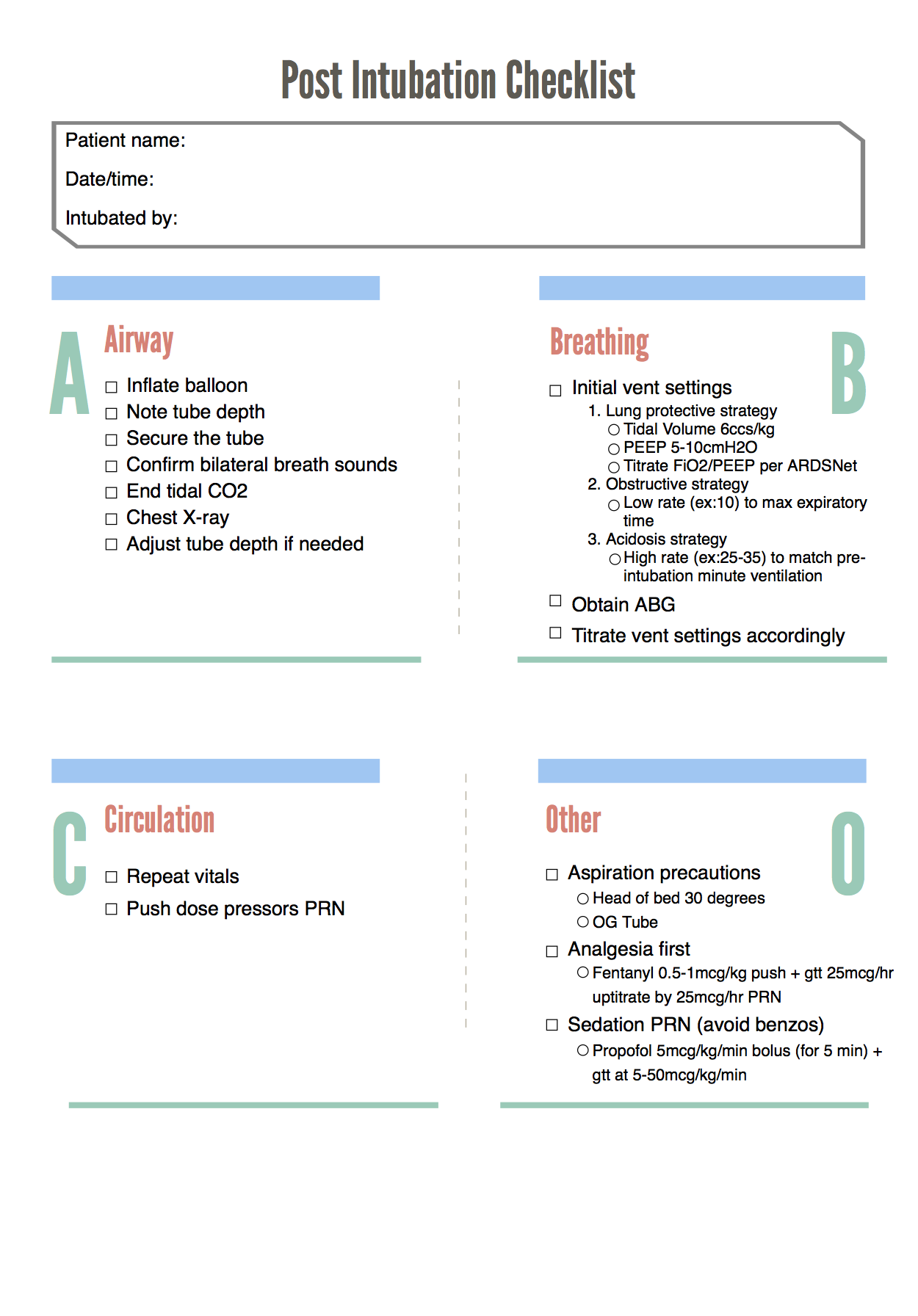
Written by:  William Ford, MD, MBA (NUEM PGY-4) Edited by: Simiao Li-Sauerwine, MD (NUEM ‘18) Expert commentary by: Matthew Levine, MD


Introduction

Ankle injuries are commonly seen in emergency medicine, and serious injuries can be found in the setting of a negative X-ray. The “high ankle sprain” involves the structure of the ankle called the syndesmosis. Isolated ligamentous disruption to the syndesmosis is uncommon, though when it occurs, it is frequently missed. This can lead to serious consequences, including long-term ankle dysfunction and the need for surgery [1]. Identifying these injuries in the ED can improve recovery and facilitate prompt follow-up.

What is the Syndesmosis?

The syndesmosis is the distal articulation of the tibia and the fibula and it keeps the joint stable. For the purposes of this discussion, the focus will be on the ligamentous parts of the syndesmosis not seen on an ankle X-ray.

There are four syndesmotic ligaments: the interosseous ligament (IOL), the anterior inferior tibiofibular ligament (AITFL), the posterior inferior tibiofibular ligament (PITFL), and the inferior transverse tibiofibular ligament (ITTFL) [2].

How Does Syndesmotic Injury Occur?

The motion often involved in syndesmotic injury is external rotation of the foot. Commonly, it is a combination of external rotation of the foot and excessive dorsiflexion of the ankle [3-6]. This is frequently encountered in high-speed collisions, uneven terrain, and cutting and jumping sports [1]. In most cases, syndesmotic injury will occur concurrently with a fracture, but sometimes, ligamentous injury may occur in isolation [7-8].

How Can I Diagnose Syndesmotic Injury?

Since this diagnosis is difficult to make without an MRI, providers must rely on a high index of suspicion. First, ask yourself if the mechanism of injury is consistent with syndesmosis disruption. Localizing the areas of tenderness on physical exam can also be helpful. Anterolateral or posteromedial ankle tenderness, as opposed to direct lateral or medial pain inferior to the malleoli, can suggest syndesmotic involvement. Finally, in accordance with the name “high ankle sprain”, more proximal pain may be indicative of a syndesmotic injury [1].

There are a few provocative tests described for diagnosing syndesmotic injuries. None are slam-dunk maneuvers, but a constellation of positive findings can be helpful in making the diagnosis.

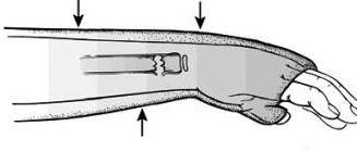
  1. Squeeze test: With the patient sitting at the edge of the bed and knee bent at 90 degrees, a strong compressive force is applied to the tibia and fibula proximal to the mid-calf. Pain indicates a positive finding.

  2. External rotation stress test: If the pain is reproduced with manual external rotation of the foot and ankle relative to the tibia, this test is positive. It is important to make sure the tibia is stabilized while performing this test.

  3. Cotton test: This test is performed by attempting to translate the talus laterally under the tibia. Increased translation compared to the contralateral side or increased pain with this maneuver is a positive finding.

  4. Fibular translation test: Stabilize the tibiotalar joint with one hand, translate the fibula anteriorly and posteriorly with the other hand. Increased pain and translation compared to the contralateral side equals a positive test.

X-ray diagnosis can also be difficult, except in the presence of frank tibiofibular diastasis [1].

Summary

Not all ankle sprains are created equal. High ankle sprains involving the syndesmosis can mean triple the recovery time of a regular sprain, chronic instability, or definitive treatment with surgery. Increased awareness and knowledge of how to diagnose these injuries is important to ensure quality care for patients.


Expert Commentary

Ankle injuries are the most common orthopedic complaint we see in the ED.  While most of these presentations are simple sprains, there are other more severe injuries that will resemble uncomplicated sprains.  Given the sheer volume of ankle injuries we see, we undoubtedly have all missed some of these more complicated injuries.  A high ankle sprain is one of these injuries.  Another classic injury that resembles an ankle sprain is the Snowboarder’s fracture. This is a fracture of the lateral process of the talus that occurs from ankle inversion and dorsiflexion.  Snowboarder’s fractures clinically resemble ankle sprains and x rays miss up to 40% of these fractures!  I have seen radiology miss some of these and then found the fracture by going back and magnifying the area of the lateral talus distal to the lateral malleolus on the AP and mortise views. 

Regardless, plain films will not detect all Snowboarder’s fractures or high ankle sprains.  It is tempting to quickly wrap and discharge all ankle “sprains” after a negative x ray.  It is important, however, to discuss with these patients what is a normal healing progression and timeline.  Emphasize that if 10 days go by and there is still significant pain, functional impairment, or reliance on the crutches or air cast, they need to follow up with orthopedics.  Provide them with the means to arrange this follow up. It is not realistic to be able to diagnose every ankle sprain mimic in the ED, but it is our duty to provide every patient (not just ankle sprain patients) with the proper instructions for follow up in case we have missed something.

Matt_Levine-33 (2).png
 

Matthew Levine, MD

Assistant Professor of Emergency Medicine

Northwestern Medicine


How To Cite This Post

[Peer-Reviewed, Web Publication] Ford W, Li-Sauerwine S. (2019, May 27). Not All Ankle Sprains are Created Equal. [NUEM Blog. Expert Commentary by Levine M]. Retrieved from http://www.nuemblog.com/blog/high-ankle-sprain.


Other Posts You May Enjoy


References

  1. Hunt KJ, Phisitkul P, Pirolo J, et al. High Ankle Sprains and Syndesmotic Injuries in
    Athletes. Journal of the American Academy of Orthopaedic Surgeons. 2015;23(11): 661-73.

  2. Fibular translation test: Stabilize the tibiotalar joint with one hand, translate the fibula anteriorly and posteriorly with the other hand. Increased pain and translation compared to the contralateral side equals a positive test.

  3. Ogilvie-Harris DJ, Reed SC, Hedman TP. Disruption of the Ankle Syndesmosis: Biomechanical Study of the Ligamentous Restraints. Arthroscopy: The Journal of Arthroscopic & Related Surgery. 1994;10(5): 558-60.

  4. Lin C-F, Gross MT, Weinhold P. Ankle Syndesmosis Injuries: Anatomy, Biomechanics, Mechanism of Injury, and Intervention. J. Orthop. Sport. Phys. Ther. 2006;36(6): 372–384. doi:10.2519/jospt.2006.2195.

  5. Dattani R, Patnaik S, Kantak A, et al. Injuries to the tibiofibular syndesmosis. J. Bone Jt. Surg. 2008;90–B(4): 405–410. doi:10.1302/0301-620X.90B4.19750.

  6. Brosky T, Nyland J, Nitz A, et al. The Ankle Ligaments: Consideration of Syndesmotic Injury and Implications for Rehabilitation. J. Orthop. Sport. Phys. Ther. 1995;21(4).

  7. Hopkinson WJ, Pierre P St., Ryan JB, et al. Syndesmosis Sprains of the Ankle. Foot Ankle. 1990;10(6): 325–330.

  8. Miller CD, Shelton WR, Barrett GR, et al. Deltoid and Syndesmosis Ligament of the Ankle without Fracture Injury. Am. J. Sports Med. 1995;23(6): 746–750.

  9. Hunt KJ. Syndesmosis injuries. Curr. Rev. Musculoskelet. Med. 2013;6: 304–312. doi:10.1007/s12178-013-9184-9.

Posted on May 27, 2019 and filed under Orthopedics.

Corneal Abrasions

Screen Shot 2019-05-07 at 10.55.04 AM.png

Written by: Julian Richardson, MD, MBA (NUEM PGY-2) Edited by: Andrew Moore, MD (NUEM ‘18) Expert commentary by: Brad Sobolewski, MD, MEd


A 40 yo construction worker comes to Emergency Department with a foreign body sensation in his left eye for two days. He states that he forgot to wear his eye protection while sanding a plank of wood the other day and felt like something has been scratching his eye ever since. Upon entering the room, his left eye is hyperemic and the patient appears to be in discomfort.


Approach to the painful red eye with foreign body sensation

The initial differential diagnosis to a painful red eye is broad and includes entities such as keratitis, corneal abrasions, corneal ulceration, acute angle glaucoma, iritis, chemical burn, scleritis, subconjunctival hemorrhage, and conjunctivitis. The patient’s history is particularly concerning for corneal abrasion, corneal ulceration, or globe rupture. A simple test to distinguish these diagnoses is the fluorescein exam.


Fluorescein exam

Fluorescein has been used in ophthalmology since the 1880s. This exam should be included for all patients where there is a suspicion of abrasion, foreign body, or infection. Fluorescein absorbs light in blue-wavelengths and emits energy in green wavelengths. It fluoresces in alkaline environments, for example Bowman’s membrane which is located below the corneal epithelium. It does not fluoresce in acidic environments such as the tear film over intact cornea.  Because of this, defects in the cornea increase fluorescein uptake and assist in locating corneal damage.

The eye should first be numbed usually with the use of a topical anesthetic drop, such as tetracaine. Next, take a fluorescein strip and place one drop of saline or local anesthetic to the strip. Place this strip inside the lower lid, remove, and ask the patient to blink. The key to a good exam is to produce a thin layer covering the surface of the eye. If too much is applied, the excess can easily be removed by asking the patient to blot their eye while closed with a tissue. The eye should then be examined using a Wood’s lamp, blue filter of a slit lamp, or penlight with a blue filter

Warning! this dye will permanently stain soft contact lenses and clothing. Be sure to remove any contacts and have plenty of gauze or other absorbant material available prior to instillation. Irrigating the excess dye out the eye after examination will help minimize staining the patient’s clothing.


Corneal Abrasion


Definition

  • Scratch to the epithelium that comprises the cornea and exposes the basement membrane. Patients generally complain of a foreign body sensation, pain, photophobia, and some vision loss. On physical exam, the clinician may find injected conjunctiva, and decreased visual acuity (if the defect is large or lies in the visual axis).

 Fluorescein Exam

  • Typically, abrasions are seen at the central part of the cornea due to limited protection of closure of the patient’s eyelids. The margins are sharp and linear in the first 24hrs. Circular defects suggests an embedded foreign body is present and may persist for greater than 48hrs. Foreign bodies can also produce vertical linear lesions and the upper lid should be lifter up to look for a foreign body under the eyelid

Management

  • Treatment with antibiotics have become the standard of care. Antibiotics are particularly indicated for abrasions caused by contacts (cover for pseudomonas), foreign bodies, or history of trauma with infectious or vegetative matter due to a higher risk of infection. Ophthalmic antibiotic therapy include: erythromycin ointment or sulfacetamide 10%, polymyxin/trimethoprim, ciprofloxacin, or ofloxacin drops (4 times a day for 3-5 days). Pain relief can be provided with oral or topical pain meds. Topical NSAIDs include .1% indomethacin, .03% flubiprofen, .5% ketorolac, 1% indomethacin, and .1% diclofenac.  If symptoms persist greater than 24 hours after treatment the patient should follow-up with a physician. If the abrasion has not healed in 3-4 days the patient should be evaluated by an ophthalmologist.


Corneal Ulceration

after ulceration.png
 

Definition

  • When a defect in the corneal epithelium becomes infected with bacteria or fungi it is defined as a corneal ulceration. This is a common complication of corneal abrasions and if left untreated can result in a corneal perforation.

Fluorescein Exam

  • Corneal staining with infiltrate or opacification around the lesion should raise suspicion for ulceration. Contact lens wearers raise the suspicion of a Pseudomonal infection. Many Pseudomonal organisms fluoresce when exposed to UV light and fluoresce prior to fluorescein application.

Management

  • Concern for ulceration requires an urgent ophthalmology consultation within 24hrs. Discharged patients should be treated with antibiotic drops or ointment.

Globe Rupture

Definition

  • Full thickness injury to the cornea, sclera, or both secondary to penetrating of blunt trauma.

Fluorescein Exam

  • Seidel test: instill a large amount of fluorescein onto eye and looking for small stream of fluorescent blue or green fluid leaking from the globe.

Management

  • Once suspected, avoid further examination or manipulation, make the patient NPO, and place an emergent ophthalmology consultation. These patients also require broad spectrum IV antibiotic coverage with a 3rd generation cephalosporin or aminoglycoside and vancomycin to prevent post-traumatic endophthalmitis.

In summary, all patients with eye pain, particularly with a foreign body sensation, warrant a fluorescein exam. A wealth of information can be gained by this simple test and will guide the management of the patient.


Expert Commentary

This is a very comprehensive review of a common complaint in the Emergency Department. You correctly identified that one must be careful to avoid instilling too much fluorescein so as to cause a false positive result. Though a drop of tetracaine or saline dilutes the fluorescein from the strip somewhat the quantity is hard to control at times – especially in noncompliant patients (like the children I usually examine in the Pediatric Emergency Department). Excess fluorescein can collect across the eye making identification of small abrasions challenging. If you put too much in rinse the eye and try again. It is also incredibly important to not sent the patient home with tetracaine drops, as too frequent use may lead to further corneal injury. The evidence is based on animal models and case series and is far from complete. Read more on this great R.E.B.E.L. EM post (link: http://rebelem.com/topical-anesthetic-use-corneal-abrasions/).

One of the main pitfalls to the use of fluorescein strips is the risk of actually causing an abrasion. The method noted in this article – placing the strip inside the lower lid margin and asking the patient to blink – can cause an abrasion if the edge of the strip touches the cornea. This is particularly challenging to do in children, since even with proper restraint the blink reflex and their tendency to recoil is high. Therefore I recommend doing one of the following:

  1. Hold the patient’s eyelids open. Drip the tetracaine or saline down the strip and allow it to drip into the eye, being careful to avoid touching the strip to the eye.

  2. Make a fluorescein dropper. This is well detailed in the Tricks of the Trade: Fluorescein application techniques for the eye form Academic Life in Emergency Medicine (link: https://www.aliem.com/2015/06/tricks-of-the-trade-fluorescein-eye/). The Angiocath dropper allows for better control of droplet size and makes it easier to instill fluorescein into the squinting eye without the risk of touching the cornea.

You could just touch the fluorescein strip to the patient's inner eyelid when trying to diagnose a corneal abrasion. But then you could cause a corneal abrasion, which is an ouroboros of terribleness from which there is no escape. So why don't you try making a dropper instead, as detailed in this PEMBLOG SHORTS video.


Brad Sobolewski, MD, MEd

Associate Professor, Assistant Director - Pediatric Residency Training Program

Division of Emergency Medicine

Cincinnati Children's Hospital Medical Center


How To Cite This Post

[Peer-Reviewed, Web Publication] Richardson J, Moore A. (2019, May 20). Corneal Abrasions [NUEM Blog. Expert Commentary by Sobolewski]. Retrieved from http://www.nuemblog.com/blog/corneal-abrasion


Other Posts You May Enjoy


References

  1. Images courtesy of http://www.tedmontgomery.com/the_eye/

  2. Marx, J. A., & Rosen, P. (2014). Rosen's emergency medicine: Concepts and clinical practice (8th ed.) Ch. 71. Opthamology. Philadelphia, PA: Elsevier/Saunders

  3. Yanoff, M., Duker, J. S., & Augsburger, J. J. (2009). Ophthalmology. Ch 4, Corneal anatomy, physiology, and wound healing. Edinburgh: Mosby Elsevier.

  4. Roberts, J. R., In Custalow, C. B., In Thomsen, T. W., & In Hedges, J. R. (2014). Roberts and Hedges' clinical procedures in emergency medicine. Ch 62. Opthalmologic procedures.

  5. Gardiner, M. F. Overview of eye injuries in the emergency department. Retrieved September 16, 2017, from https://www.uptodate.com/contents/overview-of-eye-injuries-in-the-emergency-department.

  6. Jacobs, D. S. Corneal abrasions and corneal foreign bodies: management. Retrieved September 16, 2017, from https://www.uptodate.com/contents/corneal-abrasions-and-corneal-foreign-bodies-management.

  7. Waldman, N., Winrow, B., Denise, I., Gray, A., McMAster, S., Giddings, G., & Meanley, J. (2017). An observational study to determine whether routinely sending patients home with a 24-hour supply of topical tetracaine from the emergency department for simple corneal abrasion pain is potentially safe. Annals of Emergency Medicine, 02(016).


Posted on May 20, 2019 and filed under Ophthalmology.

C-spine Clearance with Negative CT: Are We There Yet?

Screen Shot 2019-05-07 at 10.46.34 AM.png

Written by: M. Terese Whipple, MD (NUEM PGY-3) Edited by: Quentin Reuter, MD (NUEM ‘18) Expert commentary by: Matthew Levine, MD


We have excellent decision rules for clinically clearing cervical spine injury in low risk patients without imaging. However, a frustrating situation arises when a CT of their c-spine is obtained and negative, but they are having persistent midline pain. What do we do then? Are we forced to order an MR of the c-spine even when they have no neurological deficits and our gestalt tells us there is no clinically significant injury? MR often means admission, worsening of already overwhelming ED crowding, and unhappy patients when they cannot remove the c-collar for at least several more hours. Recent data and recommendations suggest that this may not be the case; a negative CT may be enough to rule out clinically significant injury. This blog post will explore some of the historical and recent data on the subject of cervical spine clearance after CT scan alone.  


There has been great historical debate over the best management for patients with persistent midline pain after negative CT, however that evidence is beyond the scope of this post. Current common practice and the recommendation of the American College of Radiology leads down the path of cervical spine MR when this situation arises [1]. Due to the cumbersome logistics of MR, much work has been done to determine if MR truly adds value to the patient’s workup. Is MR catching clinically significant injury missed by CT that changes clinical management? The majority of studies have concluded that the answer to that question is no.  

In 2015 the Eastern Association for the Surgery of Trauma (EAST) sought to tackle this question by reviewing all studies to date examining C-spine evaluation in obtunded patients [2]. They evaluated 11 studies with a total of 1718 obtunded patients who underwent C-Spine imaging with CT. None were ultimately found to have unstable fractures or unstable ligamentous injury missed by CT. There was a 9% incidence of stable injuries missed on CT and found on follow up MR, flex-x, upright XR, or clinical follow up. They found a cumulative 100% NPV for unstable C-Spine injury with CT and 91% NPV for stable injury. They did rate the quality of evidence as low for various reasons, including non-comparable imaging protocols, inconsistently reported and variable outcomes, publication bias, and an overall inability to perform a meta-analysis with the data.  However, they rated the data from which they derived the NPV as moderate quality as the NPV was consistently 100% throughout all of the trials. Based on their analysis they provided the following recommendation for obtunded blunt trauma patients:

“We conditionally recommend cervical collar removal after negative high-quality c-spine CT scan results alone.”

 They went on to further clarify,   

“It should be acknowledged that cervical collar removal can result in neurological change and even paralysis, although this may be underreported in the literature. However we cannot continue indiscriminate two-stage sequential screening for C-spine injuries if the injury rate is near 0% for the first test and the second adjunctive test results in false positives and inconsistent treatment plans.”

But the real question that is more pertinent to us as EM physicians (obtunded MR’s are usually dealt with upstairs), is:  if we can remove the c-collars of obtunded patients after negative CT, why couldn’t that be extrapolated to awake patients?  Well, they commented on that too:   

“Therefore, if collars are to be removed in a high risk obtunded population […] cervical collar removal can be logically argued for any population-obtunded or not.” [2]

 They finally call for multicenter prospective research on the subject, again citing the low quality of evidence that they used for their recommendation. That call was answered in 2017 by the Western Trauma Association. The group completed a multi institution trial with 10,000 patients who were getting a CT for evaluation of cervical spine injury prospectively enrolled at 17 centers [3]. They found only 3 CT scans that missed clinically significant injury (.03%). All of those patients had focal neurological abnormalities on exam. There was no clinically significant injury missed by CT and exam combined. CT scan alone had an NPV of 99.97%, and an NPV of 100% when combined with clinical exam. Therefore, they proposed this diagnostic algorithm:

algorithm.png


Most trials have found similar results, with a few exceptions. Two trials prior to the publication of the Western Trauma Association (WTA) paper found that CT missed a few clinically significant injuries in patients with no neurological symptoms. Both trials enrolled significantly fewer patients than the WTA paper, and only enrolled patients with negative CT who would be evaluated with MR, meaning they couldn’t comment on the overall sensitivity of CT in unstable c-spine injury. The ReConect trial in 2016 found 5 of 767 patients (.6%) with injuries requiring surgical intervention that were missed on CT [4].  Another study with similar methods published in Annals of Emergency Medicine in 2011 evaluated those who had a negative CT but were MR’ed for persistent midline C-Spine tenderness [5]. They found that out of 178 patients, 5 had injury requiring operative management that was missed on CT but found on subsequent MR (2.8%) [5].  The Annals paper is certainly an outlier, with a considerably higher rate of missed clinically significant injury than the remainder of the literature, with rates usually between 0-1% [6-18]. The authors believe this may be due to more stringent methodology.  For instance, they required MR to be performed within 48 hours when it is the most sensitive for edema, and only enrolled patients with midline tenderness rather than subjective pain [5].  While this may be true, the results have not been replicated in subsequent studies.


With the publication of the WTA paper, evidence certainly seems to be tipping in favor of CT clearance of cervical spine in neurologically intact patients. However, a few questions remain. In every study discussed here, MR resulted in discharge with hard collar in a portion of patients. Indications ranged from stable injury to persistent pain with no evidence of injury on MR. It is unclear whether hard collar placement makes a difference in the clinical course of these patients, if their stable injuries would have become unstable without it, or if it has any long term impact on outcomes such as chronic pain. This is an important question not yet adequately addressed in the literature.  The majority of these trials were also completed at trauma centers with radiologists well trained in reading c-spine imaging and high quality CT scanners. It could be difficult to generalize this data to centers with older scanners or whose radiology departments are not as expert in trauma radiology.

Incredibly high quality and reproducible evidence is required to change practice when high stakes, such as potentially missed cervical spine injury, are involved. So far we have multiple trials showing an NPV of close to 100% when CT and good neurological exam are combined, and the conditional recommendation by the EAST group. Time will tell if recommendations in the future remove the “conditional” portion as CT technology continues to improve, further studies with stringent methodologies are conducted, and the results of the WTA paper are hopefully replicated.


Expert Commentary

Thank you Dr Whipple for that really practical review of a real-life common clinical question we face all the time: Can we remove the collar?  Some important takeaways are:

  1. There is a robust and growing body of evidence that removing the collar after a negative high-quality CT is safe if the patient is neurologically intact.

  2. This practice is endorsed by two major trauma organizations, EAST and WTA. 

The endorsement by respected major trauma societies is important in translating evidence into practice.  It seems like all that is left at this point for widespread implementation is overcoming culture.  This would likely require addressing the outlier studies listed by Dr Whipple to win over those still skeptical.  Part of overcoming culture would involve buy-in from neurosurgical societies.  What do neurosurgical societies say regarding clearing these patients?  There are many instances in which a patient is discharged with recommendations from the neurosurgeon to wear a hard collar despite a negative CT and MRI.  On the surface this seems like defensive medicine and impractical for the patient.  Is the patient really going to comply with this until follow up?  Is this collar really protecting them and preventing further injury which, after negative CT and MRI and with a normal neuro exam, seems exceedingly unlikely?  Does evidence support this practice?

In the end, decision rules should be used when you want evidence to support your clinical decisions, such as removing the C collar after negative imaging in a neurologically intact patient.  Do not use decision rules, however, to overturn or replace sound clinical judgement.  If there is something about a case that makes you still feel like you could be missing an outlier injury by removing the collar, listen to that voice inside of you. It is that sound clinical judgement that will guide you through your career, not decision rules.

Matt_Levine-33 (1).png
 

Matthew Levine, MD

Northwestern Medicine, Assistant Professor of Emergency Medicine


How To Cite This Post

[Peer-Reviewed, Web Publication] Whipple T, Reuter Q. (2019, May 13). C-spine clearance with negative CT: Are we there yet? [NUEM Blog. Expert Commentary by Levine M]. Retrieved from http://www.nuemblog.com/blog/cspine-clearance-ct


Other Posts You May Enjoy


References:

  1. American College of Radiology. ACR appropriateness criteria on suspected spine trauma. Available at: http://www.acr.org.

  2. Patel MB, et al. Cervical spine collar clearance in the obtunded adult blunt trauma patient: A systematic review and practice management guideline from the Eastern Association for the Surgery of Trauma. J Acute Care Trauma Surgery. 789(2): 432-441.

  3. Inaba, K et al. Cervical Spine Clearance: A Prospective Western Trauma Association Multi-Institutional Trial. J Trauma Acute Care Surg. 2016 Dec: 81(6): 1122-1130.doi: 10.1097/TA.0000000000001194

  4. Maung A, et al. Cervical spine MRI in patients with negative CT: A prospective, multicenter study of the Research Consortium of New England Centers for Trauma (ReCONECT). J Trauma Acute Care Surg. 82 (2): 263-269.

  5. Ackland HM, et al. Cervical Spine Magnetic Resonance Imaging in Alert, Neurologically Intact Trauma Patients With Persistent Midline Tenderness and Negative Computed Tomography Results. Ann of Em Med. 2011 Dec. 58 (6): 521-530.

  6. Chew B, et al. Cervical spine clearance in the traumatically injured patient: is multidector CT scanning sufficient alone? J Neurosurg Spine. 2013. 19: 576-581

  7. Bush L, et al. Evaluation of cervical spine clearance by computed tomographic scan alone in intoxicated patients with blunt trauma. JAMA Surg. 2016; 151 (9): 807-813

  8. D’Alise  et al. Magnetic resonance imaging for the evaluation of the cervical spine in the comatose or obtunded trauma patient. J Neurosurgery  (Spine 1) 1999; 91:54-59.

  9. Resnick S, et al. Clinical relevance of magnetic resonance imaging in cervical spine clearance: a prospective study. JAMA Surg. 2014; 149 (9): 934-9.

  10. Menaker J, Philp A, Boswell S, Scalea TM. Computed tomography alone for cervical spine clearance in the unreliable patient--are we there yet? J Trauma. 2008; 64(4):898–903.

  11. Chew BG, Swartz C, Quigley MR, Altman DT, Daffner RH, Wilberger JE. Cervical spine clearance in the traumatically injured patient: is multidetector CT scanning sufficient alone? Clinical article. J Neurosurg Spine. 2013; 19(5):576–81.

  12. Como JJ, Leukhardt WH, Anderson JS, Wilczewski PA, Samia H, Claridge JA. Computed tomography alone may clear the cervical spine in obtunded blunt trauma patients: a prospective evaluation of a revised protocol. J Trauma. 2011; 70(2):345–9. discussion 9-51.

  13. Khanna P, Chau C, Dublin A, Kim K, Wisner D. The value of cervical magnetic resonance imaging in the evaluation of the obtunded or comatose patient with cervical trauma, no other abnormal neurological findings, and a normal cervical computed tomography. J Trauma Acute Care Surg. 2012; 72(3):699–702.

  14. Schuster R, Waxman K, Sanchez B, Becerra S, Chung R, Conner S, Jones T. Magnetic resonance imaging is not needed to clear cervical spines in blunt trauma patients with normal computed tomographic results and no motor deficits. Arch Surg. 2005; 140(8):762–6.

  15. Anekstein Y, Jeroukhimov I, Bar-Ziv Y, Shalmon E, Cohen N, Mirovsky Y, Masharawi Y. The use of dynamic CT surview for cervical spine clearance in comatose trauma patients: a pilot prospective study. Injury. 2008; 39(3):339–46.

  16. Brohi K, Healy M, Fotheringham T, Chan O, Aylwin C, Whitley S, Walsh M. Helical computed tomographic scanning for the evaluation of the cervical spine in the unconscious, intubated trauma patient. J Trauma. 2005; 58(5):897–901.

  17. Harris TJ, Blackmore CC, Mirza SK, Jurkovich GJ. Clearing the cervical spine in obtunded patients. Spine (Phila Pa 1976). 2008; 33(14):1547–1553.

  18. Steigelman M, Lopez P, Dent D, Myers J, Corneille M, Stewart R, Cohn S. Screening cervical spine MRI after normal cervical spine CT scans in patients in whom cervical spine injury cannot be excluded by physical examination. Am J Surg. 2008; 196(6):857–862.

Posted on May 13, 2019 and filed under Trauma.

Anticoagulation in Distal DVT

Screen Shot 2018-09-27 at 11.21.37 AM.png

Anticoagulation in Distal DVT

Written by: William LaPlant, MD (NUEM PGY-3) Edited by: William Ford, MD (NUEM PGY-4) Expert commentary by: Kelsea Caruso, PharmD


Currently there is significant heterogeneity in the treatment of distal deep vein thromboses (DDVTs), which are DVTs that occur distal to the popliteal fossa. What is the best course of action when a reasonably healthy patient has a new calf DVT?

The CACTUS trial was a randomized, placebo-controlled, double-blinded trial prospectively assessing anticoagulation for calf DVTs [1]. Notably, the trial was met with slow enrollment resulting in termination of the study prior to enrollment of the prespecified sample size, meaning that it was underpowered to detect a significant difference between the groups. It only enrolled about half of the participants it had intended to, with an initial 90% power to detect a 70% risk reduction in the composite outcome: development of a proximal DVT or symptomatic PE by day 42. This power study assumed a 10% incidence of the primary outcome in the placebo group.

Picture1.png

As you can see, in both groups the progression to proximal DVT or PE was quite low. As there were only 12 total composite events (DVT extension, PE), making a comparison between groups with any degree of certainty impossible. This study met neither their enrollment goals (only enrolling half the participants) nor the predicted incidence of the composite outcome (half the projected amount), so it was quite significantly underpowered to detect a difference.

Notably, the study also did not enroll pregnant women, patients with a previous DVT, recent PE, or recent malignancy. These patients were likely not enrolled due to their higher risk of progression, which may have biased the results towards treatment. As such, the results of this trial could never be applied to these groups.

Unfortunately, given the slow recruitment in the CACTUS trial as well as the low event rate of the composite outcome, the likelihood that this will be studied again in a prospective, double-blinded manner is unlikely. As such, we will need to put the CACTUS trial into context of retrospective evidence to try to identify an ideal practice pattern.

With regard to the retrospective data available:

In a 2016 metanalysis, the incidence of PE from DDVT was 0% to 6.2% [median 1.1%] [2], which is in line with the results from the CACTUS trail. As you can see in the chart below, there is some heterogeneity to the results, likely due to the wide variety in study methodology.

Based on the above evidence, what are the best management options? A recent review article (which includes data from CACTUS) offers two suggestions [3]:

  1. The most conservative management will still be anticoagulation of the isolated DDVT. This should be standard for patients with cancer or other pro-thrombotic state that would place the patient at high risk for the development of DVT.

  2. Deferred anticoagulation with follow up ultrasound can be used for patients without significant thrombotic risk factors. You can engage in shared decision making and discuss the risks for bleeding in your patient against the 4-5% risk for clot progression and 1% risk for PE (based on the CACTUS trail as well as retrospective data).

If you decide against anticoagulation in the emergency department, follow up imaging would be recommended in 1-2 weeks to evaluate for progression. This tight follow up window would help to ensure clot progression is identified early and would allow the patient to readdress anticoagulation with his/her primary care physician.


Expert Commentary

Thank you for this really great summary! I think you are bringing up a dilemma that we see quite often in the Emergency Department and one that, unfortunately, still doesn’t have much data to help guide our treatment recommendations. The CHEST guidelines for VTE disease were most recently updated in 2016 and do not include the results of the CACTUS trial or the results of the mentioned meta-analysis. Their recommendation is to treat an unprovoked DVT (distal or proximal) with anticoagulation for at least 3 months.

 The CACTUS trial questioned if this is required for all patients, primarily focusing on those with an isolated calf DVT. The CACTUS trial had some interesting results, but, like you mentioned, a huge patient population was excluded.. We can only extrapolate this data to a very small group of people: young (average age ~50 years old in the trial), healthy and without any risk factors for VTE. Also, the study drug utilized in the trial was nadroparin, a low molecular weight heparin, which is not available in the US. It would have been more applicable to clinical practice if this trial had utilized a direct-oral anticoagulant like rivaroxaban or apixaban. If these were used the safety results could have potentially been reduced.

So the question remains… to anticoagulate or not to anticoagulate? Here are my final thoughts:

  1. The patients you can think about deferring anticoagulation are those without any VTE risk factors (cancer, obesity, immobility, those on estrogen therapy etc.).

    • As mentioned, this should be a discussion with the patient so they understand the risk of deferring anticoagulation. They should have adequate follow-up and understand the signs and symptoms of a pulmonary embolism.

  2. The patients that should be anticoagulated are those that have any risk factor for VTE or patients from the previous point that choose treatment.

    • The anticoagulant should be selected by keeping patient specific factors in mind like past medical history, renal function and current medications

    • Unlike the CACTUS trial, try and prescribe oral therapy. Your patients will thank you if they don’t have to inject themselves daily.

kelsea caruso.png
 

Kelsea Caruso, PharmD

Emergency Medicine Clinical Pharmacist

Northwestern Memorial Hospital


How To Cite This Post

[Peer-Reviewed, Web Publication] LaPlant W, Ford W. (2019, May 6). Anticoagulation in distal DVT. [NUEM Blog. Expert Commentary by Caruso K]. Retrieved from http://www.nuemblog.com/blog/distal-dvt


Other Posts You May Enjoy


Resources

  1. Righini M, Galanaud J, Guenneguez H, et al. Anticoagulant therapy for symptomatic calf deep vein thrombosis ( CACTUS ): a randomised , double-blind , placebo-controlled trial. 2017;3(December 2016). doi:10.1016/S2352-3026(16)30131-4.

  2. Wu AR, Garry J, Labropoulos N, Brook S. Incidence of pulmonary embolism in patients with isolated calf deep vein thrombosis. J Vasc Surg. 2016;5(2):274-279. doi:10.1016/j.jvsv.2016.09.005.

  3. Robert-ebadi H, Righini M. Management of distal deep vein thrombosis ☆. Thromb Res. 2017;149:48-55. doi:10.1016/j.thromres.2016.11.009.

Posted on May 6, 2019 and filed under Hematology.

Ultrasound in Pediatric Distal Forearm Fractures

Screen Shot 2019-04-02 at 1.02.55 PM (1).png

Written by: Jason Chodakowski, MD (NUEM PGY-3) Edited by: Logan Weygandt, MD (NUEM ‘17) Expert commentary by: Rachel Haney, MD (NUEM ‘17)


Why use Ultrasound?

Distal forearm fractures are common fractures in the pediatric population. Although plain radiographs of the forearm are still considered the gold standard for definitive diagnosis, there is growing interest in using ultrasound for diagnosis because it provides zero radiation exposure, it can be used to guide local pain control, and it can confirm reduction success at the bedside. Ultrasound is easy to teach and provides value under circumstances when plain radiography might be unavailable (pre-hospital environment, disaster areas, or in developing countries).

A recent meta-analysis of 12 studies, which included 951 children 18 and younger, found that physician performed bedside ultrasound detected distal forearm fractures with a pooled sensitivity of 98% and a specificity of 96% when compared with the gold standard plain radiographs.[1] The pain associated with ultrasound use was also significantly less.[2]


How do I use Ultrasound?

To evaluate musculoskeletal pathology use the high-frequency linear array transducer employing the six-view ultrasound technique as shown below. You may detect a fracture as an apparent discontinuity or irregularity (divots, step-offs, distortion) of the hyperechoic and continuous bony cortex. Disruptions as small as 1mm can be detected.

Six-view technique (Herren et. al. 2015)

Six-view technique (Herren et. al. 2015)

Normal Cortex (Crosby et. al. 2014)

Normal Cortex (Crosby et. al. 2014)

Distal radius fracture (emergencyultrasoundteaching.com)

Distal radius fracture (emergencyultrasoundteaching.com)

Distal radius fracture (acep.org)

Distal radius fracture (acep.org)

Pitfalls

In children the evaluation of bones is complicated by the open physes, which may be mistaken for fractures. The difference is that physes will appear as smooth, downward-sloping curves unlike fractures, which will have abrupt step-offs.

Normal open tibial physis (Crosby et. al. 2014)

Normal open tibial physis (Crosby et. al. 2014)

What else is Ultrasound Good For?

  • Confirming reductions

    Ultrasound is also utilized by emergency physicians to determine successful realignment of pediatric distal forearm fractures after closed reduction.[4]

Fracture reduction (Socranksy et. al. 2016)

Fracture reduction (Socranksy et. al. 2016)

  •  Achieving adequate pain control

    Ultrasound can also be used to guide hematoma blocks. The hematoma block is a technique wherein the physician injects an anesthetic solution into the hematoma between the fractured bone fragments (see image below). It has been shown to be effective, safe, faster, and uses fewer resources with no significant difference in pain scores when compared to procedural sedation in both adults and children with distal forearm fractures.[6,7]

Clean skin and place a sterile cover over the transducer. Using 5-10cc of 1-2% lidocaine inject into the hematoma between the fractured bone fragments using an 18-22 gauge needle.

Visualization of needle (N) entering between fracture bone fragments (U) (emdocs.net)

Visualization of needle (N) entering between fracture bone fragments (U) (emdocs.net)

Take Home Points

  • Ultrasound is most useful in evaluating long bone fractures such as the femur, clavicle, ribs, or distal radius and ulna.

  • A reliable alternative to the plain radiograph is the proper six-view method it, with the advantages of being portable and radiation-free.

  • Ultrasound can also be reliably used to confirm fracture reduction, as well as for guiding forearm fracture hematoma blocks. 


Expert Commentary

Thank you for providing a concise summary of the utility of Point-of-care Ultrasound (POCUS) for pediatric forearm fractures.  

I’d like to mention a few key points regarding the use of POCUS for pediatric fracture assessment.

  • If you do a lot of adult scanning and not much pediatric scanning it is important to keep in mind that children may not be as cooperative (or stationary) as adults.

    • Smaller children may be afraid of the transducer therefore introducing the transducer to the patient as an object that will not hurt them is key. You should hand the probe to the patient, allow them to touch it and even scan themselves initially in order to get them more comfortable with the probe.

  • While the 6-view scan you describe will certainly improve sensitivity, adequate sensitivity can be achieved with a 2-view approach. Additionally, the 6-view technique may be prohibitively time-intensive in a busy Emergency Department.

    • In order to increase sensitivity with the 2-view approach, always start imaging at the point of maximal tenderness, initially in the longitudinal plane with the cortex of the bone parallel to the probe surface. Slide distal and proximal to the point of tenderness. Then rotate the probe 90 degrees to view the cortex in the transverse plane. Fractures are noted as cortical disruptions or step-offs. Fractures are most visible on POCUS when the fracture line is perpendicular to the angle of insonation.

  • Another key pearl is to use copious gel in order to optimize the focal point of the image. The focal zone on the screen is the part of the image with the highest resolution secondary to convergence of the US beams. The focal point can be changed depending upon your machine, but is typically no more shallow than about 1-cm below the probe surface, therefore if you place a good layer of gel about 1cm thick, you will place the cortext of the bone at the optimal focal point. Using copious gel is also important in reducing any potential discomfort caused by pressure from the probe.

    • If gel is a limited resource, you can use a water bath as well.

  • While POCUS is a wonderful tool, especially for fracture detection, I want you to keep in mind that the sensitivity of POCUS for fractures is the highest (low-mid 90s) for the diaphysis of long bones (femur, humerus, radius and ulna). Sensitivity is significantly lower for detecting fractures of other bones and fractures near joint lines secondary to the curvilinear nature of the metaphysis as well as the presence of cartilaginous epiphyseal plates in children.

    • While POCUS can supplant the use of radiography in austere environments, in a well-resourced emergency department, POCUS should be an adjunct to radiography. In this setting, POCUS can have utility in patients in whom you suspect occult fracture despite negative XRs or for real-time fracture reduction assessment before sedation wears off. Unless you are a pediatric POCUS expert, I would order XR’s as usual for a pediatric patient you suspect has a fracture. In the meantime- continue scanning patients with normal anatomy and documented fractures in order to develop your POCUS expertise! Happy Scanning!

Rachel Haney.png
 

Rachel Haney, MD

NUEM ‘17

Ultrasound Fellow at Massachusetts General Hospital


How To Cite This Post

[Peer-Reviewed, Web Publication] Chodakowski J, Weygandt L. (2019, April 28). Ultrasound in pediatric distal forearm fractures. [NUEM Blog. Expert Commentary by Haney R]. Retrieved from http://www.nuemblog.com/blog/us-for-fracture


Other Posts You May Enjoy


References

  1. Douma-den Hamer, Djoke, et al. "Ultrasound for Distal Forearm Fracture: A Systematic Review and Diagnostic Meta-Analysis." PloS one 11.5 (2016): e0155659.

  2. Chaar-Alvarez FM, Warkentine F, Cross K, et al. Bedside ultrasound diagnosis of nonangulated distal forearm fractures in the pediatric emergency department. Pediatr Emerg Care 2011; 27:1027.

  3. Herren C, Sobottke R, Ringe MJ, et al. Ultrasound-guided diagnosis of fractures of the distal forearm in children. Orthop Traumatol Surg Res 2015; 101:501.

  4. Dubrovsky, Alexander Sasha, et al. "Accuracy of ultrasonography for determining successful realignment of pediatric forearm fractures." Annals of emergency medicine 65.3 (2015): 260-265.

  5. Socransky, Steve, et al. "Ultrasound-Assisted Distal Radius Fracture Reduction." Cureus 8.7 (2016).

  6. Fathi, M. et al. Ultrasound-guided hematoma block in distal radius fracture reduction: a randomized clinical trial. Emerg Med J. 2014 Jul 12.

  7. Bear, David M., et al. "Hematoma block versus sedation for the reduction of distal radius fractures in children." The Journal of hand surgery 40.1 (2015): 57-61.

Posted on April 29, 2019 and filed under Ultrasound.

Top 5 Blog Posts of 2018

NUEM_blog logo_top5.png

As the long and cold Chicago winter wanes and Spring begins, let’s take a look at the most popular NUEM blogs from 2018. While we value all of our residents’ and experts’ hard work, we are highlighting these 5 posts which earned the most pageviews in calendar year 2018:

5) Auricular Hematoma Drainage

Andrew Berg, Elizabeth Byrne, and Chris Beach take us through the intimidating but fortunately uncommonly needed auricular hematoma drainage.

4) Hand Exam

Terese Wipple, Victor Gappmaier, and Avi Giladi (current Hand surgeon and my former college roommate who got me through organic chem) provide a usable guide to both basic and comprehensive hand exams, and important and common test for an important body part. You’ve really got to respect what they did here.


3) Beta Blocker Toxicity

Spencer Lang, Rachel Haney, and Patrick Lank review beta-blocker toxicity, including but far from limited to the fancier topics of high insulin euglycemic therapy and intralipids.

2) Migraine Cocktail

Headaches can be, well, a headache in the ED. Vidya Eswaran and Danielle Miller put together a comprehensive and digestible infographic collating key points of ED headache management, replete with tips & references. Also fun for me to have space to rant about my practice.


1) HiNTS Exam

Dizziness is one of the most frustrating entities we see in EM, with a handful of needles in a frustrating haystack with few tools to separate the two without turning our hospitals into full time MRI centers. The HiNTS exam gained popularity in the past few years, but has important limitations: easily applied to the wrong dizzy patients and can also be technically difficult to perform in already-dizzy patients. Will LaPlant, Mitali Parmar and Phillip Chang give a comprehensive but concise review of the key points – including how to document your findings. A well-deserved top spot for 2018, and sorry everyone, but it wasn’t even close.


Seth Trueger, MD, MPH, FACEP

Assistant Professor, Emergency Medicine Northwestern University


Posted on April 22, 2019 and filed under Top 5 2018.

Canthotomy

Screen Shot 2019-03-31 at 11.24.38 AM.png

Written by: Alex Ireland, MD (NUEM PGY-3) Edited by: Gabrielle Ahlzadeh, MD (NUEM PGY-4) Expert commentary by: Gary Lissner, MD


Expert  Commentary

The author has written an excellent illustrated review of the technique of lateral canthotomy and cantholysis for traumatic orbital compartment syndrome due to a significant retrobulbar hemorrhage. As with any procedure it is crucial to understand the indications and contraindications of the procedure. In these cases, the indication is the saving or restoring of vision lost due to excessive intraorbital pressure, and the contraindication is generally an opened globe.  This is a difficult situation because there is usually limited information and there is a limited amount of time to relieve the orbital compartment syndrome. The timing of the onset of the retrobulbar hemorrhage that created the critical pressure to cause loss of vision is often unknown.  The decision to decompress is usually based solely on a clinical examination without having time to obtain orbital radiologic studies and before an ophthalmologist can arrive to evaluate the patient. However, the clinical exam could be limited if the patient is not cooperative or is not conscious.

Initially a determination has to be made if the globe is lacerated or ruptured which could preclude the performance of the canthotomy and cantholysis .  Either a ruptured globe or an orbital compartment syndrome can cause loss of vision.  A ruptured globe can occur along with a retrobulbar hemorrhage. If the eyeball is opened, no additional external pressure should be placed on the lids or globe in order to prevent additional damage to the eye. An opened globe could be determined by seeing a laceration of the globe, seeing protruding intraocular contents, finding a very soft eye, and/or finding a distorted eye. If the eyeball is opened, a sturdy shield should be placed resting on the surrounding orbital bone to protect the eye from external pressure, and the aide of an ophthalmologist should be sought.

If it is determined that the globe is intact, a decision has to be made on clinical examination findings if a significant retrobulbar hemorrhage exists that would create enough pressure in the orbit to cause  loss of vision. There is usually not enough time to wait for imaging studies to be done. The trauma could create a large enough orbital fracture that could decompress the orbital pressure and the orbital compartment syndrome. On the other hand, an orbital fracture can create orbital emphysema. Blowing of the nose by the patient can increase the emphysema and further increase the intraorbital pressure, creating a sight-threatening orbital compartment syndrome.  

Acute vision loss is the key clue to a significant orbital compartment syndrome, but many trauma patients may not be able to cooperate for visual testing.  The reaction of the pupil to light can be used as a sign of visual loss even in the uncooperative patient. With an orbital compartment syndrome, the pupil of the involved eye will not react well to a bright light and will have an afferent defect. However, if the patient was given narcotics the pupils can become miotic and pupil testing becomes difficult.  Visual loss is not diagnostic of an orbital compartment syndrome because the trauma can create other damage that can cause visual loss including intraocular bleeding, retinal disorders, traumatic optic neuropathy, or a lacerated or ruptured globe.  Trauma can cause edema and ecchymosis of the lids, but tense proptosis with very firm retropulsion of the involved eye is a key diagnostic finding of a significant retrobulbar hemorrhage. As the orbital bleeding continues, the eye is pushed forward against the tight eye lids and the intraocular pressure increases.  A significant intraocular pressure increase can be used as another sign of an orbital compartmental syndrome.  Checking the intraocular pressure can be difficult in the presence of massive eyelid swelling.  Pulling the lids opened especially if the patient is squeezing can transmit the external pulling pressure to the eye, creating a false reading of a raised intraocular pressure.  Using curved instruments, or the blunt end of bent paper clips as shown by the Blog’s author, can help open the lids. Using a topical anesthetic to reduce eye discomfort, or using injectable local anesthetic to relax the orbicularis muscle action can also help to more easily open the eyelids to obtain a more accurate intraocular pressure. Orbital compartment syndrome will limit extraocular motility on the involved side, but the unconscious patient’s motility cannot be easily tested. A significant increase of the orbital pressure and the intraocular pressure can cause pulsation of the retina arteries and full retina veins which can help with the diagnosis of an orbital compartment syndrome if the fundus is viewed.

The Blog gives a precise pictorial and written description of the lateral canthotomy and cantholyisis technique.  However, unlike the author’s demonstration photos, most cases with significant traumatic orbital compartment syndrome have massive lid ecchymosis and subconjunctival hemorrhage and a proptotic  eye that is pushed forward tightly against the lids. Therefore, there is frequently little room to insert the instruments at the lateral canthal angle. Extreme care must be taken to avoid unwanted damage.  The author’s “tip” to insert a Morgan lens (a sclera shell if available can also be used) onto the patient’s eye is a good idea to help protect the eye, but the tight space in some cases can prevent the insertion of the lens.  It is important that when working in the  tight space to always work with the instruments pointing away from the eye and orbit to prevent injury to the globe, lateral rectus, lacrimal gland, or deeper orbital tissues.  Always aim anteriorly toward the anterior orbital boney rim during the canthotomy.  As the author suggests, the lids should be pulled or lifted away from the eyeball. Pulling the eyelid nasally and anteriorly helps tighten the crus of the lateral canthal tendon, thus making it easier to feel or strum the crus and cut it during the cantholysis. Always keep the tip of the scissors pointed away from the globe.

An orbital compartment syndrome can also occur with retrobulbar hemorrhage after surgery in the region. In such cases it can be advisable to first open the surgical wounds to determine if release of blood and clots from the depths of the wounds relieves the problem and thus eliminating the need for the canthotomy and cantholysis.  In cases of retrobulbar hemorrhage after sinus or nasal surgery, the removal of nasal or sinus packing could release the blood and relieve the orbital compartment syndrome.

The Blog’s author presents a good list of potential complications from the canthotomy and cantholysis procedure. Many of the patients with significant retrobulbar hemorrhage are elderly patients who fall on their face.  This group of patients can be on anticoagulants that could have potentiated the initial orbital hemorrhage and could create a problem of continued bleeding. The patients have to be observed after the canthotomy and cantholysis for continued or recurrent orbital bleeding and also for the possibility of a newly created surgical site eyelid bleeding that may not stop spontaneously. Additional surgery could be needed to stop the bleeding.  Also to be considered as a complication is the fact that the release of the lateral canthal tendon lid support could cause lower lid ectropion, lid retraction, or lateral canthal deformity. Such deformities sometimes have to be surgically repaired.

In conclusion, the author has written a Blog which gives an excellent guide to perform a canthotomy and cantholysis.  It can be difficult to make the decision if the procedure is needed to be done to prevent permanent loss of vision of an eye. The decision has to be based on clinical examination findings and the procedure if needed should not be delayed. After the procedure the patient needs to be observed and to have an evaluation by an ophthalmologist.


Gary S. Lissner, MD

Associate Professor, Chief Ophthalmic Plastics Service, Department of Ophthalmology, Northwestern University Feinberg School of Medicine


References

  1. Yung CW, Moorthy RS, Lindley D, Ringle M, Nunery WR.  Efficacy of lateral canthotomy and cantholysis in orbital hemorrhage.  Ophthalmic Plast Reconstr Surg. 1994 June; 10(2):137-41.

  2. Lima V, Burt B, Leibovitch I, Prabhakaran V, Goldberg RA, Selva D.  Orbital compartment syndrome: the ophthalmic surgical emergency.  Surv Ophthalmol. 2009 Jul-Aug; 54(4): 441-9.

  3. Kent TL, Morris CL, Scott IU, Fekrat S. Evaluation and management of orbital hemorrhage.  Eye Net magazine. 2018 July.

  4. Jaksha AF, Justin GA, Davies BW, Ryan DS, Weichel ED, Colyer MH. Lateral canthotomy and cantholysis in operations Iraqi Freedom and Enduring Freedom: 2001-2011.  Ophthalmic Plast Reconstr Surg. 2018 Jul 3. [Epub ahead of print].


How To Cite This Post

[Peer-Reviewed, Web Publication] Ireland A, Ahlzadeh G. (2019, April 15). Canthotomy [NUEM Blog. Expert Commentary by Lissner G]. Retrieved from http://www.nuemblog.com/blog/canthotomy


Other Posts You May Enjoy


Posted on April 15, 2019 and filed under Ophthalmology.

Pediatric ECMO: Beyond the Basics of Pediatric Resuscitation

Screen Shot 2019-03-31 at 11.08.36 AM.png

Written by: Steve Chukwulebe, MD (NUEM PGY-4) Edited by: Spenser Lang, MD (NUEM ‘17) Expert commentary by: Leah Harris, MD and Kiona Allen, MD


Case

A 38-year-old G8P6 female at 39-weeks gestation presents to your emergency department in active labor. The patient has gotten all her prenatal care at your institution, but has not made it to her due date. She reports her water breaking about 2 hours ago. She describes that the ruptured fluid was not clear but dark yellow/green, and she is now experiencing contractions every 2 minutes.

Your bedside doppler reveals reassuring fetal heart tones at a rate of 152. On your initial exam, the cervix is fully dilated and effaced…and you see the baby’s head.  As your obstetric colleagues are several minutes away, you check your pulse, dust off the radiant warmer, and prepare your resources for an ED delivery. 

The mother delivers within the next few minutes with your OB colleagues still en-route. While she has minimal complications, the newborn is now not doing as well.


Neonatal Exam

Constitutional: Full term boy, covered in meconium

Skin: Appears blue gray, cool to touch

Respirations: For the most part apneic and at times grunting

CV: Heart rate 100

Tone: Flaccid


You take over the resuscitation of the neonate immediately as he is delivered given the above exam. Attempts at stimulation, drying, oral suctioning, and warming still yields a 1-minute APGAR of 3.


NRP/PALS

As per the algorithm below, with continued apnea and SpO2 in the 50’s you begin positive pressure ventilation and continued stimulation [1]. At 15 minutes, despite your attempts with continued PPV, suctioning, and stimulations, the SpO2 continues to be in the 60’s and the heart rate begins trending towards from 100 to the 60’s as well. You decide to intubate the patient, and the tube is passed successfully. Tube placement is confirmed and the x-ray shows fluffy infiltrates bilaterally in both lung fields but no evidence of barotrauma. Attempts at bagging yields minimal resistance, however the highest SpO2 the patient reaches is in the low 80’s at 40 minutes, with improved heart rate to the 120’s. At this point, your pediatric colleagues have found their way to the ED and are ready to take the neonate to the NICU. Two hours later into your shift, you read that the neonate was placed on veno-venous extracorporeal membrane oxygenation (ECMO) and is doing better. After your shift is over, you find your way over to the NICU and you see a pink sedated baby under a radiant warmer and connected to a large ECMO circuit.


Diagnosis: meconium aspiration syndrome complicated by pulmonary vascular hemorrhage and ARDS.  

Screen Shot 2019-04-07 at 10.19.14 PM.png

Had this neonate been in your ED, and your resuscitative measures have been exhausted, at what point do you consider ECMO in the neonatal or pediatric patient?  I will review briefly the emergence of ECMO in the neonatal and pediatric literature, as well as its indications and the limited data on its utility in the emergency department setting.


From theory to practice

The first cardiac bypass circuit was created by John Gibbon in 1936 in the operating suite. ECMO is defined as a mechanical circuit outside the body where blood oxygenation and carbon dioxide removal can occur for patients with reversible cardiac or respiratory failure [2].  Through the 1950-1970s, many researchers studied various materials and techniques to further improve membrane oxygenation and attempted to increase the length of time a patient could remain on bypass. However, it was not until 1976 in which Bartlett et al. reported on the first neonate suffering from meconium aspiration syndrome to be successfully treated ECMO [3]. Since then, ECMO has grown and been used in various forms as treatment of respiratory failure in the neonatal and pediatric ICU settings.  ECMO is to be considered for any reversible pathologic process that is impeding adequate tissue oxygenation and ventilation.


Indications for ECMO in children

Neonates:

  1. Primary pulmonary hypertension of the newborn

  2. Meconium aspiration syndrome

  3. Persistent fetal circulation

  4. Congenital diaphragmatic hernia


Pediatrics:

  1. Respiratory failure

    1. Bronchiolitis

    2. RSV

    3. Pneumonia

    4. ARDS

    5. Aspiration

    6. Status asthmaticus

  2. Sepsis

  3. Cardiac arrest

    1. with reversible conditions or amenable to heart transplantation

    2. with favorable neurologic outcomes, some studies show better outcomes with CPR initiated in up to 95 minutes [5], key being excellent CPR with ETCO2 > 10 and early recognition and initiation of eCPR (extracorporeal cardiopulmonary resuscitation)

  4. Hypothermia (cold water drowning)


In general, pediatric ECMO has been found useful in the above settings, however Gehrmann et al. reviewed the indications across various institutions when patients have been refractory to medical therapies. These indications are shown in the table below.

Types

ECMO comes in two types: venoarterial (VA) and venovenous (VV). While the early VA ECMO circuits utilized the roller pump to provide non-pulsatile flow of oxygenated blood, its limitations for infants included the necessity to cannulate the artery via either an open sternotomy or open cannulation of the carotids with the risk of losing a unilateral carotid artery due to ligation [4]. VV ECMO is the newer of the two modalities and rather than accessing a major vein and a major artery, the circuit requires access to either 2 major veins or a single major vein using a double-lumen catheter.  However, each modality has its associated risks and benefits as described in the chart below [2].

Outcome Data

Neonates:

Neonates generally have better survival and outcomes on ECMO than older children and adults. One single center retrospective study in the journal of pediatric critical care published that of the 264 neonates placed on ECMO, 211 (80%) survived to discharge and 10-year survival was 71% [5]. The study had 2 cohorts and followed both neonatal and pediatric patients, collecting both demographic and survival data from patients placed on ECMO from 1987 to December 2013. Of those neonates that were alive at 90 days, at a 10-year follow up (ie.10-year conditional survival) 93% were alive. Of the common indications for neonatal ECMO, the 90-day survival and 10-year conditional survival are as follows respectively:

  • meconium aspiration syndrome (99% and 100%)

  • congenital heart anomalies (58% and 86%)

  • congenital diaphragmatic hernia (68% and 74%)

  • neonatal infection (72% and 100%)

Furthermore, patients who underwent VV ECMO had better 90-day survival and 10-year conditional survival rates than VA ECMO, 92% vs. 69%, and 98% vs. 87% respectively.


Pediatrics:

The same study followed 136 pediatric patients as well showing that the 90-day survival rate was 66% (90 patients) and that the 10-year conditional survival rate was 89%.  The study divides the 90-day survival rates for ECMO by splitting it between pneumonia (bacterial, viral, and aspiration, 66, 76, and 83% respectively), other causes of sepsis (33%), trauma (100%), and other respiratory etiology (63%). Again, patients who underwent VV ECMO had better 90-day survival rates than VA ECMO, 78% vs. 50% respectively.

Other retrospective studies report similar survival rates to discharge when ECMO is used for severe respiratory failure, with rates from 39% to 83% depending on the pulmonary diagnosis, with status asthmaticus having survival rates of 83-100% on ECMO [2,6].


Data in ED

While data on emergency department utilization of pediatric ECMO is limited, one study mentions the outcomes from its two cases [7]. The first case was a 3-year-old previously healthy girl who presented with nausea and developed cardiogenic shock in the department from viral myocarditis. This patient was placed on ECMO in the ED and transferred to the cardiac intensive care unit. She was taken off the circuit after 4 days with improved contractility, and eventually discharged home 10 days later neurologically intact. The second case was similar in etiology: a 17-year-old male who presented in respiratory distress and suffered a PEA arrest. This patient achieved ROSC, was placed on ECMO in the ED and was eventually discharged home 31 days later with a favorable neurological outcome.


Conclusions

While some centers continue to grow their extracorporeal membrane oxygenation programs to expand their resuscitative efforts, still few places have the equipment to start the circuit or even at times an on-call staff to prime the machine. In general, few studies report the usage of ECMO in the emergency departments as a means for cardiopulmonary resuscitation of a reversible etiology, and only case reports exist in the pediatric literature. However, even if the ability to start neonatal/pediatric ECMO may not be ever be present in one’s emergency department, prompt recognition and early disposition to tertiary care centers that offer these services could potentially be lifesaving in many appropriate cases.


Expert Commentary

While ECMO (extracorporeal membrane oxygenation) may seem a foreign concept for ED management, it is important to recognize that as recently as the 2009 H1N1 epidemic, utilizing this intervention as a rescue therapy to treat acute severe hypoxic respiratory failure associated with influenza resulted in cases of ECMO initiation in emergency rooms across the world. Your review introduces meconium aspiration – a frequent clinical indication – as a platform to review this technology. Critical to the understanding of ECMO is first to be able to determine the appropriate candidates for this support, then to pick the correct mode (veno-arterial versus veno-veno) and to learn your own institution’s interpretations of inclusion and exclusion criteria.   For the purposes of this commentary, we will break down the review into five sections sections: types of ECMO, who goes on ECMO, how to measure and trend hypoxemia, what can you do in the ED to maximize success on ECMO and new advances in ECMO.

ECMO modes:

ECMO is temporary cardiopulmonary bypass used to support a patient with either heart or lung failure. Originally developed from modifications of cardiopulmonary bypass and subsequently adapted for prolonged support, an ECMO circuit has both an oxygenator and a pump and a rate by which the blood passes across the oxygenator filter and has carbon dioxide removed.   Children with heart or lung failure who do not respond to conventional medical therapies may be candidates for ECMO support. The ECMO circuit drains blood from the body, exchanges oxygen and carbon dioxide on the red blood cell, warms the blood and pumps it back into the body. Placing a child on ECMO allows time for the heart and/or lungs to improve or to serve as a bridge to other long-term extracorporeal support or transplant.

In this very simple model, the oxygenator functions as the lungs, the pump takes over the work of a beating heart and the flow of the blood across the oxygenator mimics a respiratory rate and removes the carbon dioxide. If a patient’s heart is still adequately functioning as a pump and the indications for ECMO are due to oxygenation and/or ventilation defects, then utilizing ECMO as an isolated pulmonary bypass is preferred and veno-venous cannulation is warranted. Blood is therefore removed from a vein, red blood cells are oxygenated and carbon dioxide removed, and then blood is returned to a vein for the heart to pump. This approach allows for an adequate mixed venous saturation and tissue oxygenation. More importantly it allows for the heart to perfuse the coronary arteries with antegrade flow, does not require that an artery be sacrificed for access, permits a pulsatile waveform to be sensed by organs and can often be performed with a single dual-lumen catheter.  Most frequent sites are internal jugular vein or femoral vein. Cannulation can be accomplished by either using 2 separate cannulae or by utilizing a dual lumen internal jugular cannula that spans from superior vena cava (SVC) to inferior vena cava (IVC) in order to remove blood via end and side ports in the cavae and return blood via a side port of the second lumen that is positioned in the right atrium and directed towards the tricuspid valve.

If the patient has had any indications of cardiac injury including but not limited to cardiac arrest, arrhythmias, severe pulmonary hypertension, or evidence of significant cardiac dysfunction, then venoarterial cannulation is indicated as the patient’s heart can no longer be relied upon to pump blood.

ECMO Candidates:

The review elegantly lists criteria for candidacy but there are few others to be considered including mediastinal masses with airway compression and complex airway disease. Usually ECMO is on stand-by for these cases in the operating room. 

Picture2.png

Measurement of hypoxemia:

While many are using P/F ratio (as Steve commented on in his review), the gold standard remains calculating and trending oxygenation index as a surrogate marker for severity of hypoxemia. Patients with an OI > 25 x 6 hours are demonstrating refractory severe lung failure and should be considered for ECMO.

Picture4.png


Pre-ECMO Studies:

If you have a patient scenario develop in the Emergency Department with a high likelihood of progression to ECMO, there are handful of tests/treatments to be ordered prior to initiating ECMO that will make the decision to proceed with ECMO easier for the inpatient team:

1.           Is there any evidence of a CNS abnormality or bleed? Order a head ultrasound on the infant or a head CT on the child if able – you want to rule out any CNS bleed that would be worsened by anti-coagulation.  

2.           Does the child have an underlying congenital heart lesion? Order an ECG and ECHO on every patient – you want to rule out any congenital cardiac lesions and to quantify pulmonary hypertension.

3.           Is there any evidence of pulmonary hypertension – regardless of whether this is acquired or idiopathic? If “yes” – then start iNO – you can presume that all mechanisms of respiratory failure are coupled with worsening pulmonary hypertension in the child.

4.           Order a type and screen and a CBC. While not ideal, a baby with a hemoglobin of 14 g/dL can be placed on a saline-primed ECMO circuit emergently with the recognition that this will dilute and drop their Hg and interfere with oxygen carrying capacity. For smaller children where the total volume of the ECMO circuit can exceed their own intravascular volume, best to be able to prime the ECMO circuit with blood before placing the patient on the ECMO pump.

New Advances in ECMO:

The ECMO world is pushing the envelope and recognizing that preventing deconditioning and minimizing de-recruitment of both lung tissue and muscle mass is key to recovery or to eventual organ transplantation. Centers across the US and Europe now have their patients walking on ECMO. Who knows, one may walk into an ER near you some day!!

allenkiona18-epi.png
 

Kiona Allen, MD

Assistant Professor of Pediatrics, Cardiology

harriszena_epi.png
 

Leah Harris, MD

Professor of Pediatrics, Pediatric Clinical Care


How To Cite This Post

[Peer-Reviewed, Web Publication] Chukwulebe S, Lang S. (2019, April 8). Pediatric ECMO: Beyond the basics of pediatric resuscitation [NUEM Blog. Expert Commentary by Harris L & Allen K]. Retrieved from http://www.nuemblog.com/blog/pediatric-ecmo


Other Posts You May Enjoy


Resources

  1.  Pediatrics AA, Association AH. Textbook of neonatal resuscitation. 2006.

  2. Gehrmann LP, Hafner JW, Montgomery DL, Buckley KW, Fortuna RS. Pediatric Extracorporeal Membrane Oxygenation: An Introduction for Emergency Medicine Physicians. J Emerg Med. 2015;49(4):552-60.

  3. Bartlett RH, Gazzaniga AB, Jefferies MR, Huxtable RF, Haiduc NJ, Fong SW. Extracorporeal membrane oxygenation (ECMO) cardiopulmonary support in infancy. Trans Am Soc Artif Intern Organs 1976;22:80—93.

  4. Mok YH, Lee JH, Cheifetz IM. Neonatal Extracorporeal Membrane Oxygenation: Update on Management Strategies and Long-Term Outcomes. Adv Neonatal Care. 2016;16(1):26-36.

  5. Alsoufi B, Osman A, Nazer R, et al. Survival outcomes after rescue extracorporeal cardiopulmonary resuscitation in pediatric patients with refractory cardiac arrest. J Thorac Cardiovasc Surg 2007; 134:952–9.

  6. Swaniker F, Kolla S, Moler F, et al. Extracorporeal life support outcome for 128 pediatric patients with respiratory failure. J Pediatr Surg. 2000;35(2):197-202.

  7. Nevet A, Polak T, Dagan O, Waisman Y. Extracorporeal Membrane Oxygenation as a Resuscitation Measure in the Pediatric Emergency Department. Isr Med Assoc J. 2015;17(10):639-41.

Posted on April 8, 2019 and filed under Pediatrics.

Visual Guide to Splinting

Screen Shot 2019-03-28 at 7.41.31 PM.png

Written by: Danielle Miller, MD (NUEM PGY-4) Edited by: John Sarwark, MD (NUEM ‘16) Expert commentary by: Matthew Pirotte, MD


Visual Guide to Splinting HR Simplified with Lines Trueger 5-2-19.jpg

Expert Commentary

Great work by the authors to create such a handy guide to splinting. Like so many procedures in the emergency department, splinting is like snowboarding – easy to learn difficult to master! My love for emergency ortho has led me to really work on my own splinting techniques and to have a healthy respect for this skill.

The only quibbles that I would have for the otherwise excellent chart (and they are minor) are the following:

1.           Mid-shaft humerus: Rare will the patient be who can manage this with just a sling. There is tons of painful fracture movement to deal with here. The appropriate splint (a coaptation splint) is extremely challenging to place but worth it for the patient with such a painful fracture

2.           Distal radius fractures: After reduction I really like a double sugar-tong splint as opposed to a single. I worry about elbow extension (even involuntary at night) degrading the integrity of my splint and therefore my reduction.

My biggest teaching point with respect to splinting in the ED is the under-appreciated art of molding splints. Getting the plaster or fiberglass correctly placed, padded, and wrapped is really the easy part. The emergency provider must grasp the critical orthopedic concept that “crooked splints make straight bones.” Your splint needs to be working for you not just sitting there observing! If you have made a reduction your splint generally needs to be keeping in in place. This sounds more complicated than it is, follow my 3-step plan to learn how to make and mold splints:

Step 1: understand the concept of a 3-point mold.

 The concept of the 3-point mold creates a fulcrum proximal to the fracture site and bends the splint to keep the reduction from falling away. The 3-point mold looks like this.

Credit: Orthobullets

Credit: Orthobullets


Sharp-eyed readers might have noted that this is a pediatric fracture but have no fear, the splinting and molding is very similar to that of an adult. As you can see above the splint needs to be pushed and molded while it dries to keep that distal fragment from falling back. Start going into rooms with ortho residents and you’ll grasp this concept very quickly.


Step 2: start doing your own distal radius fracture reductions and splinting

Quentin Reuter MD (NUEM ’18) splinting a DRF

Quentin Reuter MD (NUEM ’18) splinting a DRF

Unless there is nerve entrapment or some other complicating factor there is rarely a need to consult on DRF fractures in the emergency department. I basically taught myself to splint by really learning how to manage distal radius fractures. There are any number of high-quality videos out there. This is a good one that demonstrates my favorite molding technique as well. The nice thing about a DRF is that with a good hematoma block you can take the time to get it right. A pair of finger traps thrown in your shift bag is a good trick, they are a high-theft item so watch out for prowling residents. The nice thing about reducing DRFs is that you end up needing to place and mold a relatively complex splint. I like a double sugar tong followed by a good mold incorporating 3-points along with some mild flexion and ulnar deviation at the wrist. You also need to pay attention to what is going on at the elbow. Putting in all together for your mold you must manage the position of the elbow, wrist flexion, and wrist ulnar deviation all while putting good pressure on your mold points., It takes a bit of time to get right but your patients (and consultants) will thank you!

Step 3: build on what you’ve learned and apply it to other common fractures

Once you understand the principles of reduction, splinting, and molding you are ready to tackle a host of other fractures. Bimalleolar/trimalleolar ankle fractures, both bone midshaft forearm fractures in school aged kids, and boxer’s fractures are all great places to start. This is a fun and rewarding part of our practice that any emergency provider can do with a bit of practice.

Always remember to check and document your nerve function after splint placement!


Matthew Pirotte, MD

Assistant Program Director, Northwestern Emergency Medicine


How To Cite This Post

[Peer-Reviewed, Web Publication] Miller D, Sarwark J. (2019, April 1). Visual Guide to Splinting [NUEM Blog. Expert Commentary by Pirotte M]. Retrieved from http://www.nuemblog.com/blog/splinting


Other Posts You May Enjoy


Posted on April 1, 2019 and filed under Orthopedics.

The Seriousness of Deliriousness: Delirium in the ED

Screen Shot 2019-02-27 at 5.44.23 PM (1).png

Written by: Nery Porras, MD (NUEM PGY-2) Edited by: Katie Colton, MD (NUEM PGY-4) Expert commentary by: Lee Lindquist, MD


Introduction:

Imagine: you arrive to your shift, surrounded by the beeping and buzzing of monitors, an intoxicated patient already on the bed in front of your computer.  You gamely walk in to the room of your first patient, a 74 year-old man for whom the tracking board reads “something is not right.”  You see a quiet, subdued elderly man who looks at first glance generally well.  The family member in the room starts talking immediately, concerned that something is just not right with this man – he seems depressed, and has been forgetting names.  As she talks you hear a commotion from the next room and poke your head around the divider to see if you can help.  There you see an older woman fighting with EMS and her EKG leads, surrounded by nurses trying to keep her from jumping off the stretcher.  These widely disparate presentations potentially represent ends of the spectrum of the important but often missed diagnosis of acute delirium.

Emergency physicians are trained in the diagnosis and initial management of life and limb-threatening medical conditions.  Delirium is an under-recognized but highly morbid condition that we are under-prepared to diagnose and treat in a busy ED.  Trained in pattern recognition, we currently lack an easy and reliable method to diagnose acute delirium.  The DSM-V identifies 5 key features that characterize this diagnosis:

  •  Disturbance in attention and awareness

  • Develops over a short period of time, represents a change from baseline, and tends to fluctuate during the course of the day

  • An additional disturbance in cognition

  • The disturbances are not better explained by another preexisting, evolving or established neurocognitive disorder

  • There is evidence that the disturbance is caused by a medical condition, substance intoxication or withdrawal, or medication side effect


What’s the big deal with a somewhat confused elderly patients?

As the US population ages, elderly Americans are becoming the predominant users of healthcare with over 20 million older American visits to the ED each year (Han, et al 2013); these older patients are preferentially affected by delirium.  One study notes delirium to be present in 8-17% of all elderly ED patients and in 40% of nursing home patients (Inouye 2014).  Acute delirium can be the presentation of potentially life-threatening conditions including metabolic derangements, sepsis, and hypoxia.  Interestingly one study showed that patients in their eighties with myocardial infarction are more likely to present with delirium than chest pain (Inouye 2014).   

The diagnosis of delirium itself confers morbidity beyond the underlying condition that it can reflect.  When comparing groups of elderly patients in the ED, those with delirium were found to be markedly more likely to die within 6 months than those without delirium – 36% versus 10%, respectively (Han 2013).  Another study found that elderly patients diagnosed with delirium had 12-month mortality of 10-26%, rates comparable to sepsis or acute MI (Gower 2012).  Despite the remarkable danger of this condition, emergency physicians are bad at making this diagnosis – per one report missing 57-83% of cases of delirium (Han 2013).


What does a patient with delirium look like?

The fluctuating presentation of delirium makes it difficult to recognize but we should be attentive to certain hallmarks, including alterations in attention and awareness and acute changes in cognition.  These can be associated with hallucinations or other perceptual disturbances.  Collateral information and family input can be critical in detecting changes from baseline function and cognition.  The more acute temporal course of delirium is important to distinguish from underlying dementia, which is itself one of the most important risk factors for delirium.  The most common presentation, the hypoactive form, is a quiet, subdued, withdrawn state. This is distinguished from the hyperactive form notable for hypervigilance, irritability and restlessness. Hypoactive and mixed-type deliriums are the most common presentations in the ED, representing up to 96% of acute cases in the ED (Han 2013). Unfortunately, the hypoactive form is both more commonly missed and associated with a worse prognosis (Inouye 2014).


So, how do I diagnose delirium? Can this be done in the ED? 

As the personal, medical, and financial risks of delirium are increasingly recognized, research has focused on creating better tools for recognition and diagnosis.  Traditionally this diagnosis has required an extended interview and evaluation, usually performed by a psychiatrist or geriatrician; these methods are rarely available in the ED or inpatient setting.  Inouye and her colleagues built upon the DSM definition of delirium to create the Confusion Assessment Method (CAM) tool to quickly and easily screen for delirium.  

The CAM algorithm for diagnosis of delirium, using standardized psychiatric evaluation for delirium for validation, showed a sensitivity of 94%-100% and a specificity of 90%-95% for the diagnosis of delirium (Inouye 1990).  The CAM was later adapted and validated for use in the ED (Lewis 1995) and most recently that CAM has been modified to be even more ED friendly.  In 2013 the Geriatric Emergency Department Guidelines were put together by ACEP, SAEM, AGS (American Geriatric Society) and ENA (Emergency Nurses Association in which they delineate a real-world adaptation of the CAM, the bCAM. In conjunction with the Delirium Triage Screen, these two tools are quick, easy and practical method of assessing for delirium in the ED.  

What should be in your differential diagnosis when you suspect delirium?

 You have made the step of recognizing that your elderly patient is off their baseline; what should you consider as part of your work-up?  Consider the acronym “I WATCH DEATH” (below), a helpful reminder of the broad (and overlapping) differential of diagnoses that exist for acute delirium.  For a more nimble tool particularly useful in the ED evaluation of the delirious elderly patient, the “ABCDEF” acronym (below) was developed by Rosen et al (Rosen 2016) to identify overlooked precipitating causes.

pic3.png


What can I do to acutely de-escalate a delirious patient?

As always, consider non-invasive measures first – recruiting family for this can be critical.  Alleviating pain and discomfort and frequent re-orientation can help calm an acutely agitated patient.

Pharmacological interventions should be reserved for the agitated patient with overt psychotic manifestations. The use of restraints should be avoided if possible as the use of restraints themselves can worsen delirium. If medications are required, low doses of haloperidol of 0.5-1 mg can be used, although caution must be exercised with regard to QTc prolongation in patients with polypharmacy (Gower 2012).  Atypical antipsychotics can be considered as well; the chart below details one potential protocol for pharmacologic management of acute delirium.

pic5.png


Take Home Points:

  • Delirium is under-recognized and under-diagnosed but carries marked increases in morbidity and mortality in the elderly population.

  • This diagnosis can and should be made in the ED; consider diagnostics aids like the CAM.

  • Delirium is often the presenting symptom of an underlying pathology and a broad differential should be consider.

  • Pharmacological interventions are a last resort; if needed low does of haloperidol or other atypical antipsychotics can be considered.


Expert Commentary

I love that delirium in the emergency department is being discussed! As a geriatrician, so many of my older adult patients are brought into the ED with these symptoms and it is vital that delirium is recognized and treated effectively. Delirium is a serious problem among older adults – and their families and supporters. It carries a high risk of extended hospitalizations, morbidity, and mortality. Katie and Nery did an excellent job describing the full spectrum of Delirium – ranging from hypo to hyperactive delirium. The hypoactive delirium symptoms are frequently missed because older adults affected by this are very quiet and often appear distracted.  With “the squeaky wheel getting the grease”, we are more likely to recognize and respond to the older adult with hyperactive delirium instead. 

I fully concur that collateral information is important. I would emphasize that we need to encourage patient families and supporters to stay with the patient – even after the patient is being admitted and moved to the inpatient setting. What can be most frustrating is that families will sometimes leave once the patient is called from the waiting room or after they have been seen by one staff member. We need to reaffirm with families that they need to remain at bedside and continue to remain at bedside when the patient is admitted. Having a loved one nearby will help both physicians in obtaining more information as well as the patient is reorienting and soothing them. I have seen too often that family members will leave from the ED, since the patient is being admitted, and subsequently, the patient’s agitation worsens. Important lesson:  Please tell families to follow the patients to the inpatient floors/rooms and continue to stay with them.  

 Besides the family, collateral information should be obtained from the patient’s prior residence. So many older adult patients are transferred from skilled nursing facilities (SNF), or senior residences/communities (e.g. assisted living, memory care) for worsening confusion and delirium. I would suggest that ED teams contact the SNF nurses to determine collateral information. It would also be helpful to become familiar with the places that house seniors in their area (e.g. set up a tour, ask for clinical/nurse station phone numbers) so that communication can flow more easily.

From a clinical practice, the most common causes of delirium that I see are medication-related (withdrawal, polypharmacy) and dehydration. Doing a medication/brown bag review is always my first step. What I see is that older adults will sometimes start an OTC medication (e.g. diphenhydramine, sleep aid, or a pain medicine/sleep aid combo like a “acetaminophen PM”) and that will cause the acute confusion.   Dehydration is also a common cause for delirium among older adults since we lose our thirst mechanism as we age and do not feel the need to drink. With summer weather, it is easy for a senior to become dehydrated with the first symptoms being delirium.

I would like to point out that UTIs are frequently misdiagnosed as a cause for delirium. What does that mean? We have been taught that UTIs can be the culprit for delirium - but it is usually not.  Actually, checking a UA for confusion alone is no longer recommended by the CDC. The CDC recommends that urinary symptoms (e.g. frequency, dysuria) are present for UA testing. Why? Many older adults have asymptomatic bacteriuria – so much so that UTIs are frequently over-diagnosed in the ED as the culprit for delirium. When the culture comes back days later, there is a lack of evidence for the UTI. Even on the graphic presented by Katie and Nery on infections causing delirium – UTI is not listed.  What I will do is ask for collateral information about any prior urinary symptoms from the families, fevers/chills, leukocystosis, and check for suprapubic tenderness on exam to assess urinary issues as a culprit for delirium.

Lastly, avoid benzodiazepines to treat delirium in older adults. I realize that it is listed on the infographic but this class of medicines are likely to exacerbate the delirium. Benzos can be used for alcohol withdrawal symptoms/delirium tremens but that is it. The ABIM Choosing Wisely Campaign has noted this “Don’t use benzodiazepines or other sedative-hypnotics in older adults as first choice for insomnia, agitation or delirium.”   Non-pharmacologic methods are by far the best to treat delirium.  

Delirium is not fun for our older adults or their families. The quicker we can recognize the symptoms of delirium and ameliorate the cause in the ED, the better everyone’s lives are. I congratulate Nery and Katie for a great discussion of delirium and I hope that it will impact how we treat our seniors with delirium!


Resources: 

  1. ABIM Choosing Wisely – Geriatrics. Available at: http://www.choosingwisely.org/societies/american-geriatrics-society/

  2. Mody L, Juthani-Mehta M: Urinary tract infections in older women: a clinical review. Jama 2014, 311(8):844-854.

  3. McKenzie R, Stewart MT, Bellantoni MF, Finucane TE: Bacteriuria in individuals who become delirious. Am J Med 2014, 127(4):255-257.

  4. Nicolle L.E., Bradley S., Colgan R., Rice J.C., Schaeffer A., Hooton T.M. Infectious Diseases Society of America Guideline for the diagnosis and treatment of asymptomatic bacteriuria in adults. Clin. Infect. Dis. 2005;40:643–654.


Lee Lindquist, MD, MPH

Associate Professor of Medicine

General Internal Medicine and Geriatrics


How To Cite This Post

[Peer-Reviewed, Web Publication] Porras N, Lindquist L. (2019, March 25). The seriousness of deliriousness: delirium in the ED [NUEM Blog. Expert Commentary by Lindquist L]. Retrieved from http://www.nuemblog.com/blog/delirium


Other Posts You May Enjoy


Resources

  1. Gower, Lynn, et al. “Emergency Department Management of Delirium in the Elderly.” Western Journal of Emergency Medicine, vol. 13, no. 2 Jan. 2013, pp. 194-201.

  2. Grossmann, Florian F. et al. “Screening, detection and management of delirium in the emergency department – a pilot study on the feasibility of a new algorithm for use in older emergency department patients: the modified Confusion Assessment Method for the Emergency Department (MCAM-ED).” Scandinavian Journal of Trauma, Resuscitation and Emergency Medicine, vol. 22, no. 1, 2014, p. 19.

  3. Geriatric Emergency Department Guidelines. (2014). Annals of Emergency Medicine, 63(5). doi:10.1016/j.annemergmed.2014.02.008

  4. Han, Jin H., et al. “Delirium in the Older Emergency Department Patient: A Quiet Epidemic.” Emergency Medicine Clinics of North America, vol. 28, no. 3, 2010, pp. 611-631.

  5. Inouye, Sharon K, et al. “Delirium in elderly people” The Lancet, vol. 383, no. 9934, 2014, pp. 911-922.

  6. Inouye, Sharon K. “Clarifying confusion: The Confusion Assessment Method.” Annals of Internal Medicine, vol. 113, no. 12, 1990, pp. 941-948.

  7. Inouye SK. The Confusion Assessment Method (CAM):Training Manual and Coding Guide. 2003; New Haven: Yale University School of Medicine.

  8. Lewis LM et al. “Unrecognized delirium in ED geriatric patients.” American Journal of Emergency Medicine. vol. 13, 1995, pp. 142-45.

  9. Monette, Johanne, et al. “Evaluation of the confusion assessment method (CAM) as a screening tool for delirium in the emergency room.” General Hospital Psychiatry, vol. 23, no. 1 , 2001, pp. 20-25.

  10. Rosen, Toney, et al. “Assessment and Management of Delirium in Older Adults in the Emergency Department.” Advance Emergency Nursing Journal, vol. 37, no. 3, 2015

Posted on March 25, 2019 and filed under Neurology.

Lovenox in NSTEMI

Screen Shot 2019-02-27 at 5.32.26 PM (1).png

Written by: Terese Whipple, MD (NUEM PGY-3) Edited by: Andrew Ketterer, MD (NUEM Alum ‘17) Expert commentary by: Michael Macias (NUEM Alum ‘17)


The Case

 A 64 year old female with history of diabetes and hypertension is brought to your emergency department (ED) by EMS for “acid reflux.” She has new T wave inversions in leads II, III, and aVF with a troponin of 0.12.  The patient is given aspirin, nitroglycerine, and ticagrelor, but before signing your heparin drip order, you ask yourself:

 Would enoxaparin (LMWH) be a better option?

 The Recommendation:

 The AHA and ACC guidelines state, “In patients with NSTE-ACS, anticoagulation, in addition to antiplatelet therapy, is recommended for all patients irrespective of initial treatment strategy. Treatment options include:

  • Enoxaparin: 1 mg/kg subcutaneous every 12 hours, continued for the duration of hospitalization or until PCI is performed. An initial IV loading dose is 30 mg (Level of evidence A)

  • Unfractionated heparin (UFH) IV: initial loading dose 60 IU/kg (max 4000 IU) plus 12 IU/kg/h (max 1000 IU/h) adjusted per activated PTT in according to specific hospital protocol (Level of evidence B)”[2]

  • Bivalirudin/Fondaparinux- These are other options outside the scope of this post


The Evidence:

So why is the evidence for enoxaparin Level A and UFH level B? Multiple randomized controlled trials have examined this issue:

Screen Shot 2019-03-06 at 7.21.31 PM.png
  • The ESSENCE (Efficacy and Safety of Subcutaneous Enoxaparin in Non-Q-Wave Coronary Events) trial was the first major RCT to demonstrate the efficacy of enoxaparin over UFH. Although it was conducted in the late 90s, before GIIb/IIIa inhibitors or early invasive intervention were commonplace, it demonstrated a statistically significant decrease in the risk of death, MI, or recurrent angina in those randomized to treatment with enoxaparin (16.6%) v. UFH (19.8%) (p=.019). Incidence of major bleeding was similar in both groups.

  • TIMI-II: Enoxaparin was found to be superior with no increase in hemorrhage. Outcomes measured were death, MI, and urgent revascularization at 8 and 43 days, respectively, for enoxaparin (12.4%, 17.3%) and UFH (14.5%, 19.7%) (P=.048).

  • SYNERGY: In patients undergoing early PCI, enoxaparin was not inferior to UFH in the treatment of NSTEMI. However, enoxaparin was associated with more major bleeding (3.7% v 2.5%, p=.028). Both bleeding and efficacy were potentially confounded by crossover from LMWH before randomization to UFH upon randomization and vice versa … more below.

  • A to Z: Studied the efficacy and safety of enoxaparin and tirofiban compared w/ UFH and tirofiban. Incidence of death, MI, refractory ischemia with enoxaparin (8.4%) v. UFH (9.4%) were similar with similar bleeding incidences. Enoxaparin was found to be not inferior to UFH.

  • A systematic review conducted in 2004 of approximately 22,000 patients extending across the evolution of ACS treatment from conservative management to early PCI, demonstrated that enoxaparin is more effective than UFH in preventing MI and death.

It may seem that as treatment of NSTEMI has evolved to include antiplatelet therapy and early invasive intervention, the superiority of enoxaparin has been negated. However, both the SYNERGY and A to Z trials were potentially confounded by the fact that a majority of patients received pre-randomization therapy. A subgroup analysis performed on those patients who received only enoxaparin in the SYNERGY trial and no pre-treatment anticoagulation demonstrated the superiority once again of enoxaparin over UFH in regards to the combined outcome of death and MI (13.3% vs. 15.9% , p= 0.004).  The bleeding risk also seemed to be increased by pre-randomization therapy. The subgroup analysis showed no significant difference in major bleeding between UFH and enoxaparin when the patients received enoxaparin only (OR 1.04, CI 0.83-1.3).


If changing anticoagulation potentially increases bleeding risk, what about those destined for PCI?

The practices and procedures involved in PCI are beyond the scope of emergency medical practice, however the medications that we choose in the ED have downstream effects on patient care. Therefore, if we chose to use enoxaparin in the ED, we need to make sure that it won’t interfere with the ability of the patient to undergo PCI, and that it won’t increase their risk of adverse outcomes. Fortunately, this has been evaluated in controlled trials:

  •  STEEPLE (Safety and Efficacy of Enoxaparin in PCI patients, an international randomized evaluation). This trial examined the safety and efficacy of IV bolus (0.5 mg/kg or 0.75 mg/kg) of LMWH at time of PCI.[7]

    • Bleeding: Incidence of non-CABG-related bleeding complications in first 48 hours - 5.9% with 0.5 mg/kg enoxaparin (p=0.01 vs. UFH), 6.5% with 0.75 mg/kg enoxaparin (p= 0.051 vs. UFH), and 8.5% with UFH.

    • Conclusion: IV bolus 0.5 mg/kg enoxaparin associated with reduced rates of bleeding, 0.75 mg/kg associated with similar bleeding risk to UFH. For the combined end point of bleeding and ischemic events, both doses of LMWH were non-inferior to UFH.

    • 1-year mortality rates were comparable between patients receiving enoxaparin and UFH (2.3% for 0.5 mg/kg, 2.2% for 0.75 mg/kg, 1.9% for UFH)

  • CRUSADE (Can rapid risk stratification of unstable angina patients suppress adverse outcomes with early implementation of ACC/AHA guidelines). This trial studied the efficacy and safety of LMWH compared with UFH in high risk NSTEMI patients also receiving early GPIIb/IIIa inhibitor therapy.

    • In patients who underwent PCI within 48 hours the ORs for risk of death and reinfarction were similar for LMWH compared to UFH (OR 0.93, CI 0.67-1.31).

    • In patients who underwent PCI >48 hours into hospitalization, LMWH therapy was associated with reduced rates of death or reinfarction (OR 0.57, 95% CI = 0.44-0.73) and transfusion (OR 0.66, 95% CI = 0.52-0.84).

    • Conclusion:  Early invasive management with LMWH and GP IIb/IIIa inhibitor in NSTEMI is safe and doesn’t result in increased bleeding complications. In fact, it actually improves outcomes for those who don’t undergo PCI within 48 hours. 9


Take Home Points:

  1. Enoxaparin has been proven to be at least non-inferior and likely even superior to UFH when it comes to reducing the risk of death and MI in the setting of NSTEMI.

  2. Bleeding risk with enoxaparin compared to UFH appears to be equal (with the exception of the Synergy trial).

  3. Enoxaparin is safe and efficacious for use during PCI.

  4. Dialogue should occur between ED providers and interventional cardiologists to ensure their comfort with enoxaparin use and to prevent bleeding complications. If everyone is on board with using enoxaparin, it will likely get your patient anticoagulated more expediently than they otherwise would while waiting for pharmacy to mix up your heparin drip.


Expert Commentary

 Dr. Whipple, thank you for an excellent review of the literature supporting the use of lower molecular weight heparin (LMWH) for Non-ST-Elevation Myocardial Infarction (NSTEMI). There are two important points to discuss here before we even talk about LMWH, specifically: (1) What is the evidence behind good ole unfractionated heparin (UFH) in patients with NSTEMI? (2) Based on the evidence for UFH, is there a subgroup of patients who are likely to benefit more than others?


What is the evidence behind good ole unfractionated heparin (UFH) in patients with NSTEMI?

If you look at the AHA/ACC guidelines, UFH is listed as a Class I recommendation. Like any recommendations in guidelines, it is always important to look back at the data behind it. Specifically with respect to UFH, this data is weak but unfortunately is the best we have. Many of studies supporting the use of UFH in NSTEMI involved patients with “unstable angina” in the era before modern laboratory diagnostics (most studies used creatinine kinase), dual anti-platelet therapy (DAPT),  GpII/IIIa inhibitors, early invasive strategies, and revascularization [10]. This is a very different population than NSTEMI patients today. In general, these studies did find a strong trend in reduction of composite endpoints (mainly recurrent angina, death or MI) however this was only during hospitalization (short term endpoints) and this is the main crux of the AHA/ACC guideline recommendations. Further in the guidelines favor, the Cochrane review also concluded that UFH in NSTEMI reduces the rate of MI with a relative risk of 0.4 (0.25-0.63) [11]. However what both the guidelines and Cochrane review failed to consider is the benefits of UFH to our patients at a later time point. Deeper analysis of the Cochrane review reveals that the majority of their data points came from the FRISC study, which used only a six-day end point for their outcomes. A meta-analysis performed by Oler in 1996, took into account a time period beyond the UFH treatment duration (2-12 weeks) and found no significant difference in outcomes:

“Because the anticoagulant effects of heparin are brief, any benefit of therapy is unlikely to last beyond the duration of treatment.  Consistent with this theory, we found no reduction in the risk of MI or death between 2 and 12 weeks following randomization in patients with unstable angina who received heparin and aspirin compared to those who received aspirin alone.  This result underscores that heparin is a short-acting, temporizing therapy, and not an intervention that alters underlying atherosclerotic disease. - Oler et al. 1996”

 Not only do the studies that the guidelines base their data off fail to consider more later end points, they also include a different patient population, in a different era of acute coronary syndrome management. Since heparin has now become the standard of care for management of NSTEMI, no further placebo- controlled trials of heparin will ever exist.


Based on the evidence for UFH, is there a subgroup of patients who are likely to benefit more than others?

Based on the discussion above, there isn’t strong evidence to support UFH use in all comers with NSTEMI however based on its mechanism of action, it is likely to benefit those with high risk NSTEMI (TIMI Risk Score), or those who will undergo coronary intervention or revascularization. Intuitively this makes sense and jives with the evidence in the AHA/ACC guidelines. Patients who are placed on UFH and bridged to PCI/revascularization will benefit from the theoretical plaque stabilization. Those who are observed without any intervention will not, as once heparin is discontinued, their ongoing plaque burden and coronary anatomy will be unchanged, placing them at risk for a “rebound” event.

Screen Shot 2019-03-06 at 7.31.51 PM.png

In the emergency department we are burdened with making decisions with minimal initial information and only a few hours of observation. That being said, making a confident decision about which NSTEMI patients will need PCI/revascularization (and therefore will benefit from UFH) may prove very difficult. Therefore it may be more prudent to consider when certain situations make intervention less likely. These situations may include:

  • Patients without ACS symptoms but with elevated troponin:

    • Renal disease

    • hypotension

    • Sepsis

    • Toxic ingestion

    • Significant metabolic derangement

    • Anemia

    • Heart failure (without suspicion for ischemic etiology)

    • Supraventricular tachycardia (SVT)

    • Other presentations where troponin elevation is suspected to be related to a supply/demand issue

  • Patients with atypical history and high bleeding risk

The best course of action in these situations is to discuss the utility of UFH with the consulting cardiologist or admitting hospitalist about what is right for your patient based on the risk and benefits of anticoagulation, as well as your clinical suspicion for true acute coronary syndrome.


UFH or LMWH?

Now that we have a better understanding of the utility of UFH in NSTEMI, the use of LMWH becomes more clear. The same considerations just discussed should be similarly applied to the use of LMWH. As your literature review demonstrates, LMWH appears to be as good (if not better) than UFH with a similar bleeding risk profile. It is also easier to administer, requires less monitoring and has a lower risk of accidental supra-therapeutic anticoagulation. While it seems that it may be an obvious decision to switch to LMWH, remember that there is always a significant time lag between evidence and its incorporation into clinical practice. Therefore as you mentioned in your take home points, clear communication with other services is key. Before you go rogue giving LMWH to all your NSTEMI patients, I recommend having a evidence based discussion with your cardiologists and hospitalists to ensure everyone is on the same page.

Mike_Macias-01.jpg
 

Michael Macias, MD

NUEM Alumus 2017

University of California, San Diego


How To Cite This Post

[Peer-Reviewed, Web Publication] Whipple T, Ketterer A. (2019, March 18). Lovenox in NSTEMI [NUEM Blog. Expert Commentary by Macias M]. Retrieved from http://www.nuemblog.com/blog/lovenox-NSTEMI


Other Posts You May Enjoy


Resources:

  1. Antman EA, et al. Enoxaparin prevents death and cardiac ischemic events in unstable angina/non-Q-wave myocardial infarction results of the TIMI 11B trial. Circulation. 1999; 100: 1593-1601.

  2. Amsterdam EA, et al. 2014 AHA/ACC Guideline for the Management of Patients With Non–ST-Elevation Acute Coronary Syndromes. Circulation. 2014;000:000–000. DOI:10.1161/CIR.0000000000000134

  3. Blazing MA, et al. Safety and efficacy of enoxaparin v unfractionated heparin in patients with no-ST-segment elevation acute coronary syndromes who receive tirofiban and aspirin: a randomized controlled trial. JAMA. 2004; 292: 55-64.

  4. Cohen et al. A comparison of Low-Molecular-Weight Heparin with Unfractionated Heparin for unstable coronary artery disease.  N Engl J Med. 1997; 337: 447-52.

  5. Harvey et al. Efficacy and safety of enoxaparin compared with unfractionated heparin in high-risk patients with non-ST-segment elevation acute coronary syndrome undergoing percutaneous coronary intervention in the Superior Yield of the New Strategy of Enoxaparin, Revascularization, and Glycoprotein IIb/IIIa inhibitors (SYNERGY) trial. Am Heart J 2006; 152: 1042-50. Doi: 10.1016/j-ahj.2006. 08.002

  6. Montalescot et al. Enoxaparin versus unfractionated heparin in elective percutaneous coronary intervention. N Engl J Med. 2006; 355: 1006-1017.  DOI: 10.1056/NEJMoa052711

  7. Montalescot, et al. Enoxaparin versus unfractionated heparin in elective percutaneous coronary intervention: 1 year results from the STEEPLE Trial. J Am Coll Cardiol Intv 2009; 2: 1083-91.

  8. Peterson JL, et al. Efficacy and bleeding complications among patients randomized to enoxaparin or unfractionated heparin for antithrombin therapy in non-ST-Segment Elevation Acute Coronary Syndromes. JAMA. 2004;292:89-96.

  9. Singh KP, et al. Low-molecular-weight heparin compared with unfractionated heparin for patients with non-ST-segment elevation acute coronary syndromes treated with glycoprotein IIb/IIIa inhibitors: results from the CRUSADE initiative J Thromb Thrombolysis. 2006; 21:211–220

  10. Oler A, et al. Adding heparin to aspirin reduces the incidence of myocardial infarction and death in patients with unstable angina. A meta-analysis. JAMA. 1996; 276(10):811-5.

  11. Andrade-Castellanos CA, et al. Heparin versus placebo for non-ST elevation acute coronary syndromes. Cochrane Database. 2014; (6):CD003462.

  12. Antman EM, et al.The TIMI risk score for unstable angina/non-ST elevation MI: A method for prognostication and therapeutic decision making. JAMA. 2000; 284(7):835-42.

Posted on March 18, 2019 and filed under Cardiovascular.

Optic Neuritis

Screen Shot 2019-02-27 at 5.24.53 PM.png

Written by: Steve Chukwulebe, MD (NUEM PGY-4) Edited by: Victor Gappmaier, MD (NUEM Alum ‘18) Expert commentary by: Shira Simon, MD, MBA


Case:

31-year-old female, with no significant past medical history, presents to the emergency department with a mild headache and blurry, decreased vision in the right eye for the past 2 days. Other historical elements include that the patient has been experiencing pain with eye movement.  However, she denies difference in color perception. She also denies any trauma to the eye, recent fevers, chills, malaise, exposures, or travel. The left eye is unaffected.


Exam:

Normal vitals and a well appearing female of stated age

Ocular exam reveals normal appearing eyes without chemosis, proptosis, conjunctival injection, scleritis, icterus, or foreign bodies

Non-dilated funduscopic exam is unremarkable

Visual acuities are OD: 20/40 and OS: 20/20 

Tonometry reveals right and left eye pressures are < 20 mmHg 

Slit lamp exam is without cells or flare 

Fluorescein stain is without any corneal uptake

CN 3-12 are intact, and no focal neurologic deficits on exam


Differential Diagnosis [1,2]: 

Painless Vision Loss

  • Central retinal artery occlusion

  • Central retinal vein occlusion

  • Retinal detachment

  • Vitreous detachment/vitreous hemorrhage

  • Tumor/Mass

  • Stroke 

Painful Vision Loss

  • Acute angle closure glaucoma

  • Scleritis

  • Anterior uveitis (iritis)

  • Optic neuritis

  • Keratitis

  • Corneal abrasion/trauma

  • Temporal arteritis

The challenge in diagnosing optic neuritis is to exclude other causes of acute monocular vision loss.  Therefore, a normal appearing eye, slit lamp exam, fluorescence stain, and intraocular pressures eliminates scleritis, uveitis, keratitis/abrasion, and glaucoma respectively [5].  Additionally, the history is less suggestive of other urgent and emergent ocular pathologies such as retinal detachment and central retinal artery occlusion.

Some physical exam findings more specific to optic neuritis include:

  • Afferent pupillary defect

  • Optic disk swelling and papilledema, which can be seen by ultrasound [6]

  • Decreased perception in the saturation of deep red colors

  • Decreased visual acuities ranging from 20/25 to 20/190, and even no light perception[4]

  • Eye pain, especially with eye motion, is seen in 92 percent of patients

  • Loss of color vision out of proportion to the decrease in visual acuity

  • Retro-orbital headache

Further diagnostics may include an MRI of the brain and orbits, but is not necessary for the diagnosis of optic neuritis [5].  MRI can help characterize the disease burden and assess the risk for the development of multiple sclerosis [7].

Given the patient’s age and constellation of symptoms, both neurology and ophthalmology consultation were performed with the leading concerning diagnosis being optic neuritis. The patient was admitted for further evaluation with a dilated ocular exam, MRs of the brain, and treatment.


Pathophysiology and Clinical Disease:

Optic neuritis is an inflammatory and demyelinating process that usually presents with monocular vision loss [3-4].  While there are many causes for optic neuritis, the demyelinating lesions seen in optic neuritis are similar to those that have been associated with multiple sclerosis.  Patients are typically women between the ages of 20-40.  Patients often develop progressive symptoms over a period of a few hours to several days.


Treatment and Timing:

Typical treatment is high dose intravenous steroids for three days. Another treatment regimen that has been described is intravenous methylprednisolone (250 mg four times per day) for three days, followed by oral prednisone (1 mg/kg per day) for 11 days, and then a four-day taper [8, 9]. 

If the diagnosis is uncertain, then is it acceptable to delay treatment for an MRI or specialist consultation the following morning?

The Optic Neuritis Treatment Trial described treatment of optic neuritis with high dose intravenous methylprednisolone.  The initial multicenter trial enrolled 457 patients from July 1, 1988, through June 30, 1991.  They ultimately randomized 389 patients with acute optic neuritis (and without known multiple sclerosis) to receive intravenous methylprednisolone (250 mg every six hours) for 3 days followed by oral prednisone (1 mg per kilogram of body weight) for 11 days, oral prednisone (1 mg per kilogram) alone for 14 days, or placebo for 14 days.  They then assessed the neurologic status of the subjects for a period of two to four years and published their results in 1993.  Also by following the subjects longitudinally, the authors were able to reanalyze the data and publish again in 2003. From the initial study, it was learned that high the steroids hastened the recovery of visual function, but did not affect long term visual outcomes when compared to both placebo and oral prednisone [10, 11, 12] after six months, and ten-year follow-up.  It was found that a 3-day course of methylprednisolone reduced the rate of development of multiple sclerosis over a 2-year period [12]. Multiple sclerosis developed within the first two years in 7.5 percent of the IV methylprednisolone group vs. 14.7 percent of the oral prednisone group vs. 16.7 percent of the placebo group.  The adjusted rate ratio for the development of definite multiple sclerosis within two years in the intravenous methylprednisolone group was 0.34 as compared with the placebo group and 0.38 as compared with the oral-prednisone group.  However, after a 5-year period the treatment effect was no longer significant for the same group of patients.

Therefore, in a scenario when a provider may delay high dose steroids for a neurology and ophthalmology consultation, or for an MRI, it can be inferred that this delay will not affect long term outcomes for the patient. However, given that early treatment hastens symptom recovery and delays progression to multiple sclerosis, it may be beneficial to start high dose IV steroids in the emergency department in patients if optic neuritis is the suspected diagnosis.


Expert Commentary

This is a very good review of optic neuritis. There are just a few, additional nuanced points I’d like to mention, as well as some tips on counseling the patient.

  • Visual field changes: A central visual field deficit is commonly seen with this entity. It is most easily detected on formal visual field testing in the ophthalmology clinic (Humphrey or Goldmann visual fields), though some patients may report this on their own in the acute care setting.

  • Optic nerve swelling: In the ONTT, only 35% of patients had disc swelling (65% had normal appearing optic nerve heads on fundoscopy), so evaluating with ultrasound would not be revealing in most cases.

  • MRI for diagnosis: MRI is actually very important for the diagnosis of optic neuritis, as inflammation of a different part of the optic nerve (other than the nerve head) can only be visualized radiographically. MRI of the orbits and brain should be obtained with thin slices through the orbits, with fat suppression, and gadolinium (in addition to FLAIR sequences to look for demyelination).

  • Differential diagnosis: While this article talks about optic neuritis from demyelination related to possible MS, it’s important to bear in mind that there are other causes of optic neuritis: autoimmune disease (e.g., lupus), infections (e.g., syphilis or Lyme), other inflammatory conditions (e.g., sarcoidosis), other demyelinating processes (like neuromyelitis optica), or it can be idiopathic.

When counseling the patient about prognosis, it may be helpful to remember the “10-20-40-60” rule. I had been taught this, and it helps quickly summarize the ONTT prognostic statistics (that are outlined very nicely in this article). The “10-20-40-60 rule” mnemonic reminds us that at 10 years: there is a 20% chance of developing MS after an episode of idiopathic optic neuritis if there were no white matter lesions found on MRI; a 40% chance regardless of MRI findings; and, a 60% chance if 1+ white matter lesions are found on MRI.

It is also helpful to let the patient know that visual recovery is often excellent. Most patients return fairly close to their baseline vision within 3-5 weeks, although they may note some lingering difficulties with contrast sensitivity and color vision. An afferent pupillary defect may also persist.


Shira Simon, MD, MBA

Assistant Professor of Ophthalmology and Neurology

Northwestern Medicine


How to Cite This Post

[Peer-Reviewed, Web Publication] Chukwulebe S, Gappmaier V. (2019, March 11). Optic Neuritis. [NUEM Blog. Expert Commentary by Simon S]. Retrieved from http://www.nuemblog.com/blog/optic-neuritis


Other Posts You May Enjoy


Resources

  1. Dargin JM, Lowenstein RA. The painful eye. Emerg Med Clin North Am. 2008;26(1):199-216, viii.

  2. Vortmann M, Schneider JI. Acute monocular visual loss. Emerg Med Clin North Am. 2008;26(1):73-96, vi.

  3. The clinical profile of optic neuritis. Experience of the Optic Neuritis Treatment Trial. Optic Neuritis Study Group. Arch Ophthalmol. 1991;109(12):1673-8.

  4. UpToDate

  5. Germann CA, Baumann MR, Hamzavi S. Ophthalmic diagnoses in the ED: optic neuritis. Am J Emerg Med. 2007;25(7):834-7.

  6. Teismann N, Lenaghan P, Nolan R, Stein J, Green A. Point-of-care ocular ultrasound to detect optic disc swelling. Acad Emerg Med. 2013;20(9):920-5.

  7. Beck RW, Arrington J, Murtagh FR, Cleary PA, Kaufman DI. Brain magnetic resonance imaging in acute optic neuritis. Experience of the Optic Neuritis Study Group. Arch Neurol. 1993;50(8):841-6.

  8. Beck RW, Cleary PA, Anderson MM, et al. A randomized, controlled trial of corticosteroids in the treatment of acute optic neuritis. The Optic Neuritis Study Group. N Engl J Med. 1992;326(9):581-8.

  9. Sellebjerg F, Nielsen HS, Frederiksen JL, Olesen J. A randomized, controlled trial of oral high-dose methylprednisolone in acute optic neuritis. Neurology. 1999;52(7):1479-84.

  10. Beck RW, Gal RL, Bhatti MT, et al. Visual function more than 10 years after optic neuritis: experience of the optic neuritis treatment trial. Am J Ophthalmol. 2004;137(1):77-83.

  11. Beck RW, Trobe JD, Moke PS, et al. High- and low-risk profiles for the development of multiple sclerosis within 10 years after optic neuritis: experience of the optic neuritis treatment trial. Arch Ophthalmol. 2003;121(7):944-9.

  12. Beck RW, Cleary PA, Trobe JD, et al. The effect of corticosteroids for acute optic neuritis on the subsequent development of multiple sclerosis. The Optic Neuritis Study Group. N Engl J Med. 1993;329(24):1764-9.

Posted on March 11, 2019 and filed under Ophthalmology.

Verbal De-escalation in the ED

Blog Post Thumbnails_2-19.png

Written by: Vidya Eswaran, MD (NUEM PGY-3), Zach Schmitz, MD (NUEM PGY-2), Abiye Ibiebele, MD (NUEM PGY-2) Edited by: Michael Macias, MD and Arthur Moore, MD (NUEM Alum ‘17) Expert commentary by: John Bailitz, MD


Introduction

I’ve been suffering in that waiting room for hours!

That worthless tech stabbed me with a needle ten times!! 

If you don’t give what I want right now, I am going to hurt someone!!!

Every emergency physician can recall a time when they hear these words or similar phrases echoing throughout the Emergency Department (ED). As a patient becomes increasingly agitated, nearby staff and visitors become distracted, and ultimately concerned about everyone’s safety. Emergency Physicians can help restore the calm amidst the chaos by recognizing subtle clues and intervening early.

According to the Bureau of Labor Statistics, those who work in the healthcare sector had three times the rate of illness and injury from violence compared to all private industries [1]. In a survey of emergency medicine residents, 65.6% reported an experience of physical violence by a patient, 96.6% reported verbal harassment from a patient, and 52.1% reported sexual harassment by a patient. Often these patients were under the influence of drugs or alcohol, had a psychiatric disease, or an organic cause of their agitation, such as dementia [2]. In Australian emergency departments, 3 of every 1000 ED visits are associated with an episode of violence or acute behavioral disturbance [3].

When violence by a patient is imminent or has already occurred, the patient requires immediate restraint by either chemical or physical means [4]. ACEP guidelines for patient restraints supports the careful and appropriate use of restraints or seclusion if “careful assessment establishes that the patient is a danger to self or others by virtue of a medical or psychiatric condition and when verbal de-escalation is not successful [5].” Verbal “de-escalation techniques aim to stop the ascension of aggression to violence, and the use of physically restrictive practices, via a range of psychosocial techniques [6],” and should be tried before one simply grabs “5 and 2.”


Clinical Approach to Agitation 

How do you determine which patients would benefit from verbal de-escalation?

Look for the agitated patient, the one who has an angry demeanor, using loud and aggressive speech, seems tense and is grasping their bed rails or clenching their fists, the one who is pacing or fidgeting [4]. These patients are in the pre-violent stage, and may have the potential to be successfully talked down. If a patient has already engaged in violent acts, it is too late to intervene with verbal de-escalation methods.


What principles should be used to assist with verbal de-escalation of the agitated patient?

In 2012 the American Association for Emergency Psychiatry offered a consensus statement on verbal de-escalation and created ten key domains to guide care of agitated patients. These domains offer a great framework for how to approach the agitated patient before the situation escalates [7].


Domain 1: Respect the patient’s and your personal space. First and foremost in any patient encounter, especially one with an agitated patient, is your safety. Aim to keep at least 2 arms lengths distance between you and the patient. This offers you room for your own personal safety, and is seen as non-confrontational and non-threatening in the eyes of the patient. Ensure that both you and the patient have an unobstructed pathway to the exits and that you do not stand in the way of the patient’s path to leaving the room.


Domain 2: Do not be provocative. The majority of our interpersonal interactions is communicated not by the words we say, but how we say them. Our body language is crucial and we must be mindful of it when attempting to calm a patient. Keep your hands visible and at ease, bend your knees slightly, avoid excessive direct eye contact and approach your patient from an angle instead of head-on. These positions convey a non-threatening demeanor.


Domain 3: Establish verbal contact. The first person to interact with the patient should be the one who leads the verbal de-escalation. Introduce yourself and tell the patient your role and orient them to their surroundings. Then, ask the patient what they would like to be called. This gives the patient the impression that you believe he or she is important and has some control over the situation.


Domain 4: Be concise and caring. Use short sentences and simple vocabulary to get your point across. Give your patients time to process what you have said and to respond before continuing. You should be prepared to repeat your message multiple times until your patient understands.


Domain 5: Identify wants and feelings. In order to provide empathetic care, you must understand your patient’s perspective. Listen carefully to what your patient says to pick up clues and respond to their desires. One tactic is to say “I really need to know what you expected when you came to the ED today. Even if I can’t provide it, I would like to know so that we can work on it together.”


Domain 6: Listen closely to what the patient is saying. This is closely related to Domain 5. Practice closed loop communication, repeat what the patient has told you to ensure you have understood them correctly.


Domain 7: Agree, or agree to disagree. Look for something that you can agree with in what the patient is saying. “I agree, waiting can be frustrating” or “Yes, I understand that the nurse has stuck you three times.” However, if you can’t find something to agree with the patient about, do not lie - agree to disagree.


Domain 8: Lay down the law and set clear limits. Draw a line which the patient must not cross. Let him or her know that harming himself or others is unacceptable and will result in specific consequences (seclusion, arrest, prosecution). This should not be portrayed as a threat, but rather be conveyed in a respectful manner. You can preface it with “Your behavior is making our staff uncomfortable and that makes it difficult for us to help you.”


Domain 9: Coach the patient on how to stay in control. Give the patient tactics they can use to help de-escalate the situation. “If you sit down we can discuss why you are here.”


Domain 10: Be optimistic and provide hope that the patient will be able to get to a favorable outcome. “I don’t want you to stay here longer than you need to. Let’s work together to help you get out of here feeling better.” Be sure to debrief with the patient and staff after the de-escalation, so that there is a strategy in place if this were to happen again.


Other strategies include recruiting the patient’s friends and family to help and to employ the three Fs technique - feel, felt, found [8]. “I understand that you feel X. Others in the same situation have felt that way to. Most have found that doing Y can help [4].”


Think of verbal de-escalation as a procedure just like intubations or central lines - with practice, comes mastery. Rehearse what you plan to say, and do mental run-throughs of de-escalations. Watch and learn from others who do this well. The English Modified De-Escalating Aggressive Behaviour Scale (EMDABS) is a validated tool you can use to assess your performance during de-escalations [9].


Summary

Unfortunately, physical and chemical restraint of the severely agitated patient is sometimes needed to protect ED staff, visitors, and the patient themselves. [10,11]. But by recognizing subtle signs of agitation early, we can often utilize effective verbal-de-escalation techniques to create safety for everyone!

Check out our infographic for an easy to use mnemonic which summaries key points.


Expert Commentary

Thank you for this outstanding review of an incredibly important topic topic with tragically little supporting evidence to guide best practice. Colleagues and patients in multiple different practice settings throughout Chicago have repeatedly taught me several key pearls and pitfalls when dealing with the agitated patient.


For the mildly agitated patient, always open with an apology that empathically validates the patient or visitor emotions. “I am very sorry that you have been suffering in the waiting room for so long. That must have been very frustrating. My sincere apologies.” Never reply with a sharp justification or any explanation. Just acknowledge their frustration, own it, and the patient will quickly move past the emotion. Often times, this first connection forms the foundation of a healthy patient physician relationship and even a thank you letter to your ED Director.


For the patient whose agitation is escalating despite appropriate efforts by trained ED staff, call security or hit the duress button for back up before stepping into any potentially risky situation. If you are single covered at 3 AM, you cannot become another patient. When help arrives, ask security to simply stand in the hallway as you begin your DEFUSE techniques. Politely but purposefully excuse inexperienced staff members who have themselves become agitated and entered into a shouting match with patients or visitors. Always keep your own emotions in check and debrief with everyone involved after each learning opportunity.


Finally, when the sedation is needed for patient and staff safety, assemble your team, and proceed with the utmost professionalism. As a physician, you have no role in the physical restraint process unless absolutely necessary. Your security staff has been trained in proper physical restraints while chemical sedation catches up with the dangerous patient. If the patient is acutely agitated without concerns for respiratory depression, utilize 5 mg of Haldol + 5 mg of versed instead of the traditional 2 mg of Ativan. Everyone in the ED will become safer more quickly and the patient will wake up more rapidly for reassessment.


Just remember, “Patients First” always starts with everyone’s safety first!


John Bailitz, MD

Program Director, Northwestern Emergency Medicine


How To Cite This Post

[Peer-Reviewed, Web Publication] Eswaran V, Schmitz Z, Ibiebele A, Macias M, Moore A. (2019, March 4). Verbal De-escalation in the ED. [NUEM Blog. Expert Commentary by Bailitz J]. Retrieved from http://www.nuemblog.com/blog/verbal-deescalation


Other Posts You May Enjoy


References

  1. Nonfatal Occupational Injuries and Illnesses Requiring Days Away From Work, 2014.Bureau of Labor and Statistics News Release. 2015. http://www.bls.gov/news.release/archives/osh2_11192015.pdf

  2. Schnapp et al. Workplace Violence and Harassment Against Emergency Medicine Residents, WJEM 2016: 17(5) 567-573. https://www-ncbi-nlm-nih-gov.ezproxy.galter.northwestern.edu/pmc/articles/PMC5017841/pdf/wjem-17-567.pdf

  3. Downes et al. Structured team approach to the agitated patient in the emergency department. Emergency Medicine Australasia (2009) 21, 196-202 http://onlinelibrary.wiley.com.ezproxy.galter.northwestern.edu/doi/10.1111/j.1742-6723.2009.01182.x/epdf

  4. Moore, G and Pfaff, J. Assessment and emergency management of the acutely agitated or violent adult.Up to Date. https://www-uptodate-com.ezproxy.galter.northwestern.edu/contents/assessment-and-emergency-management-of-the-acutely-agitated-or-violent-adult?source=search_result&search=Assessment%20and%20emergency%20management%20of%20the%20acutely%20agitated%20or%20violent%20adult&selectedTitle=1~150#H17

  5. ACEP Policy on Patient Restraints https://www.acep.org/Clinical---Practice-Management/Use-of-Patient-Restraints/

  6. Price et al. Learning and performance outcomes of mental health staff training in de-escalation techniques for the management of violence and aggression. The British Journal of Psychiatry (2015) 206, 447-455 http://bjp.rcpsych.org.ezproxy.galter.northwestern.edu/content/bjprcpsych/206/6/447.full.pdf

  7. JS Richmond et al. Verbal De-escalation of the Agitated Patient: Consensus Statement of the American Association for Emergency Psychiatry Project BETA De-escalation Workgroup. Western Journal of Emergency Medicine: Integrating Emergency Care with Population Health 2012: 13(1) 17-25. http://escholarship.org/uc/item/55g994m6

  8. Nickson, C. De-Escalation. Life in the Fast Lane. 2014. http://lifeinthefastlane.com/ccc/de-escalation/

  9. Mavandadi et al. Effective ingredients of verbal de-escalation: validating an English modified version of the ‘De-Escalating Aggressive Behaviour Scale’. Journal of Psychiatric and Mental Health Nursing, 2016, 23, 357-368. http://onlinelibrary.wiley.com.ezproxy.galter.northwestern.edu/doi/10.1111/jpm.12310/epdf

  10. Weingart, S. Podcast 060 – On Human Bondage and the Art of the Chemical Takedown. EMCrit. 2011. http://emcrit.org/podcasts/human-bondage-chemical-takedown/

  11. Nickson, C. Chemical Restraint. Life in the Fast Lane. 2014. http://lifeinthefastlane.com/ccc/chemical-restraint/

Posted on March 4, 2019 and filed under Psychiatry.

Intraosseous Lines

Written by: Dana Loke, MD (NUEM PGY-3) Edited by: Victor Gappmaier, MD (NUEM Alum ‘18) Expert commentary by: John Sarwark, MD


Since its development in the 1940s, intraosseous (IO) access has been a mainstay of resuscitation in both pediatric and adult patients[1]. IO access has been used in medical emergencies requiring immediate access when peripheral IV access is either not possible or time-prohibiting, such as cardiac arrest, status epilepticus, shock, trauma, and burns[2]. There is mounting evidence supporting the continued use of IO lines both in-hospital and pre-hospital. Studies have demonstrated similar outcomes between IO and IV access with return of spontaneous circulation, further supporting their use during cardiac arrest3. In fact, the International Liaison Committee on Resuscitation (ILCOR) stated in 2010 that “Delivery of drugs via an endotracheal tube is no longer recommended – if IV access cannot be achieved, drugs should be given by the IO route”[4]. Currently, IO access is achievable in two ways: manually or with commercially-available drill-inserted devices (such as the EZ-IO).


Why use an IO instead of a central line during a resuscitation?

Besides the obvious advantage that IO insertion is quicker, it is also cheaper than central line insertion. On average IO lines cost about $100 compared to $300 for central lines[4]. Additionally, there are far fewer complications associated with IO lines compared to central lines. All medications that can be given via central line can also be given via IO line. Blood can be drawn and sent for lab analysis just as with IV access. Although an IO line only has one lumen, there are multiple possible insertion sites, and multiple IO lines can be placed in the same patient simultaneously. Furthermore, IO lines are easy to remove when more definitive vascular access has been obtained. 

But perhaps one of the biggest advantages of an IO is that it requires less training and experience to insert than a central line. Their ease of use allows trained RNs, techs, and novice providers to obtain vascular access without interrupting assessment by senior providers. In fact, a recent study showed that IO access can effectively be taught as a four-step approach to medical students[5].


Contraindications

There are a few important contraindications to IO access for the EM physician to be aware of. The first is any fracture at or proximal to the insertion site, as this can cause extravasation of fluids or medication and increases the risk of compartment syndrome[2]. The second is cellulitis or evidence of other infection such as abscess at the insertion site, similar to other procedures like central lines and PIV access[2]. Sites of prior attempts should not be used for a second attempt. Lastly, patients at risk for fracture (for instance, osteogenesis imperfecta or osteopetrosis) should not be given an IO line[4].

Technique

Although manual insertion of an IO line is possible using a Cook or Jamshidi needle, commercial devices such as the EZ-IO are widely available and should always be used for IO insertion if available. The EZ-IO kit includes the IO needle set (with 45 mm, 25 mm, and 15 mm needles), power driver, IO tubing, and adhesive dressing as shown in the image below[6]. The 25 mm needle is the standard needled used for adults. If the humerus is used or there is excessive tissue (i.e. muscular or obese patients), then the 45 mm needle should be used. The 15 mm is generally used in children or any adults <40 kg.

Picture1.png

There are multiple sites in which IO access can be obtained as shown in the image below [4,5]:

  • Proximal humerus – insert about 1 cm above the surgical neck (which can be found by sliding a finger up the anterior humeral shaft until the greater tubercle is felt)

    • Of note, this is the preferred insertion site during resuscitation, especially those involving abdominal trauma, as medications and fluids enter the circulation faster as they reach the central circulation via the SVC and bypass the pelvic and abdominal vessels.

  • Proximal tibia – insert 2 finger breadths below the patella and 1-2 cm medial to the tibial tuberosity in adults

  • Distal tibia – insert 3 cm proximal to the most prominent aspect of the medial malleolus

  • Femur, iliac crest, sternum – not routinely used and may require a special device

Picture2.png

After choosing the correct needle and site, clean the site with alcohol swabs or chlorhexidine, and connect the needle set to the power driver (i.e. the drill). Remove the needle cap and insert the needle by drilling with a consistent downward pressure perpendicular to the bone surface. It is customary to feel a sudden loss of resistance, as the needle “gives” when entering the medullary space. Remove the power driver from the needle and secure the site with the adhesive dressing. At this point, you can aspirate blood for lab analysis and then connect primed IO tubing to flush the IO line. Now the line is ready for use to primed IV tubing. Make sure to document the time of insertion as IO lines should not be left in for more than 24 hours.


 Medications

Any medication that can be given intravenously can also be given intraosseously[2]. This includes contrast media and blood transfusions[7]. Some amount of hemolysis of transfused blood has been documented when given intraosseously, although this is of unclear clinical significance[4]. According to multiple studies, the IO route is pharmacokinetically equivalent to the IV route and therefore there is no need to change medication doses when administering via IO8.


Labs

While any lab can be sent from an IO blood sample, it is important to be cognizant of the correlation between certain labs when obtained via IO vs IV. Labs that have good correlation include hemoglobin/hematocrit, chloride, glucose, urea, creatinine, and albumin[4]. However, other labs including WBC, platelets, serum CO2, sodium, potassium, calcium do not correlate well or in any predictable way with venous samples4.


Removal

All IO lines should be removed within 24 hours of the insertion time or earlier if there is any sign of extravasation (i.e. progressive pain or swelling at the insertion site). After disconnecting all infusions, the EZ-IO needle and hub can be removed by attaching a 10 cc luer-locked syringe to the hub and rotating clockwise while pulling back[4]. As with all potential sharps, the device should be disposed of in a biohazard sharps container.


Complications

Although rare, there are several complications of IO access to be aware of. These include osteomyelitis, fracture, extravasation, compartment syndrome, and necrosis of the epiphyseal plate[4]. However, perhaps the most common complication is injury to self or others while obtaining access. To avoid injury, warn conscious patients about the procedure and in unconscious patients make sure to stabilize the site.


Key Points

  • IO lines are a mainstay of resuscitation and used when peripheral IV access is either not possible or time-intensive.

  • Compared to central access in resuscitations, IO access is cheaper, quicker, safer, and easier to obtain.

  • Contraindications to IO insertion include fracture at or proximal to the insertion site, cellulitis or other infection overlying the insertion site, prior attempt at the insertion site, or bone disease such as osteogenesis imperfecta or osteopetrosis.

  • Make sure to choose the appropriate needle and insertion site prior to obtaining IO access.

  • All medications that can be given intravenously can be given intraosseously at the same doses.

  • Remember that not all lab values from an IO sample correlate with IV samples. Accurate IO labs include: hemoglobin/hematocrit, chloride, glucose, urea, creatinine, and albumin.

  • IO lines should be removed within 24 hours of insertion.

  • Complications are rare but include osteomyelitis, fracture, extravasation, compartment syndrome, and necrosis of the epiphyseal plate.


Expert Commentary

Intraosseous lines in the emergency department have always made perfect sense to me so I appreciate the scrappy enthusiasm for them in this article.  In my opinion, they are an incredibly logical alternative to “crash” central lines.  I mean, seriously, in a scenario where a patient is coding or about to code, why in god’s name should we be farting around with a femoral line?  People get stuck with needles.  Arteries are inadvertently punctured.  There’s a huge mess and it’s often not sterile.  An IO goes in clean and easy within seconds and boom, you’ve got yourself access, and essentially central access at that.  You can put pretty much anything in it, although I did hear in my time around the ICU circuit in residency that the exception to this is TPN.  That’s largely a piece of meaningless chin stroking trivia but, hey, maybe it will make you sound smarter on MICU rounds tomorrow AM.

The needle sizes can be a bit confusing in a tense situation.  To avoid any issues, just remember this to keep it simple: USE THE BLUE NEEDLE.  I have yet to have a patient who was so obese they needed the yellow needle in their tibia, and the pink one is exclusively just for kids.  The best route is indeed via the humerus as the flow rate is reportedly as good as a subclavian line.  Folks tend to jump for the tibia as their go-to site, and that’s perfectly fine but if you’re doing this out of uncertainty with the landmarks of the humerus it’s easier than you think.  One way I was taught was to place your hands in an inverted “V” over the proximal humerus, and where your thumbs meet over the bone there is your target for your drill. 

Not to be a Robert Downer Junior but I should remind everyone that there was an article in Annals this year entitled “Intraosseous Vascular Access Is Associated With Lower Survival and Neurologic Recovery Among Patients With Out-of-Hospital Arrest.”  The title really states it all, showing a lower rate of “favorable” modified Rankin score in the patients that got IO over IV.   Another study from Resuscitation last year titled “Intraosseous compared to intravenous drug resuscitation in out-of-hospital cardiac arrest” also showed decreased rates of ROSC and survival to hospitalization (but not to discharge) in the IO group.  Bear in mind the IO’s in these articles are placed in the field and not in the hospital, although I can’t think there’s too much difference in technique between us and our friends out in the rigs.  

I’m not going to get into the nitty gritty of each of these papers and I certainly don’t think we are actively making moves to remove them from our practice in our specialty but keep your eyes open for more discussion and research on this in the coming months to years.  I still like the IO and it’s still a part of my practice and I encourage you to use it as well.  Drill, baby, drill!

NO_NAME-30 (1).png
 

John P Sarwark, MD

NUEM Alumnus 2016


How to Cite This Post

[Peer-Reviewed, Web Publication] Loke D, Gappmaier V (2019, February 25). Intraosseous Lines [NUEM Blog. Expert Commentary by Sarwark J]. Retrieved from http://www.nuemblog.com/blog/io


References

  1. Foëx, B.A. (2000). Discovery of the intraosseous route for fluid administration. J Accid Emerg Med, 17(2) 136-137.

  2. Faga, M., & Wolfe, B. (2016). Vascular access in hospitalized patients. Hospital Medicine Clinics, 5(1), 1-16.

  3. Clemency, B., Tanaka, K., May, P., Innes, J., Zagroba, S., Blaszak, J., Hostler, D., Cooney, D., McGee, K., & Lindstrom, H. (2017). Intravenous vs. intraosseous access and return of spontaneous circulation during out of hospital cardiac arrest. Am J Emerg Med , 35(2), 222-226.

  4. Nickson, C. (2015, June 10). Intraosseous access. Retrieved February 26, 2018, from https://lifeinthefastlane.com/ccc/intraosseous-needle-insertion/

  5. Afzali, M., Kvisselgaard, A.D., Lyngeraa, T.S., & Viggers, S. (2017). Intraosseous access can be taught to medical students using the four-step approach. BMC Med Educ, 17(1), 50.

  6. Teleflex. (2018). Intraosseous access. Retrieved February 26, 2018, from https://www.teleflex.com/usa/product-areas/vascular-access/emergency-trauma-products/intraosseous-access/

  7. Winkler, M., Talley, C., Woodward, C., Kingsbury, A., Appiah, F., Elbelasi, H., Landwher, K., Li, X., & Fleischmann, D. (2017). The use of intraosseous needles for injection of contrast media for computed tomographic angiography of the thoracic aorta. J Cardiovasc Comput Tomogr, 11(3), 203-207.

  8. Von Hoff, D.D., Kuhn, J.G., Burris, H.A. 3rd, & Miller, L.J. (2008). Does intraosseous equal intravenous? A pharmacokinetic study. Am J Emerg Med, 26, 31–38

Posted on February 25, 2019 and filed under Procedures.

Diagnose on Sight: Rhabdomyolysis

Screen Shot 2019-01-30 at 1.38.40 PM.png

Written by: David Kaltman, MD (NUEM PGY-3) Edited by: Charles Caffrey, MD (NUEM Alum ‘18) Expert commentary by: Josh Waitzman, MD


Case: A 25 year-old man with no significant past medical history presents to the ED with discolored urine. He states that he does not exercise regularly, but two days ago he went to a spinning class with several co-workers. He endorses increasingly severe bilateral lower extremity soreness over the past 24 hours that has not improved with ibuprofen. Overnight, he began to note discoloration of his urine, prompting his presentation. Vital signs are unremarkable. Physical exam is notable for diffuse and equivalent tenderness to palpation of his thighs; his lower extremities are neurovascularly intact distally.

 
blog_urine_credit.jpg


Clinical Questions: What should be the disposition of this patient: home vs. medical floor vs. ICU? Is the initial CK useful in this risk stratification? 

This patient has a classic case of exercise-induced rhabdomyolysis. Rhabdomyolysis is a clinical syndrome and is variously defined; most criteria use CK > 5x upper limit of normal as a cutoff. The most common etiologies in adults are:

  1. Trauma, often crush injury or prolonged downtime

  2. Prolonged exertion, particularly in hot weather and in those who are not well conditioned

  3. Toxicologic, including alcohol, sympathomimetics, PCP, carbon monoxide, statins, envenomations

  4. Viral infection

Patients often present with myalgias, weakness, and dark urine. In severe cases, nausea and mental status changes (secondary to uremia or other metabolic catastrophe) may be present. Muscle necrosis leads to the release of intracellular contents into the systemic circulation, making hyperkalemia and hypocalcemia the most immediately life-threatening problems.

Workup should include:

  1. Serial physical exams for compartment syndrome

  2. ECG, given the likelihood of electrolyte derangements

  3. Labs: Chemistry panel (including Mg and Phos), CBC, urinalysis, total creatine kinase (CK) *note: urine myoglobin is specific but not sensitive; its half-life is only 1-3 hours

  4. If very ill-appearing, consider adding DIC panel, LDH, and hepatic chemistries


Does the total initial CK level in the ED correlate with risk of acute renal failure? Not really. The risk of renal failure depends more on concurrent conditions such as sepsis, acidosis, hypovolemia, and comorbidities. However, multiple reviews have shown that the risk is higher when the total level approaches 15,000. One 2013 retrospective review of 2,000 patients did not find any significant correlation until CK levels exceeded 40,000. On the other end, one prospective study showed that no patients with CK levels <5,000 went on to develop renal failure.


Management

Initial management should include identifying and treating the underlying cause (detoxification, removing offending agent, cooling, etc.), and then treating to prevent renal failure and other long term complications. Prompt IV fluid resuscitation can generally begin with a bolus of 1-2L. Much like DKA, do not give more than this until the patient is able to urinate. Once urination is proven, titrate fluid resuscitation to achieve about 250mL/hr of UOP (3mL/kg/hr). Urinary alkalinization with a bicarbonate drip is a controversial treatment that may have a renal protective effect; no randomized controlled trials to date have demonstrated benefit. This may be a reasonable option in severe cases if the urine is very acidic (pH<6.5) and if your hospital has an ICU experienced with a urinary alkalinization protocol. Signs of renal failure should merit nephrology consultation and consideration of dialysis.


Disposition

Consider:

  1. Discharge with close follow up for patients who are generally healthy, have normal creatinine, downtrending CK, and can reliably consume lots of fluids at home

  2. Medical admission for patients with AKI, co-morbidities (pre-existing or trauma, heat injury)

  3. ICU admission for impending renal failure (unable to void, highly elevated creatinine, severe metabolic acidosis), arrhythmia, DIC, concern for early compartment syndrome


Case Conclusion

The patient is able to urinate after receiving 2L of NS. His initial CK is 45,000 but serum creatinine and potassium are within normal limits. He is admitted to the general medicine service where he receives aggressive IV fluid resuscitation. Fortunately, he does not develop acute kidney injury and is later discharged in his usual state of health with only a newfound aversion to bicycles.


Expert Commentary

The above is a really nice presentation of a mild-to-moderately severe case of rhabdomyolysis. A few additional points of emphasis:

Although we like to think about rhabdo as a disease of excess Cross Fit, the history often includes a broad variety of causes.

rhabdo.jpg

 I think the most common rhabdomyolysis history in a patient on whom nephrology is consulted is “found down after a fall.” Patients can spend hours or days with prolonged muscle compression and resulting muscle breakdown. Individuals who have this presentation tend to be older and more frail, and often have more medical comorbidities like CKD, DM and HTN that predispose them to acute kidney injury.

In addition to the nice list of causes that Dr. Kaltman provides, I’d add a few other rarer, but can’t miss conditions, including

- seizure and electrocution injuries (which break down muscle from prolonged activation)

- compartment syndrome (from burns or vascular occlusion, including ECMO cannulation)

- eating quail (called “coturnism”). I’ve never actually seen this, but please call nephrology if you do! It makes for great cocktail party chatter. 

I agree with the statements re: CK levels for diagnosis of rhabdomyolysis. There is no CK level that perfectly excludes rhabdomyolysis, but it is hard to attribute an AKI to rhabdomyolysis without a CK in the tens of thousands. CK can be added onto a mint green tube in our laboratory, which makes it an easy test to add on after you see hyperkalemia or AKI.

A very useful and rapid screening test for rhabdomyolysis is the urinalysis, which will often show large blood but few or no RBCs on microscopy. Both myoglobin and hemoglobin contain heme, which is recognized by the urine dipstick as “blood”; however free myoglobin (from rhabdomyolysis) or free hemoglobin (from hemolysis) do not appear in the urine as RBCs. The test can be done in under 30 minutes and allow for rapid diagnosis and initiation of therapy. Urine myoglobin can also be checked, but has a longer turnaround time.

Prompt treatment of the patient with aggressive (1-2L per hour to start) IV crystalloid resuscitation is critical to the management of rhabdomyolysis. I can’t overstate enough how important volume is. If a patient is failing medical management, it is often from under-resuscitation. I don’t necessarily buy into the “proving they can make urine” before giving more volume—keep giving volume and trial high dose diuresis or dialysis if the patient is showing clinical signs of volume overload.

In terms of which fluid to use, classically the recommendation has been for saline because balanced crystalloids contain potassium. However, the amount of potassium in LR is quite low, and LR has never been shown to cause hyperkalemia in patients.

If patients are acidemic and have a normal/high calcium, isotonic bicarbonate is probably the crystalloid of choice. However, if the patient is hypocalcemic prior to receiving bicarbonate, treating their acidosis with bicarbonate can in further calcium binding to albumin, lower serum ionized calcium and symptomatic hypocalcemia.

TL;DR

  • Don’t forget about rhabdomyolysis in older people who are found down.

  • In true rhabdomyolysis CK should be 10,000+ to explain a severe kidney injury.

  • The UA is a fast test that can suggest rhabdomyolysis with large blood and few RBCs.

  • Be aggressive with early isotonic fluid resuscitation. We can always take fluid off later with dialysis if we need to.

  • LR or NS are reasonable fluid choices. Isotonic bicarbonate is great as long as the patient is both acidemic and does not have a low calcium.

  • Call nephrology if you are worried about your patient or if they say that they ate quail.

josh_waitzman.png
 

Josh Waitzman, MD

Nephrology Fellow

Northwestern Medicine


How to Cite This Post

[Peer-Reviewed, Web Publication] Kaltman D, Caffrey C (2019, February 18). Diagnose on Sight: Rhabdomyolysis [NUEM Blog. Expert Commentary by Waitzman J]. Retrieved from http://www.nuemblog.com/blog/rhabdomyolysis


Other Posts You May Enjoy


References

  1. Bosch, X., et al. (2009). "Rhabdomyolysis and acute kidney injury." N Engl J Med 361(1): 62-72.

  2. Brown, C. V., et al. (2004). "Preventing renal failure in patients with rhabdomyolysis: do bicarbonate and mannitol make a difference?" J Trauma 56(6): 1191-1196.

  3. Chatzizisis, Y. S., et al. (2008). "The syndrome of rhabdomyolysis: complications and treatment." Eur J Intern Med 19(8): 568-574.

  4. Chavez, L. O., et al. (2016). "Beyond muscle destruction: a systematic review of rhabdomyolysis for clinical practice." Crit Care 20(1): 135.

  5. McMahon, G. M., et al. (2013). "A risk prediction score for kidney failure or mortality in rhabdomyolysis." JAMA Intern Med 173(19): 1821-1828.

  6. Parekh, R. (2014). Chapter 127: Rhabdomyolysis. Rosen's Emergency Medicine. R. H. John Marx, Ron Walls, Saunders: 1667-1675.


Posted on February 18, 2019 and filed under Renal.

Post-Intubation Checklist

Screen Shot 2019-01-30 at 1.18.39 PM.png

Written by: Andra Farcas, MD (NUEM PGY-2) Edited by: Paul Trinquero, MD (NUEM PGY-4) Expert commentary by: Andrew Pirotte, MD


Developing a Post-Intubation Checklist

Multiple studies have shown checklists in medicine can be beneficial. They have been used to reduce rates of catheter-related blood stream infections and ventilator associated pneumonias and to improve team performance in various settings.  

In the ED setting, a peri-intubation checklist for trauma patients resulted in more use of rapid sequence intubation and a trend towards improvement in post-intubation sedation rates.[1] This checklist included meds for pre-intubation (pre-treatment, induction, paralytics) and information about which intubation device was used but had only one line for post-intubation medications and did not include other post-intubation safety measures.  

One of the few studies that we could find specifically focused on a post-intubation checklist was a MICU study by McConnell, et al. They looked at the proportion of patients who had an ABG drawn within 60 minutes of mechanical ventilation initiation, as well as rates of respiratory acidosis and acidemia. They found that after initiating a post-intubation checklist and timeout, the rates of ABGs increased, which led to earlier recognition of inappropriate ventilation settings.  

There are a lot of pre-intubation checklists available for public use. For example, a great podcast/blogpost by Scott Weingart on the topic was developed into a checklist by Jeffrey Siegler and Christ Huntley. Their versions can be found at https://emcrit.org/emcrit/post-intubation-package/.

Our goal was to design a checklist specifically for the post-intubation setting that could potentially be implemented in our emergency department. We took ideas from aforementioned studies and existing checklists, as well as personal experience. In addition to covering a broad array of post-intubation tasks, we wanted to focus especially on post-intubation sedation and initial vent settings. In regards to these important tasks, what we do in the ED matters. Not only are the first few hours a critical period in the course of illness, but there is significant downstream momentum associated with choices made in the Emergency Department.  

The SPICE trial showed a link between deep early sedation and prolonged ventilation and increased mortality.[6] Conversely, an analgesia only, no-sedation approach has been shown to reduce time on the ventilator.[7] Consequently, we advocate for an analgesia-first approach. Fentanyl is a commonly used opioid for this purpose because of its rapid onset and short half-life. An easy starting point is a 0.5 - 1mcg/kg fentanyl push, followed by a drip starting at 25mcg/hr and uptitrated by 25mcg every 15-30 minutes (concurrent with another bolus as needed to control pain).

If pain is under control and additional sedation is needed, there are many options. Propofol is commonly used and is easily titratable. Start with a bolus of 5 mcg/kg/min (for 5 min) and start the drip at 5 to 10 mcg/kg/min, increasing by 5-10mcg/kg/min intervals every 5 min as needed (usual range 5-50mcg/kg/min). In the case of hypotension precluding the use of propofol, consider ketamine. Try to avoid benzodiazepines as these have been shown to increase risk of delirium.

Similarly, the initial vent settings that we chose in the ED matter and they can affect duration of ventilation, ICU length of stay, hospital length of stay, and other patient-oriented outcomes.[2] Not all illnesses requiring intubation and mechanical ventilation are the same and consequently vent-settings are not a one size fits all selection. Try to tailor settings to the individual patient and illness and choose one of the following broad strategies[9]:

  1. Lung Protective Strategy (ARDS, lung injury, default for most patients): goal is to minimize additional injury via volutrauma or barotrauma. Set the tidal volume at 6-8cc per kg (of ideal body weight). Soon after intubation, drop Fio2 to 30% and PEEP to 5cm then titrate according to ARDSNet strategy for goal oxygen saturation 88-95%.

  2. Obstructive Strategy (asthma or COPD): goal is to minimize air trapping by maximizing expiration time. Hence, set a low rate (perhaps 10) which will minimize I:E ratio (perhaps 1:4). Tidal volume can be standard 8cc/kg. This strategy may require permissive hypercapnea.

  3. Severe acidosis (DKA, severe sepsis, etc.): Goal is to mimic the pre-intubation minute ventilation. Set the respiratory rate to match pre-intubation rate (usually at least 25-30).

Below is our designed post-intubation checklist:

Farcas_Checklist_v2.png

Expert Commentary

This column highlights the need for optimized post-intubation management.  This process requires attention to detail and patient needs.  Effective management not only involves delivery of adequate analgesia and sedation, but also efficient titration of the ventilator.  Each of these aspects of post-intubation management can be multi-faceted and challenging.  To assist with these processes and to simplify tasks, a checklist can be of great value.

Checklists can help create a stepwise clinical approach and trigger timely delivery of individual tasks.  Checklists can also help prevent omission of vital steps.  A task as simple as a chest X-ray to confirm endotracheal tube placement and positioning can be overlooked in an emergent situation.  The checklist provided in the review provides a simple, direct pathway to assist with post-intubation management, and avoid task omission.  In addition, this checklist can help emphasize strategies in the post-intubation period.  For example, the use of an “analgesic first” pathway for patient comfort following intubation.

As stated in the blog post, evidence now suggests “analgesic first” pathways improve patient outcomes.  The clinician should strive to enhance analgesia prior to escalating sedation.  Sedation has its role in post-intubation management, but should be employed only if escalated analgesic efforts fail.  “Analgesic first” pathways decrease ICU length of stay, decrease complications, and improve outcomes.  In addition to managing patient comfort, the clinician must also focus on optimizing ventilation and oxygenation.

Successful ventilator management requires attention to detail and the clinical scenario.  Every patient has different ventilation and oxygenation needs.  In addition to frequently reevaluating the patient clinically, a common and effective strategy for optimizing a ventilated patient is use of frequent blood gas measurement.  Titration of ventilation and oxygenation can be aided greatly with serial blood gas monitoring.  The use of blood gas data can also guide the provider utilizing a specific ventilation strategy (eg Lung-protective strategy).  Common problems in early post-intubation management include excessive oxygen delivery and hypoventilation.  Both of these can be identified by blood gas sampling.  Once optimal ventilation and oxygenation is achieved, the clinician can proceed with further diagnostic and stabilization pathways.

 Within the airway community, much focus is placed on optimized laryngoscopy and endotracheal tube delivery, no desaturation during intubation, interesting new equipment, etc.  However, managing an airway does not conclude with delivery of the endotracheal tube.  All clinicians managing airways would benefit greatly from accompanying this enthusiasm for intubation with focused and detailed care (often supplemented by checklists) in the post-intubation period. 

Special thanks to Dr. Jordan Kaylor and Dr. Matthew Pirotte

andrew_pirotte.png
 

Andrew Pirotte, MD

Department of Emergency Medicine, University of Kansas Hospital

Clinical Assistant Professor, University of Kansas Medical Center


How To Cite This Post

[Peer-Reviewed, Web Publication] Farcas A, Trinquero P (2019, February 11). Post-Intubation Checklist [NUEM Blog. Expert Commentary by Pirotte A]. Retrieved from http://www.nuemblog.com/blog/post-intubation


Other Posts You Might Enjoy


References

  1. Conroy, M.J., Weingart, G.S., Carlson, J.N. Impact of checklists on peri-intubation care in ED trauma patients. American Journal of Emergency Medicine, 2014; 32:541-544.

  2. Fuller, B.M., Ferguson, I.T., Mohr, N.M., Drewery, A.M., Palmer, C., Wessman, B.T. et al. Lung-Protective Ventilation Initiated in the Emergency Department (LOV-ED): A Quasi-Experimental, Before-After Trial. Annals of Emergency Medicine, 2017; 70(3):406-418.  

  3. Guthrie, K., Rippey, J. Emergency Department Post-Intubation Checklist. Agency for Clinical Innovation, 2013. https://www.aci.health.nsw.gov.au/__data/assets/pdf_file/0003/273792/emergency-department-post-intubation-checklist-charles-gairdner.pdf. Accessed May 26, 2018.

  4. McConnell, R.A., Kerlin, M.P., Schweickert, W.D., Ahmad, F., Patel, M.S., Fuchs, B.D. Using a Post-Intubation Checklist and Time Out to Expedite Mechanical Ventilation Monitoring: Observational study of a Quality Improvement Intervention. Respiratory Care, 2016; 61(7):902-912.

  5. Nickson, C. Post-intubation care. Life In The Fast Lane, Jan 5 2013. https://lifeinthefastlane.com/ccc/post-intubation-care/. Accessed May 26, 2018.

  6. Shehabi, Y., Bellomo, R., Reade, M., Bailey, M., Bass, F., Howe, B. et al. Early Intensive Care Sedation Predicts Long-Term Mortality in Ventilated Critically Ill Patients. American Journal of Respiratory and Critical Care Medicine, 2012; 186(8):724-731.

  7. Strøm, T., Martinussen, T., Toft, P. A protocol of no sedation for critically ill patients receiving mechanical ventilation: a randomised trial. The Lancet, 2010; 375:475-480

  8. Weingart, S. Podcast 84 – The Post-Intubation Package. EMCrit RACC, Oct 16 2012. https://emcrit.org/emcrit/post-intubation-package/. Accessed May 26, 2018.

  9. Weingart, S. Managing Initial Mechanical Ventilation in the Emergency Department. Annals Of Emergency Medicine, 2016; 68(5):614-61

Posted on February 11, 2019 and filed under Pulmonary.

Intubation Positioning: Beyond Sniffing

Screen Shot 2019-01-30 at 1.01.19 PM.png

Written by: Katie Colton, MD (NUEM PGY-4) Edited by: Charles Caffrey, MD (NUEM Alum ‘18) Expert commentary by: Andrew Pirotte, MD


The patient

On a recent Thursday night in a single-attending ED, we received a call that a patient was several minutes out with respiratory distress.  He had been enjoying his routine post-dinner cocaine insufflation and became dyspneic, per bystanders. We could hear yelling in the back of the ambulance and EMS reported that they were only able to get an oxygen saturation, which was about 70%. 


The scene

Two minutes later, EMS rushed in with a patient who looked to be in his mid-50s who was clearly struggling to breathe; it required 6 people to transfer him to our cart.  He was altered, hypoxic, approximately 500 pounds, and needing an airway in the near future.  While there are many considerations for the difficult airway, what are particular positioning options that may increase the chances of success in this patient?


“Positioning is 90% of the battle”

Beyond the technical difficulties posed by the morbidly obese patient, there are physiologic differences that complicate their oxygenation and ventilation.  Due to the weight of the chest and larger abdomen they will have a decreased functional residual capacity and total lung capacity.  Supine position can complicate pre-oxygenation, endotracheal intubation (ETI), and cause hypotension in these patients.  

Many providers are still trained almost entirely in ETI with a supine patient, but there is growing evidence that a head-up position can improve pre-oxygenation and facilitate ETI.

Some authors advocate for an aggressive ramped position, using either a pre-formed foam ramp or a stack of pillows or blankets, like in the pictures below.  I would argue that unless you have a stack of pillows at the ready and a number of spare hands this technique may be difficult in the less-controlled setting.

(Simoni 2005)

(Simoni 2005)

sniff2.png

So then what?

Newer studies – and anecdotal experience - are showing good results with upright intubation through simple manipulation of the head of the bed.  One example of this is the back-up head-elevated position as seen below[2]. First, by brief placement of the patient into Trendelenburg, the patient is brought all the way to the top of the bed (1 in image), and then the back of the bed is ramped up to at least 30 degrees above the horizontal (2 in image), with the head placed into the “sniffing” position with a towel roll (3 in image).[2] In their retrospective analysis, Khandelwal et al. found a lower rate of intubation-related complications as compared to a supine cohort, 9.3% vs 22.6%.[2]

(Khandewal 2016)How to place the patient in the advised back-up, head-elevated position.

(Khandewal 2016)

How to place the patient in the advised back-up, head-elevated position.

A team from IU showed improved intubation success with head of bed elevation in both a simulated [4] and ED setting [3].  This approach allows the patient to be positioned during preoxygenation.  Redundant tissue falls away from the face and chest, improving the patients ability to breathe for themselves and the ease of BVM, if needed.  Khandelwal et al found lower risk of aspiration and esophageal tube placement. For every 5 degree increase in head of bed angulation above the horizontal, Turner et al found increased likelihood of first pass success with ETI.

(Turner 2017)

(Turner 2017)

In short, consider using the bed to your advantage in these difficult patients.  It takes time to overcome habits but there is good evidence for changing up your positioning plan.


Case Conclusion

The patient’s head of the bed was ramped up to 45 degrees. Utilizing rapid sequence intubation, the resident took one look with a size 4 Macintosh acquiring a Grade II view, and was able to place an 8-0 tube. A follow-up chest x-ray showed appropriate placement and frank pulmonary edema.  The patient was treated for pulmonary edema and admitted to the ICU.


Expert Commentary

As with all clinical excellence, the devil is in the details.  Skillful airway management requires attention to detail, notably patient positioning.  This case and review serve as a reminder that close attention to setup and positioning can help enhance successful airway management.

Positioning is critical to airway success.  Particularly in the setting of higher body-mass index patients, optimized positioning is a critical step to safe and successful airway management.  As suggested by the review, simply placing the patient in supine positioning is not optimal and should be avoided if possible.  Improved positioning can be achieved in several ways, but often the most straight-forward is by raising the head of the bed, or stacking towels and pillows.  This position is often referred to as the “sniffing” position. 

The sniffing position refers to bringing the sternal notch and the ear into the same plain (see blog post image).  This positioning not only improves ergonomics for the clinician, but provides enhanced laryngoscopy and endotracheal tube delivery success.  In addition, sniffing positioning compliments and enhances other airway optimization strategies. 

One significant benefit of the sniffing position is preventing collapse of soft tissue and occlusion of the airway.  The relief of redundant tissue with the sniffing position likely improves high-quality mask ventilation, as the tissue collapse into the posterior oropharynx is less prominent.  Physiological benefits of sniffing position also include decreased lung atelectasis and improved delivery of oxygen during airway preparation. Considering these points, utilization of the sniffing position (rather than supine positioning), vitally strengthens the airway management pathway.

Positioning remains crucial to optimized airway delivery. The sniffing position does not require expensive equipment or great skill; it is a straight-forward, useful, and impactful strategy to enhance airway management.  As emergency airway management continues to evolve, much focus has been on enhancing laryngoscopy.  In addition, there have been great strides in technology and monitoring equipment.  But even with the best equipment and technology, simple strategies such as optimizing positioning can lead to high-yield results. 

Special thanks to Dr. Jordan Kaylor and Dr. Matthew Pirotte

andrew_pirotte.png
 

Andrew Pirotte, MD

Department of Emergency Medicine, University of Kansas Hospital

Clinical Assistant Professor, University of Kansas Medical Center


How to Cite This Post

[Peer-Reviewed, Web Publication] Colton K, Caffrey C (2019, February 4). Intubation Positioning: Beyond Sniffing [NUEM Blog. Expert Commentary by Pirotte A]. Retrieved from http://www.nuemblog.com/blog/intubation-positioning.


Other Posts You May Enjoy


Resources 

  1. RF Simoni et al. Tracheal Intubation of Morbidly Obese Patients: A Useful Device. Brazilian Journal of Anesthesiology, 2005; 55: 2: 256-260

  2. N Khandelwal et al. Head-Elevated Patient Positioning Decreases Complications of Emergent Tracheal Intubation in the Ward and Intensive Care Unit. Anesth Analg. 2016 Apr;122(4):1101-7.

  3. Turner JS et al.  Feasibility of upright patient positioning and intubation success rates at two academic emergency departments. Am J Emerg Med. 2017 Feb 5. pii: S0735-6757(17)30100-6.

  4. Turner JS et al. Cross-over study of novice intubators performing endotracheal intubation in an upright versus supine position. Intern Emerg Med. 2016 Jun 14.


Posted on February 4, 2019 and filed under Airway.

Flexor Tenosynovitis

Screen Shot 2019-01-02 at 6.01.01 PM.png

Written by: Kevin Dyer, MD (NUEM PGY-3) Edited by: Adnan Hussain (NUEM Alum ‘17) Expert commentary by: Aviram Giladi, MD


Case Presentation

A 29 year old right-handed male with no significant past medical history presents to the ED with left hand pain for the past 4 days. He reports that the pain started in the MCP joint of his left 2nd digit and was just “achy” at first, a mild 3/10. He noted associated swelling and erythema over the joint as well. Symptoms slowly became worse and he went to an urgent care facility yesterday where he was diagnosed with gout and sent home with NSAIDs and Norco. Overnight the pain became markedly worse, 8/10, and he began having subjective fevers. He denied any trauma to the area, no history of gout, no immune compromising diseases or medications, and no IV drug use.  In the ED his vitals were stable and he was afebrile. He appeared uncomfortable. His left 2nd digit had fusiform swelling, pain with passive extension, tenderness to percussion along the flexor sheath, and was held in slight flexion at rest. The MCP was noted to be erythematous with the erythema extending to the palmar surface of the hand.

Background

This patient’s exam was concerning for flexor tenosynovitis (FTS), an infection of the flexor tendon and its synovial sheath that can result in deformity, tendon necrosis and adhesions leading to loss of function, or loss of limb, especially if treatment is delayed [1]. The flexor tendon sheath consists of visceral and parietal layers and functions to provide a gliding surface and nutrition to the extrinsic tendons of the digits. Once bacteria are inoculated into the space between the two layers, the synovial fluid becomes a medium for bacterial growth and the closed nature of the sheath limits the host’s immune response to fight infection [2].

Most patients with FTS will endorse a traumatic injury occurring 2-5 days prior to ED presentation [2]. Pang et al noted that 57 of their 75 patients (76%) with flexor tenosynovitis were caused by a traumatic event [4]. Of these, 81% were caused by a puncture wound. Often times, the inciting injury may have been trivial and patients may not endorse an event. Therefore, it is incredibly important to still consider the diagnosis of FTS even if a traumatic component is missing from the patient’s history.

Patients should be asked about associated symptoms such as fever, chills, anorexia, and malaise. Additionally, questions assessing the patient’s handedness, immune status, proximal extent of the pain, and other sites of pain should also be asked.

untitled.png

Microbiology

The most likely causative bacteria for FTS are skin flora. A review of four studies published within the past 10 years showed that out of 201 cases, 92 (46%) were caused by Staphylococcus with 20 (10%) being methicillin-resistant Staphylococcus aureus(MRSA). Streptococcus species were the cause of 30 cases (15%), gram negative bacteria were the cause of 28 cases (14%), and 18 (9%) were olymicrobial. Interestingly, 49 (24%) cases of confirmed FTS were culture negative, which has been attributed to early use of intravenous antibiotics or an aggressive immune response [4].

Diagnosis

The clinical diagnosis of FTS is based on the work of Dr. Allen B. Kanavel who described the four cardinal signs as:

  1. Fusiform swelling.

  2. Pain with passive extension of the digit.

  3. Tenderness over the flexor sheath.

  4. The digit held in slight flexion at rest.

No published study has validated the sensitivity and specificity of Kanavel’s signs, nor has there been a study validating inter-observer reliability. However, several studies have looked at the presence of the individual signs in patients diagnosed with the condition. Studies published by Pang et al and Nikkah et al had a combined 91 patients [3,6]. The most common sign was fusiform swelling and was present in 89 of 91 patients (98%). The second most common was pain on passive extension (73%), followed by tenderness over the flexor sheath (67%), and finger held in slight flexion (67%). Dailiana et al reported that only 54% of their 41 patients exhibited all four of Kanavel’s signs [5]. However, all of their patients displayed tenderness over the flexor sheath, which has been described by several authors as the most important sign when distinguishing FTS from other infections of the hand [4,5,8,9].

Image from EM in 5, used with permission from Anna Pickens, MD (10).

Image from EM in 5, used with permission from Anna Pickens, MD (10).

Work up for patients with suspected FTS should include plain films to rule out a retained foreign body and fractures [2]. Laboratory studies should include white blood count (WBC), C-reactive protein (CRP), and erythrocyte sedimentation rate (ESR). Bishop et al studied 71 patients with clinically diagnosed FTS, 69 of which were confirmed by operative findings or positive intraoperative cultures [7]. All 69 patients had elevation of at least one of the three inflammatory markers, a positive predictive value of 100%. They reported negative predictive values for WBC, ESR and CRP as 4%, 3%, and 13%, respectively. The two patients without FTS were diagnosed with calcific tendinitis, both patients had normal inflammatory markers. These results suggest that a positive inflammatory marker when FTS is suspected makes the likelihood of infection extremely high. However, normal inflammatory markers cannot reliably rule out an infection.


Treatment

The two cornerstones for treating FTS are prompt administration of IV antibiotics and emergent hand surgery consultation. Antibiotic treatment should be guided by local antibiotic susceptibilities as well as the mechanism of infection. As discussed above, Staphylococcus, including MRSA, and Streptococcus species account for 61% of FTS cases whereas an additional 23% of cases are caused by gram negatives or are polymicrobial.  Therefore, broad spectrum antibiotics are required. A common approach is to use vancomycin with piperacillin/tazobactam [2]. Consultation with an infectious disease specialist or a clinical pharmacist should be considered for patients with antibiotic allergies, immunocompromise, or chronic infections.


Learning Points

  • FTS can cause a loss of hand function or a loss of limb if treatment is delayed.

  • Patients may not endorse a traumatic event and the diagnosis must still be considered despite this.

  • The clinical diagnosis of FTS is made using Kanavel’s Signs:

    • Fusiform swelling.

    • Pain with passive extension of the digit.

    • Tenderness over the flexor sheath.

    • The digit held in slight flexion at rest.

  • Staphylococcus and Streptococcus species account for 61% of infections.

  • The two cornerstones of treatment are:

    • Broad antibiotics – Vancomycin and Zosyn is sufficient

    • Emergent Hand Surgery Consultation


Expert Commentary

Thank you for putting together this nice case report.  Hand infections are common, and the challenge for the emergency provider is in deciding which patients are appropriate for a surgical consult (and for the surgery team, deciding which patients need surgery).  In the era of strong IV antibiotics, the timing and indications for intervention are shifting.  With early intervention using strict extremity elevation (hanging from the ceiling if possible) and IV antibiotics, we are avoiding surgery for some patients.  This includes some with early FTS, before all of the Kanaval ’cardinal signs’ are evident.  IV antibiotics have made it so that some patients with FTS, a condition traditionally considered to require surgery, are able to avoid surgery altogether.  Maintaining a high index of suspicion for these problems is important in seizing these opportunities. 

When specifically thinking about FTS, the notable challenge is that most patients (around 50% or more, as highlighted in the Dailiana article review) will present with only one or two ‘cardinal’ findings.  Identifying any potential inciting event – whether small puncture, cut, or even working in the garden – helps to increase your index of suspicion.  As the case highlights, many patients have a red joint, hand pain, or other presenting complaints that muddy the picture.  The obvious FTS patients are relatively easy to identify, but the other 50% or more can be very challenging to diagnose.

Many infection, gout, arthritis flare, “hand that’s swollen and red”, etc. patients have such pain that a good exam is difficult.  But, deciding on one of the four types of hand infection surgical emergencies – abscess, septic arthritis, purulent FTS, or necrotizing fasciitis – is critical.  If the patient will not tolerate passive extension of the finger, my preferred way to evaluate for FTS without being unnecessary cruel is by manually compressing the tendons in the distal 1/3 of the volar forearm.  You can try this on yourself – let your arm relax and then squeeze your forearm at the junction between the middle and distal 1/3 (where flexor tendons start to become distinct from muscles) and you can make your fingers flex; relax on the forearm and they will return to resting posture.  If that maneuver creates focal pain in the swollen finger, my concern for FTS goes up. 

Overall, high index of suspicion is critical.  Rule FTS out, not in – convince yourself the patient doesn’t have a potential surgical problem by doing whatever evaluation and early treatment you think is appropriate and following the course, rather than delaying intervention until the presentation is more obvious.  And, whenever in doubt, keep the patient NPO and consult a specialist so that a treatment plan can be put together without unnecessary delay or risk.   

golado.png
 

Aviram Giladi, MD, MS

The Curtis National Hand Center, MedStar Union Memorial Hospital


How to Cite this Post

[Peer-Reviewed, Web Publication] Dyer K, Hussain A (2019, January 28). Flexor Tenosynovitis [NUEM Blog. Expert Commentary by Giladi A]. Retrieved from http://www.nuemblog.com/blog/flexor-tenosynovitis.


Other Posts You May Enjoy


References

  1. Kennedy CD, Huang JI, Hanel DP. In brief: Kanavel’s signs and pyogenic flexor tenosynovitis.ClinOrthopRelat Res 2016;474:280–4.

  2. Hyatt MT, Bagg MR. Flexor Tenosynovitis.OrthopClin N Am 2017;48:217-27

  3. Pang HN, Teoh LC, Yam AK, et al. Factors affecting the prognosis of pyogenic flexor tenosynovitis. J Bone Joint Surg Am 2007; 89:1742.

  4. Draeger RW, Bynum DK Jr. Flexor tendon sheath infections of the hand. J Am AcadOrthopSurg 2012;20:373–82.

  5. Dailiana ZH, Rigopoulos N, Varitimidis S, et al. Purulent flexor tenosynovitis: factors influencing the functional outcome. J Hand SurgEurVol 2008;33:280–5.

  6. Nikkhah D, Rodrigues J, Osman K, Dejager L. Pyogenic flexor tenosynovitis: one year’s experience at a UK hand unit and a review of the current literature. Hand Surg 2012; 17:199.

  7. Bishop GB, Born T, Kakar S, et al. The diagnostic accuracy of inflammatory blood markers for purulent flexor tenosynovitis. J Hand Surg Am 2013;38:2208–11.

  8. Boles SD, Schmidt CC. Pyogenic flexor tenosynovitis.Hand Clin 1998;14:567–78.

  9. Pollen AG. Acute infection of the tendon sheaths. Hand 1974;6:21–5.

  10. Pickens, Anna. "Flexor Tenosynovitis." EM in 5. N.p., 20 Apr. 2014. Web. 10 May 2017.


Posted on January 28, 2019 and filed under Orthopedics.

Unstable Cervical Spine Fractures

Screen Shot 2019-01-02 at 5.45.38 PM.png

Written by: Sarah Sanders, MD (NUEM PGY-4) Edited by: Alison Marshall, MD (NUEM Alum ‘17) Expert commentary by: Steve Hodges, MD


Fractures of the cervical spine are injuries that must be approached with caution. Some are stable, some are unstable, and mismanagement can lead to life-altering sequelae. Remembering which fractures fit into which category is imperative for optimum emergency department care.

A quick review of cervical spine anatomy is a helpful starting point:

All the cervical spine anatomy images are credited to Netter, FH Atlas of Human Anatomy, Sixth Edition.

1.png
2.png

All the cervical spine anatomy diagrams are credited to Agur, AMR & Dalley, AF of Grant’s Atlas of Anatomy: Twelfth Edition.

The common mnemonic “Jefferson Bit Off A Hangman’s Thumb,” is used to remember the unstable fractures, which will be reviewed in this post.

Screen Shot 2019-01-05 at 10.59.28 PM.png
Screen Shot 2019-01-05 at 11.05.37 PM.png
Screen Shot 2019-01-05 at 11.09.01 PM.png
Screen Shot 2019-01-05 at 11.10.04 PM.png
Screen Shot 2019-01-05 at 11.10.49 PM.png

Ultimately, understanding the mechanism of injury is crucial in identifying and accurately managing these injuries. The below PV cards are organized by mechanism and tailored down to be an on-shift reference.

In conclusion, cervical spine injuries required a high index of suspicion and caution by the emergency medicine physician as their variability and potential for neurological impairment is high. Hopefully this review can provide you with additional insight and ease of memory when the next Level 1 trauma rolls through the door.  


Expert Commentary

Thanks for this insightful post.  You've done a really nice job in laying out the most salient points.  It is important to have an understanding of the anatomy and this is a great review.  Knowing the factors that put people a high risk for injury is also paramount.   Certain chronic disease states and anatomic variances, as you note, do put select patient populations at risk for specific injury.  The mechanism of injury is something we always talk about, but when it comes to neck trauma we need to really pay attention to all available history including paramedic reports, or even cell phone video, to get the best possible picture or the mechanism of injury.   I can not stress enough the importance of a detailed neurological exam including sensation(s) and reflexes.  Any asymmetric finding should raise your level of suspicion for severe injury.   Moving to advanced imaging is especially important if there is a complaint of a focal neurological deficit; be that transient, subjective or blatantly obvious. 

Athletes or motorcyclist who have suspected cervical spine injury, who have protective shoulder pads and/or helmets pose a unique challenge.  Eventually the protective devices are going to need to be removed.   There are many opinions on how best to do this and whether an x-ray should be done before attempting the removal of protective gear.   From personal experience; it can be difficult to remove protective gear;  I recommend getting "all hands on deck" and using an methodical slow organized approach.  More recent thought is that cervical spine imaging should incorporate procedures for removal of equipment before initial radiographic evaluation.[1]  Once the gear is removed a c-collar should then be applied and you can proceed with imaging.

Recommendations for imaging the cervical spine for trauma has changed quit a lot over the last several years.  The National Emergency X-Radiography Utilization Study (NEXUS) and the Canadian C-Spine Rule (CCR) have been validated and have allowed our practice to advance such that we can effectively practice clinical medicine.  However, a word of caution on using these criteria with patients who could be impaired.  Sometimes the mild dementia, delirium or subtle drug, alcohol intoxication can lead us astray when we rely solely on these criteria.   The cross table lateral films and specifically flexion/extension views have fallen out of favor.  Most patients without focal neurological complaint or deficit are imaged with plain CT.  If your patient has a focal neurological complaint or deficit, a suspected ligamentous or disc injury an MRI should be done.  Depending on the exam and risk factors I would consider either a CTA or and MRA to evaluate for vascular injury.  

You asked about c-collars specifically.  What is available to you will be somewhat hospital - vendor specific.  I prefer the Aspen or the Miami collar, they are very similar in function overall and superior to the pre-hospital EMS ones.  When a patient has to be transported to another facility; make sure that the patient has full immobilization with back board and head-side blocks with the collar and head secured to the side blocks.    An immobilized patient that requires intubation can make an easy air way difficult and a difficult airway terrifying.  Make sure you have all your equipment prepared, including a surgical method, before you intubate.  The person holding c spine immobilization needs to knows their role.....don't let go and don't move.  This is the time when a video assisted intubation should be used.  Use either the intubating bronchoscope or a video laryngoscope.   This article talks a bit about managing airway in cervical spine injury and is a nice reference.[2]

Closing thoughts: maintain a high level of suspicion for injury in the setting of a focal neurological deficit, immobilize early immobilize often and don't be shy about intubating before transferring. 

  1. https://doi.org/10.1067/mem.2001.116333  Annals of Emergency Medicine; Baldwin et. al., "Football protective gear and cervical spine imaging" July 2001 Volume 38, Issue 1, Pages 26–30

  2. 10.4103/2229-5151.128013    International Journal of Critical Illness and Injury Science; Austin et. al., "Airway management in cervical spine injury" Jan-Mar; 4(1): 50–56

hodges.png
 

Steven W. Hodges, MD, FACEP

Assistant Medical Director

Northwestern Lake Forest Hospital


How To Cite This Post

[Peer-Reviewed, Web Publication] Sanders S, Marshall A (2019, January 21). Unstable Cervical Spine Fractures [NUEM Blog. Expert Commentary by Hodges S]. Retrieved from http://www.nuemblog.com/blog/cervical-spine-fractures.


Other Posts You May Enjoy


References

  1. Adams, J. Lin, M. Mahadevan, SV. “Spine Trauma and Spinal Cord Injury.” Section VIII, Chapter 75. Emergency Medicine: Clinical Essentials. Second Edition. P652 - 657.  

  2. Agur, AMR; Dalley AF. Grant’s Atlas of Anatomy. Twelfth Edition. Chapter 4: Back. 2009.

  3. Bergenheim, AT, Forssell, A. “Vertical Odontoid Fracture. Case Report.” Journal of Neurosurgery. Vol 74 (4) p665-667. 1991.

  4. Netter, F. Atlas of Human Anatomy. Section Head & Neck. Sixth Edition. 2014.

  5. Wheeless, CR. “Cervical Spine.” Wheeless Textbook of Orthopedics by Duke University. April 26 2016. 2 Jan 2017. http://www.wheelessonline.com/ortho/cervical_spine 

Posted on January 21, 2019 and filed under Trauma.

PJP Pneumonia

Screen Shot 2019-01-02 at 5.33.54 PM.png

Written by: Julian Richardson, MD, MBA (NUEM PGY-2) Edited by: Sarah Sanders, MD (PGY-4) Expert commentary by: Michael Angarone, DO


Pneumocystis pneumonia (PCP) is one of the most common AIDS-defining opportunistic infections. It is a fungal disease that affects patients with an impaired immunity. Though it is most commonly associated with HIV and AIDS, the advent of HAART has been associated with a decreasing prevalence of PCP within the HIV population.[6] While the prevalence has been decreasing in the HIV population, it has been increasing in patients receiving immunosuppressive therapy.[2]

PCP in the non-HIV patient is becoming an important diagnosis but is less recognized in its early stages. PCP is now mostly diagnosed in the non-HIV population and is associated with higher mortality rates. When comparing PCP in the non-HIV versus HIV population at a single center, over nine years the ratio of non-HIV to HIV PCP patients increased from 1.7 to 5.6 and mortality at day fourteen was 25.9% v 1.4%, respectively.[1] Part of the explanation for the high mortality is that the non-HIV population frequently presents with less symptoms and diagnosis via microscopic examination, which is quicker, more often produces a false-negative compared to real-time polymerase-chain reaction (PCR). By lack of early recognition of PCP in the non-HIV patient and delay in diagnosis, there is a lag in treatment initiation which contributes to the higher mortality rate of the non-HIV patients with PCP.[4]

The clinical features and diagnostic work-up are non-specific and it takes a high-index of clinical suspicion to ensure these patients are treated appropriately. The symptoms of PCP are generally fever, non-productive cough, and progressive dyspnea. With regards to imaging, chest x-ray can be normal in ten percent of these patients or non-specific or inconclusive in thirty percent of these cases. The classic finding on chest x-ray is bilateral reticular infiltrates. Definitive diagnosis is identification of the Pneumocystis organism, which can be done by induced sputum, BAL or lung biopsy. Tests that can identify the organism include PJ DFA or PJ PCR. Induced sputum is just as good as BAL for those with AIDS, which may not be true for other populations. Both the induced sputum culture and BAL are time-consuming and thus PCR is becoming essential to assist with rapid diagnosis.[3,5]


Treatment

Bactrim is the mainstay of treatment for both non-HIV and HIV PCP. In the non-HIV population, Bactrim has been shown to be highly effective and reduces PCP-related mortality by 83%.[7] The prophylactic dose is 80-160mg daily  while the treatment dose is 15-20mg/kg/day (both dosed based on the trimethoprim component) divided every six to eight hours for twenty-one days. Adverse reactions are commonly seen in HIV patients and can range from rash, fever, leukopenia, thrombocytopenia, azotemia, hepatitis, and hyperkalemia. Given the efficacy of bactrim compared to alternative regimens, it is suggested that supportive care be initiated prior to initiating alternative regimens which are listed below.[5]


Alternative regimens (for treatment) all twenty-one days [5]

  • Pentamidine (4mg/kg IV daily)

  • Primaquine (30mg PO daily) + Clindamycin (900mg IV every six hours or 600mg PO every eight hours)

  • Atovaquone: 750mg PO twice a day


When should steroids be utilized?

Steroids should be given for moderate to severe disease. Moderate to severe disease is defined as PO2 <70mmHg or A-a gradient >35mmHg on room air. The steroids are dosed on a twenty-one day prednisone taper, starting at 40mg PO twice a day. PO regimens taper from 40 mg BID for 5 days to 40 mg daily for 5 days to 20 mg daily for 11 days.  If the patient is unable to tolerate PO, IV methyprednisolone can be given at seventy-five percent of the prednisone dose. When considering steroids, early initiation is important and an ABG should be obtained in the emergency department.[5]


Managing Treatment Failure

Clinical failure is defined as lack of improvement or worsening respiratory function after four to eight days of PCP treatment. In patients with mild to moderate disease, this occurs in about ten percent of cases. It should be noted that in the absence of corticosteroids administration, clinical deterioration is not uncommon. ED providers must also consider co-infection with additional microbes, while the inpatient team may discuss bronchoscopy. Depending on disease severity, options for alternative therapies include atovaquone, clinidamycin, and primaquine, however these decisions should be held in conjunction with the patient’s primary physician and/or infectious disease doctor.[5]


Expert Commentary: 

Pneumocystis jerovecii pneumonia (PJP) is a life threatening and severe infection that traditionally affected individuals with AIDS. As described in the blog piece the epidemiology of affected patient populations has changed over the past 20 years with effective HIV therapy. It is currently more common to see PJP in persons that have had an organ transplant, stem cell transplant, those on glucocorticoid therapy and those receiving therapies that deplete their T-lymphocytes. A major risk factor for the development of PJP is the use of glucocorticoid medications. On average the median dose of steroids associated with the development of PJP is 30mg/day.[1] At Northwestern Medicine we recently published on our experience with PJP in the Solid Organ Transplant recipient population. We found 15 cases of PJP over a 15 year period. Among these 15 patients, six required intensive care unit management and three (20%) died from there infection. Low absolute lymphocyte count, especially <500 cells/mm3, had the strongest association with development of PJP.[2] This data demonstrates that the key immune cells that help defend against the development of PJP are the lymphocyte and in particular the CD4+ T-lymphocyte. The presentation of PJP in non-HIV infected individuals is similar to that in HIV-infected with fever, dry cough and progressive dyspnea, but many non-HIV infected individuals may present with fulminant respiratory failure.  Fortunately trimethoprim-sulfamethoxazole remains the treatment of choice for this infection, with adjunctive steroids used for those with severe respiratory compromise.  For the emergency room provider PJP should be on the list of potential causes of pneumonia or respiratory compromise not just in HIV-infected persons, but in those with compromised immune systems. Like all infections early recognition and early treatment result in better patient outcomes.

  1. Yale SH, Limper AH. Pneumocystis carinii pneumonia in patients without acquired immunodeficiency syndrome: associated illness and prior corticosteroid therapy. Mayo Clin Proc. 1996;71(1):5.

  2. Werbel WA, Ison MG, Angarone MP, Yang A, Stosor V. Lymphopenia is associated with late onset Pneumocystis jirovecii pneumonia in solid organ transplantation. Transpl Infect Dis. 2018 Mar 7:e12876

angarone.png
 

Michael Angarone, DO

Assistant Professor of Medicine, Infectious Diseases

Northwestern Medicine


How To Cite This Post

[Peer-Reviewed, Web Publication]  Richardson J, Sanders S (2019, January 14). PJP Pneumonia [NUEM Blog. Expert Commentary by Angarone M]. Retrieved from http://www.nuemblog.com/blog/PJP


Other Posts You May Enjoy


Resources

  1. Bienvenu, Anne-Lise, et al. “Pneumocystis Pneumonia Suspected Cases in 604 Non-HIV and HIV Patients.” International Journal of Infectious Diseases, vol. 46, 2016, pp. 11–17., doi:10.1016/j.ijid.2016.03.018.

  2. “DPDx - Pneumocystis.” Centers for Disease Control and Prevention, Centers for Disease Control and Prevention, 30 Dec. 2017, www.cdc.gov/dpdx/pneumocystis/index.html.

  3. “Fungal Diseases.” Centers for Disease Control and Prevention, Centers for Disease Control and Prevention, 26 Apr. 2017, www.cdc.gov/fungal/diseases/pneumocystis-pneumonia/index.html.

  4. Liu, Yao, et al. “Risk Factors for Mortality from Pneumocystis Carinii Pneumonia (PCP) in Non-HIV Patients: a Meta-Analysis.” Oncotarget, Vol. 8, No. 35, Apr. 2017, doi:10.18632/oncotarget.19927.

  5. “PCP Adult and Adolescent Opportunistic Infection.” National Institutes of Health, U.S. Department of Health and Human Services, 25 July 2017, aidsinfo.nih.gov/guidelines/html/4/adult-and-adolescent-opportunistic-infection/321/pcp.

  6. Rosen, Peter, et al. “HIV Infection and AIDS.” Rosen's Emergency Medicine: Concepts and Clinical Practice, Elsevier, 2018.

  7. Stern, Anat, et al. “Prophylaxis for Pneumocystis Pneumonia (PCP) in Non-HIV Immunocompromised Patients.” Cochrane Database of Systematic Reviews, Jan. 2014, doi:10.1002/14651858.cd005590.pub3.

Posted on January 14, 2019 and filed under Pulmonary.