What to expect when you're expecting a concussion

Written by: Kelsey Green, MD (NUEM ‘23) Edited by: Jordan Maivelett, MD (NUEM ‘20) Expert Commentary by: Jake Stelter, MD

Written by: Kelsey Green, MD (NUEM ‘23) Edited by: Jordan Maivelett, MD (NUEM ‘20) Expert Commentary by: Jake Stelter, MD


Mild TBI Final Draft-1.png

Expert Commentary

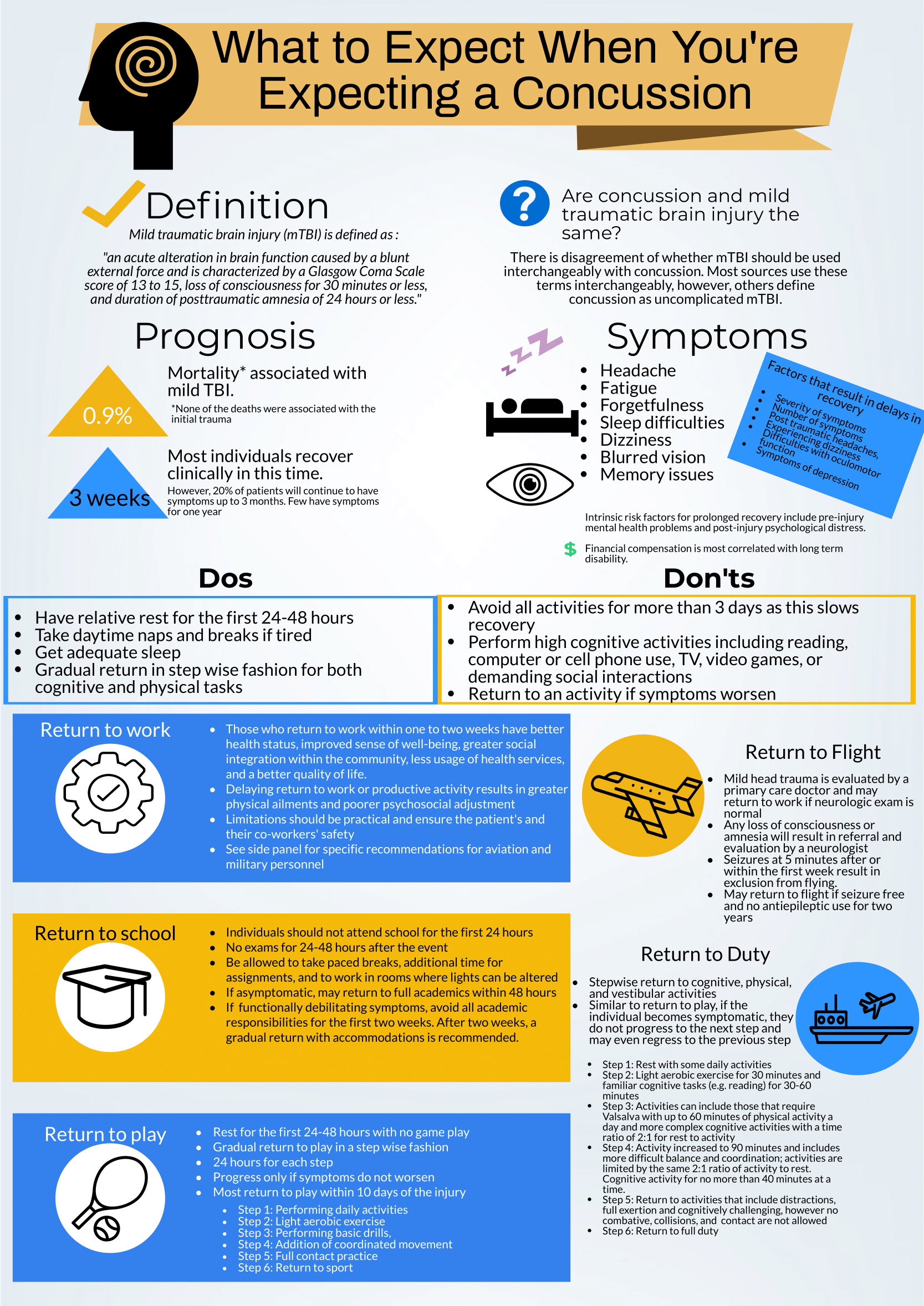
This is a great review of anticipatory guidance when counseling patients who have been diagnosed with a concussion.  As noted, “mild traumatic brain injury (mTBI)” is often used synonymously with “concussion.” A better way to conceptualize this is to view concussion as a form of mTBI, realizing that mTBI can represent a spectrum of conditions.  One of the most important treatments of concussion from the Emergency Department (ED) perspective is to counsel patients on what to expect and how to best control their symptoms.  Concussions can present with a wide range of symptoms as detailed and can be quite distressing and disruptive to patients. As correctly pointed out, the presence of vestibular symptoms (i.e. dizziness or gait instability) as well as pre-existing mental health diagnoses, such as depression or anxiety, are associated with a protracted symptom course. Setting expectations of the symptoms they may develop and the possible timeline of symptom duration is important for patients as they manage their condition.  Early conservative treatment with adequate sleep and relative cognitive and physical rest will help manage and reduce the intensity of symptoms.  In our current society, it is nearly impossible to completely avoid screens and reading.  Hence, “everything in moderation” is appropriate when counseling these patients.  If the patient has to work at a computer, advise them to take frequent breaks for at least 10 minutes for every 30 minutes of screen time.  In addition, it is recommended that patients with a concussion avoid alcohol.  It is also advisable to avoid excessive caffeine.  However, if a patient already uses caffeine on a daily basis, they should not stop completely, as that can lead to withdrawal headaches.  Over-the-counter pain relievers, such as naproxen, ibuprofen or acetaminophen are appropriate for headache treatment, provided there are no contraindications to use.

 There are multiple return-to-learn, -work and -play protocols that have been published.  This is particularly applicable to athletes who have sustained a sport-related concussion (SRC).  Most schools and athletic programs have protocols that have been developed in conjunction with athletic trainers and team physicians.  It is important to remember that as an ED provider, you should not clear a patient to return to play.  That process needs to be conducted by the school athletic trainer in collaboration with the team physician after they have had the opportunity to evaluate the patient. You should consider referring your concussion patients to a Primary Care Sports Medicine or Neurology provider for follow-up if they do not have a team physician to visit.

There are multiple free resources available to providers who are interested in learning more about concussion and educating patients.  The Sport Concussion Assessment Tool – 5th Edition (SCAT5) is an in-depth evaluation tool that is often used by Sports Medicine clinicians when evaluating the extent and severity of a patient’s concussion syndrome.  These resources are listed here:

References

American Medical Society for Sports Medicine position statement on concussion in sport:

https://bjsm.bmj.com/content/53/4/213

SCAT5:

https://bjsm.bmj.com/content/bjsports/early/2017/04/26/bjsports-2017-097506SCAT5.full.pdf

27447 (1).jpg

Jacob Stelter, MD

Emergency Medicine, Primary Care Sports Medicine

Division of Emergency Medicine

NorthShore University HealthSystem


How To Cite This Post:

[Peer-Reviewed, Web Publication] Green, K. Maivelett, J. (2021, Feb 14). What to expect when you're expecting a concussion. [NUEM Blog. Expert Commentary by Stelter, J]. Retrieved from http://www.nuemblog.com/blog/concussion.


Posted on February 15, 2021 and filed under Neurology.

Pericardiocentesis

Written by: David Feiger, MD (NUEM ‘22) Edited by: Abiye Ibiebele, MD (NUEM ‘21) Expert Commentary by: Daniel Schimmel, MD, MS

Written by: David Feiger, MD (NUEM ‘22) Edited by: Abiye Ibiebele, MD (NUEM ‘21) Expert Commentary by: Daniel Schimmel, MD, MS


Introduction

An emergent pericardiocentesis may be a life-saving procedure and is indicated in patients with pericardial effusion and associated hemodynamic instability. The degree of pericardial effusion severity lies on a continuum and when associated with hemodynamic instability, is known as cardiac tamponade. The volume and rate at which the effusion develops most affects a patient’s hemodynamics. The clinical exam findings – hypotension, distant heart sounds, and jugular venous distention (known as Beck’s Triad) – often do not occur simultaneously if cardiac tamponade is suspected and confirmed early.

Given the urgency of the situation and severity of the patient’s clinical status, blind insertion of a needle into the pericardial space using anatomic landmarks has historically been the method of choice to restore hemodynamic stability. The availability of bedside ultrasound in many emergency departments more recently has led many physicians to pursue an image-guided pericardiocentesis. Various studies have demonstrated decreased mortality and morbidity with an ultrasound-guided approach when compared to a blind approach. However, depending on the clinical context and tools available, a blind approach may be the optimal choice. 

Indications and Contraindications

Hemodynamic instability secondary to pericardial effusion, is the number one reason to perform an emergent pericardiocentesis in the emergency room. Pericardiocentesis for patients with symptomatic pericardial effusion but without hemodynamic instability may be deferred to inpatient management. 

An emergent pericardiocentesis has few contraindications. Aortic dissection or cardiac free wall rupture is sometimes considered an absolute contraindication to pericardiocentesis, but in the absence of immediately available life-saving procedures, pericardiocentesis should be strongly considered. Relative contraindications include use of anticoagulation, platelets < 50K, and uncorrected coagulopathy. Furthermore, trauma patients with hemopericardium should preferentially undergo surgical pericardial drainage or emergency thoracotomy. 

Blind Emergent Pericardiocentesis

Relevant Anatomy to Keep in Mind

Internal thoracic artery (internal mammary artery) – artery running cephalad to caudal on the anterior chest wall parallel to the sternum bilaterally

Neurovascular bundle – a collection of an intercostal vein, artery, and nerve running caudal to each rib

Materials

  • Sterile gloves, gown

  • Chlorhexidine swab

  • At least a 7 cm 18-gauge spinal needle or introducer needle if planning for continuous access to pericardial space

  • Syringes (10mL and 60-80mL)

  • Three-way stopcock

  • Plastic drainage tubing

  • Surgical clamp (optional)

1) Position the patient appropriately.

Provide respiratory support with nasal cannula or mechanical ventilation as indicated. Placing patients upright at 30 degrees to enhances comfort and allows gravity-dependent pooling of pericardial fluid.

2) Select an entry site.

Left parasternal and apical approaches are the most commonly pursued and have been shown to be superior to the classic subxiphoid approach in observational studies. When selecting an entry point, recall the location of the internal thoracic artery and subcostal neurovascular bundle. Cleanse a large area of the chest and upper abdomen with the chlorhexidine swab.

3) Insert and advance the spinal needle.

Insert the spinal needle into skin. Once the bevel is in the skin, remove the stylet, attach a three-way stopcock with a large volume syringe attached and maintain negative pressure as you advance the needle. Avoid sliding the needle laterally to prevent lacerating tissues. If a different trajectory is required, retract the needle, keeping the bevel in the skin and drive the needle at the desired angle maintaining negative pressure on the syringe. Momentary resistance may be met as the needle approaches the pericardium, but with continued advancement, a “pop” may be felt followed by aspirate in the syringe and improving patient hemodynamics. See below for more detailed instructions for each approach.

subxyphoid.PNG

Subxiphoid

Insertion: 1 cm inferior to the left xiphocostal angle, 30 degrees with the patient’s chest

Direction: Towards left mid-clavicle. If unsuccessful, retract the spinal needle and redirect 10 degrees towards the patient’s right

parasternal.PNG

Parasternal

Insertion: Fifth intercostal space at the left parasternal border no more than 1 cm lateral, cephalad to the inferior rib and perpendicular to the patient’s chest

Direction: Posterior

apical.PNG

Apical

Insertion: Fifth, sixth, or seventh left intercostal space approximately 6 cm from the parasternal border, cephalad to the inferior rib

Direction: Patient’s right shoulder

4) Draining the pericardial effusion.

Steady the needle with a surgical clamp at the needle shaft closest to the surface of the skin effectively preventing further needle advancement. Attach the plastic tubing to the stopcock allowing emptying of the syringe contents into a collecting vestibule without exchanging syringes. If planning to place a more permanent line (see “Establishing Continuous Access to the Pericardial Space”), consider aspirating just enough fluid to stabilize the patient’s hemodynamics and leave the remaining pericardial fluid to provide space for placing a line.

Tips for Pericardiocentesis with Ultrasound Guidance

Additional materials:

  • Bedside ultrasound

  • Sterile ultrasound probe cover

  • Skin marker

The safety and success of the steps above can be enhanced with bedside ultrasound. Bedside ultrasound can help prior to the procedure by finding the largest effusion nearest the skin and during the procedure by visualizing the needle trajectory in order to avoid important organs and other structures.  There are several methods of ultrasound use in pericardiocentesis.

In static guidance, ultrasound is only used for procedure planning. The subxiphoid, parasternal, and apical views can be explored to find the largest effusion and determine the optimal entry. Often, the needle entry point is marked with a skin marker and another mark is made for the planned trajectory. Distance and angle from the skin to the effusion is also noted.

In dynamic guidance, the needle is passed through the skin parallel to the 2D plane created by the ultrasound probe after finding an effusion pocket. The needle can be visualized as it is advanced towards the pericardial effusion and enters the pericardial space.

No ultrasound? Hook up an EKG!

Additional materials:

  • Continuous EKG monitor

  • Wire with alligator clips

A continuous EKG can be used to prevent inadvertent traversing of the myocardium with the needle without an ultrasound. Attach one alligator clip to the needle and the other to an anterior lead on a continuous EKG. ST-elevations will be apparent on the EKG if the myocardium is touched. If ST-elevations are noted, simply retract the needle.

Confirmation of Pericardial Access with Ultrasound Guidance

Additional materials:

  • Bedside ultrasound

  • Two 10 mL syringes (one with 4 mL of saline, one with 0.5 mL of air)

Confirming success in accessing the pericardial space can also be made injecting agitated saline and visualizing bubble artifact on ultrasound. To do so, attach the two saline syringes to the three-way stopcock. Turn off access towards the patient and rapidly push the contents from one syringe to the other until the fluid appears opacified. When all the saline is in one syringe, close off the access to the empty syringe and push the fluid towards the patient, visualizing it on the ultrasound. Confirmation is especially important when blood is aspirated and helps distinguish between pericardial versus ventricular placement.

Establishing Continuous Access to the Pericardial Space

Additional materials:

  • Flexible or curved-tip (J) guidewire

  • 6-8 Fr drainage catheter (pigtail, sheath, or central venous catheter)

  • 7 cm or longer 18-gauge introducer needle (as opposed to spinal needle)

  • Dilator

  • 11 blade scalpel

  • Suture

  • Needle driver

Many of the materials above may be found in a central venous kit. Using the Seldinger technique, a line can be placed for continuous access to the pericardial space. Ensure that an introducer needle is used when initially accessing the pericardial space. Keeping the surgical clamp and needle in place, remove the stopcock and syringe, and gently advance the guidewire just beyond the bevel of the needle. Remove the introducer needle, ensuring the guidewire does not move, and use the scalpel to make a short incision at the guidewire’s entry into the skin. Advance the dilator over the guidewire to loosen the tissue. Remove the dilator leaving the guidewire in place and advance the drainage catheter just 1 cm beyond the guidewire into the pericardial space. Retract the guidewire while maintaining the position of the catheter, aspirate fluid to confirm placement, and secure the drain’s position with sutures and placement of a sterile dressing. Further confirmation of proper placement can be made using the agitated bubble study as described in “Confirmation of Pericardial Access with Ultrasound” above.

Conclusion

There are very few contraindications for an emergent pericardiocentesis in a patient with pericardial effusion and hemodynamic instability. While ultrasound-guided pericardiocentesis have lower morbidity and mortality rates, clinical context and emergent patient decompensation may make an image-guided procedure infeasible. Apical and parasternal access with a blind procedure have fewer complications than a subxiphoid approach. A pericardiocentesis may be a life-saving intervention as even a small amount of fluid aspirated may dramatically improve a patient’s hemodynamics.


Expert Commentary

Thank you Dr. Feiger for this excellent summary of pericardiocentesis.  From center to center there can be variability in the expertise and mechanism through which pericardiocentesis is performed.  Some institutions may have an echo focused pericardiocentesis service, while some institutions may perform the bulk of their pericardiocentesis in an interventional suite with the assistance of fluoroscopic imaging.  However, there are times when pericardiocentesis must be performed as an emergency procedure with landmark guidance.  Luckily, point of care ultrasound has been very commonplace in the emergency department and intensive care units facilitating visualization of fluid pockets that can be identified for safer access and to demonstrate successful drainage at the end of the procedure.  

The relevant anatomy and associated complications from tissue injury during needle advancement for pericardiocentesis changes depending on the planned access route.

Subcostal

  • Liver laceration or puncture

  • Pneumothorax

  • Right atrial or ventricular laceration

Apical

  • Pneumothorax

  • Left or right ventricular laceration

Parasternal

  • Pneumothorax

  • Right ventricular laceration

In each location, careful use of ultrasound can avoid potential life-threatening complications.  How the ultrasound is used also varies depending on location.  Generally the ultrasound can be used to identify a path and I will have trainees hold the ultrasound the exact same way as they would hold their needle to mimic the path they will use when advancing into the pericardial space.  

Common errors that I have seen are listed below.

  1. Moving the needle side to side while it is in the body to try and find the appropriate space.  I am impressed at the body’s ability to tolerate a straight in and out movement of a needle.  But moving a needle tip back and forth creates lacerations that are difficult to heal and may result in tissue damage and uncontrollable bleeding requiring surgical intervention.

  2. Moving the needle to find it under ultrasound, rather than moving the probe to find the needle can be dangerous.  If the needle is in the wrong location, it should be moved.  Otherwise, do not bounce the needle within the body to try and identify it on ultrasound.

  3. A common subcostal error is needle path facing towards the spine through the torso.  The subcostal position can be more successful with the patient upright at 30 degrees so that the fluid layers to the bottom portion of the heart, increasing the pocket size for needle entry.  However, this position then requires the physician to aim slightly up, almost moving parallel to the ribs, to avoid needle entry being too low and passing underneath the effusion.

  4. Also from the subcostal position, the initial position angle for subcostal pericardiocentesis should be to the left middle cervical bone as Dr. Feiger mentioned.  However, many performing subcostal pericardiocentesis overcompensate initially and head too laterally to capture fluid. 

  5. From the apical position, the fluid is likely best obtained with the patient in the left lateral decubitus position.  A drop door that is often present on an sonographer’s bed for performing echocardiograms is nice but not necessary.

As mentioned, the cardiac probe is not generally used to watch needle entry but to identify the most optimal path.  However, if also equipped with a vascular probe, in the parasternal location, the vascular probe can easily watch the needle enter into the pericardial space while avoiding delicate structures like the internal thoracic artery.

Using fluoroscopic guidance, needle location can be identified in relation to the movement of the cardiac border and wire advancement can be used to identify a course consistent with the pericardial space and not limited by other cardiac structures.  A small amount of contrast can be injected into the space and seen to highlight the cardiac borders allowing confirmation of the pericardial space.   

But in the absence of fluoroscopy, and a more specific confirmation, an echo with agitated bubbles injected can verify presence of the needle, or a microcatheter, in the pericardial space.  If after injection, bubbles are seen within the cardiac chambers, the needle should be withdrawn.  If possible to obtain a pressure measurement during the procedure, this is one other guide to inform the operator of the needles location.  If a right ventricular waveform Is present, the needle has entered the ventricular space.  It may be possible to withdraw the needle until the high pressures of the ventricle reduce and if the needle aspirates, another agitated bubble injection can be performed.

An urgent pericardiocentesis with a large effusion can be easily performed at the bedside, particularly with the aid of an ultrasound and knowing the anatomy with immediate improvement in hemodynamics.  Send the fluid for analysis and exchange the needle for a drain to ensure patient stability until the evaluation is complete.

Daniel Schimmel.PNG

Daniel Schimmel, MD, MS

Interventional Cardiologist

Northwestern Memorial Hospital

Associate Professor

Feinberg School of Medicine


How To Cite This Post:

[Peer-Reviewed, Web Publication] Feiger, D. Ibiebele, A. (2021, Feb 8). Pericardiocentesis. [NUEM Blog. Expert Commentary by Schimmel, D]. Retrieved from http://www.nuemblog.com/blog/pericardiocentesis.


Other Posts You May Enjoy

References

  1. Gueria, Rajesh. “Ultrasound Guided Procedures in Emergency Medicine Practice - Pericardiocentesis.” Sonoguide, 2008, www.acep.org/sonoguide/pericardiocentesis.html.

  2. Heffner, Alan C. “Emergency Pericardiocentesis.” Edited by Allan B Wolfson et al., UpToDate, 29 May 2019, www.uptodate.com/contents/emergency-pericardiocentesis?search=pericardiocentesis&source=search_result&selectedTitle=1~65&usage_type=default&display_rank=1.

  3. Konnoff-Phillips, Kelly and Janis Provinse, directors. Agitated Saline Bubble Study. Highland Hospital Emergency Department, 2015.

  4. Maisch, Bernhard, and Sabine Pankuweit. Interventional Pericardiology: Pericardiocentesis, Pericardioscopy, Pericardial Biopsy, Balloon Pericardiotomy, and Intrapericardial Therapy. Springer, 2011.

  5. Nicks, Bret A, et al. Emergency Pericardiocentesis. New England Journal of Medicine, 22 Mar. 2012, www.nejm.org/doi/full/10.1056/NEJMvcm0907841.

Posted on February 8, 2021 and filed under Procedures.

Kawasaki Disease

Written by: Richmond Castillo, MD (NUEM ‘23) Edited by: Andra Farcas (NUEM ‘21) Expert Commentary by: Wee-Jhong Chua, MD

Written by: Richmond Castillo, MD (NUEM ‘23) Edited by: Andra Farcas (NUEM ‘21) Expert Commentary by: Wee-Jhong Chua, MD


kawasaki disease.png

Expert Commentary

A great summary of a rare, but important illness to consider in children with prolonged fevers. Kawasaki disease is a vasculitis to small and medium vessels, often occurring in childhood. While most children recover, there are important cardiac complications that need to be considered. Treatment with IVIG within the first 10 days of illness has been shown to reduce the prevalence of coronary artery aneurysms. In fact, untreated disease has been associated with an incidence of coronary artery aneurysm as high as 25%. [1]

The diagnosis of this disease process is challenging as there are no definitive testing methodologies and even the etiology of the systemic inflammatory process remains unknown. While most guidelines include 5 days of fever, the 2017 American Heart Association Scientific Statement indicates that 4 days of fever with the corresponding clinical features can be diagnostic. Moreover, in rare instances experienced clinicians have been able to make the diagnosis as early as 3 days. [1] In clinical practice, the clinical pathways at leading children’s hospitals use 4 days as a threshold for evaluation with the presence of 4 or 5 principal clinical features are present. [2,3] However, the standard practice continues to be 5 days for the classic diagnosis.

With high clinical suspicion for Kawasaki Disease, transfer to a pediatric hospital should be considered with consultations with Infectious Disease, Cardiology, and Rheumatology. It is important to have a high clinical suspicion for Kawasaki disease in children with prolonged fevers.

References

  1. McCrindle BW, Rowley AH, Newburger JW, et al. Diagnosis, treatment, and long-term management of Kawasaki disease: A scientific statement for health professionals from the American Heart Association. Circulation. 2017;135(17):e927-e999. doi:10.1161/CIR.0000000000000484

  2. D. Whitney, MD; K. Dorland, BSN; J. Beus, MD; J. Brothers, MD; L. Buckley, MD; S. Burnham MD; D. Campeggio, MSN; K. DiPasquale, MD; H. Ghanem MD; J. Hart MD; J. Lavelle, MD; C. Law PharmD; S. Natarajan, MD; J. Ronan, MD; V. Scheid, MD; S. Swami, MD; H. Ba C. Kawasaki Disease or Incomplete Kawasaki Disease Clinical Pathway — Emergency Department and Inpatient | Children’s Hospital of Philadelphia. Accessed October 20, 2020. https://www.chop.edu/clinical-pathway/kawasaki-disease-incomplete-kawasaki-disease-clinical-pathway

  3. Seattle Children’s Hospital, M Portman, M Basiaga, E Beardsley, R Engberg, K Hayward, K Kazmier, M Leu, R Migita, J Rasiah, R Sadeghian, S Sundermann, S Vora, S Zaman, 2018 February. Kawasaki Disease Pathway. Available from: http://www.seattlechildrens.org/pdf/Kawasaki-Disease-Pathway.pdf

Wee Chua.PNG

Wee-Jhong Chua, MD

Attending Physician, Pediatric Emergency Medicine

Ann & Robert H. Lurie Children's Hospital of Chicago


How To Cite This Post:

[Peer-Reviewed, Web Publication] Castillo, R. Farcas, A. (2021, Feb 1). Kawasaki Disease. [NUEM Blog. Expert Commentary by Chua, W]. Retrieved from http://www.nuemblog.com/blog/kawasaki-disease.


Other Posts You May Enjoy

Posted on February 1, 2021 and filed under Pediatrics.

Apple Heart Study

Written by: Em Wessling, MD (NUEM ‘22) Edited by: Dana Loke, MD (NUEM ‘19) Expert Commentary by: Rod Passman, MD

Written by: Em Wessling, MD (NUEM ‘22) Edited by: Dana Loke, MD (NUEM ‘19) Expert Commentary by: Rod Passman, MD


Chief Complaint: My watch thinks I have Atrial Fibrillation!

 As technology advances, medicine must continue to advance in pace. Wearable technology has been evolving for decades. The information gathered from a wide range of these devices may someday help to provide healthcare workers with valuable information about a patient’s condition. However, for now, there is limited research on their utility within the healthcare field.

 Thus far, both Apple Watch and Fitbit have been shown to correctly identify tachycardia during atrial tachyarrhythmias, but their accuracy to the heart rate varied with the type of arrhythmia (1,2). Apple Watch has been shown to be more accurate than Fitbit (1,2). The WATCH AF trial demonstrated it was possible with reasonable sensitivity (93.7%) and specificity (98.2) to use smart watches to diagnose Atrial Fibrillation (3). 

How Apple Watch is tracking atrial fibrillation:

-        Photoplethysmography: the use of light to determine volume within a structure at a given time

-        Pulse is estimated by time between peak volume seen by photoplethysmography.

-        When the pulse is highly variable between consecutive beats, irregular heart beat is suspected.


Apple Heart Study: The plan and the preliminary data (4, 5)

Study Design: Prospective, single arm pragmatic study

-        Enrolled 419,093 participant  

-        Inclusion Criteria: appropriate Apple technology, Age≥22 years, US resident, proficient in English, valid phone number and email.

-        Exclusion criteria: self-reported atrial fibrillation , atrial flutter, or anticoagulation

-        Methods:  “Irregular Pulse Notification” (indication of possible atrial fibrillation) sent to participants  if 5/6 irregular pulses within  48 hour period, at which point participant was instructed to wear EKG patch for up to 7 days.

-        Primary Outcome: Proportion of patients alerted with “Irregular Pulse Notification” who were found to have atrial fibrillation or atrial flutter on EKG patch, in the 65+ population as well as in all-comers.

-        Secondary Outcomes: Positive predictive value (PPV) of irregular heart rhythm notification; percentage of those with irregular notification who contacted a health care professional within 3 months.

Preliminary Data presented at ACC:

-        Participants who received “Irregular Pulse Notification”:  2,161 (0.52% all comers)

-        Participants age >65 who received “Irregular Pulse Notification”: >3%

-        EKG Patches sent to 658 participants; 450 returned.

  • 34% of those returned showed atrial fibrillation

  • PPV for Tachogram: 71%

  • PPV for “Irregular Pulse Notification”: 84%

-        Notification to doctor - approx. 50%

Limitations:

  • Small sample size for EKG patches, despite high enrollment

  • Self-reported data

  • Self-selecting group, i.e.may not be able to extrapolate prevalence data to those who do not wear smart watches


Potential Impact on Emergency Departments:

As more and more studies validate the accuracy of wearable technology to measure and recognize health conditions, the implications must be analyzed as well.

Prior to 2017, researchers began to predict that there would be an expansive increase in the rates of atrial fibrillation due to “worldwide aging” (6). While this review acknowledged there were “potential applications” for smart phone technology in the diagnosis, their predictions of the expanse of the epidemic of atrial fibrillation preceded definitive research showing increased diagnosis rates with wearable technology, which will likely only further expedite this growing patient population.  The mSToPS Trial showed that immediate in-home monitoring with an EKG patch had 3% greater rates of atrial fibrillation diagnosis compared to delayed EKG monitoring at 4 months. This led to increased use of anticoagulants and increased health care utilization (7). If this increase was seen with EKG patches, consider the influx of patients to primary care and cardiology clinics in addition to emergency departments that can be projected based on the rise of smart watch detection of atrial fibrillation. Researchers in Australia had begun studying this prior to the commencement of the Apple Heart Study (8). When cardiac patients were asked if they trusted smart watches to predict arrhythmia and measure their heart rate only 53% agreed; however, that did not stop 91% from reporting they would seek care if their watch alerted them about an abnormality (8). While the preliminary data from the Apple Heart study shows that a much smaller percentage of those who were not previously cardiac patients sought medical care when alerted by their Apple Watch, further study is needed to see the extent to which advances in smart watch health technology will lead to an influx in patients to the Emergency Department due to concerns of arrhythmia found by a smartwatch (5).

 While the accuracy of these methods of arrhythmia detection are still being studied, the potential for ED presentation with this chief complaint will continue to rise. In the fourth quarter of 2017 financial year, Apple alone sold greater than 8 million smart watches worldwide, making it the largest watch vender in the world (9).  With these increased sales, comes the potential for increased recognition of arrythmia by smartwatch. Healthcare organizations throughout the country must strive to develop effective and efficient clinical pathways in order to evaluate, potentially diagnose, and treat this patient population. Upon presentation to the Emergency Department, each patient should receive an EKG, telemetry monitoring while in the Emergency Department and screaming lab work: often including CBC, BMP + Mg, and troponin.  From there, the pathway may vary. Many would agree, if the patient is and has always been asymptomatic, work up is unremarkable, with normal sinus rhythm on their EKG, discharge home with an EKG patch and follow up with cardiology is reasonable. Conversely, an EKG showing atrial fibrillation would constitute a new diagnosis and further work up would proceed as with any other new diagnosis of Atrial Fibrillation. However, for those who fall in-between, the disposition is not as clear. What would you do?


Expert Commentary

More than 800 years ago, Maimonides described an irregular pulse that likely represented atrial fibrillation (AF). The development of the electrocardiogram by Einthoven 700 years later allowed surface recordings of human AF for the first time.1 With the recognition that AF is often asymptomatic and paroxysmal, the development of inexpensive, non-invasive, passive monitors for irregular rhythm identification has long been recognized as a potentially important tool for arrhythmia detection and management.

At its core (pun intended), the purpose of the Apple Heart Study was to assess the feasibility of AF screening in large populations by monitoring participants with a wrist-worn photoplethysmography (PPG) monitor.2 The PPG algorithm in the Apple Watch samples the pulse several times daily during periods of physical inactivity and increases the sampling rate if an irregular tachogram is detected. If 5 out of 6 tachograms are consistent with AF (requiring > 60 minutes of AF), the wearer receives an irregular rhythm notification. Since the version of the Apple Watch used in the study did not have the 30-second ECG feature (available in Series 4 watches and later), the Apple Heart Study protocol asked those who received the irregular rhythm notification to wear an ECG patch at a later date. 

Several important facts can be gleaned from the Apple Heart Study.  First, the study virtually enrolled 419,297 individuals in less than a year, a testament to the interest in the subject matter, the ease of remote enrollment when appropriate, and the enormous potential of digital health studies. Second, the fear that the healthcare system would be inundated with false positive AF notifications appears unfounded as 99.8% of participants under age 40 did not receive an irregular rhythm notification.  Third, the positive predictive value for the irregular rhythm notification was surprisingly high (84%) despite that fact that the patch was applied a mean of 13 days following the notification and was worn for less than 7 days on average. This last point is worth emphasizing since with paroxysmal AF, a negative monitor placed two weeks after an irregular rhythm notification may simply mean that AF was not present during both time periods.

The study also has some important caveats.  The Apple Heart Study did not report the sensitivity and specificity of the PPG algorithm for AF detection, a critical piece of missing data needed for clinical care and future research. Furthermore, only a minority of patients who received an irregular rhythm notification actually wore and returned the ECG monitor, showing that virtual enrollment doesn’t always translate into virtual protocol compliance. From a research perspective, wearable AF monitors have allowed for large-scale screening studies such as the Huawei  Heart and Heartline Studies aimed at understanding the true prevalence of AF and the risks and benefits of early detection and treatment.3,4 From a clinical perspective, a patient who says “my watch says I have AF” still requires ECG confirmation, but that too has been made easier with the new generation of wearables.

References

1. Prystowsky EN. The history of atrial fibrillation: the last 100 years. J Cardiovasc Electrophysiol. 2008;19(6):575-582. doi:10.1111/j.1540-8167.2008.01184.

2. Perez MV, Mahaffey KW, Hedlin H, et al. Large-Scale Assessment of a Smartwatch to Identify Atrial Fibrillation. N Engl J Med. 2019;381(20):1909-1917. doi:10.1056/NEJMoa1901183

3. Guo Y, Wang H, Zhang H, et al. Mobile Photoplethysmographic Technology to Detect Atrial Fibrillation. J Am Coll Cardiol. 2019;74(19):2365-2375. doi:10.1016/j.jacc.2019.08.019

4. www.heartline.com

WbGJlUXI_400x400.jpg

Rod Passman, MD

Professor, Feinberg School of Medicine

Cardiac Electrophysiology

Northwestern Memorial Hospital


How To Cite This Post:

[Peer-Reviewed, Web Publication] Wessling, E. Loke, D. (2021, Jan 25). Apple Heart Study. [NUEM Blog. Expert Commentary by Passman, R]. Retrieved from http://www.nuemblog.com/apple-heart.


Other Posts You May Enjoy

References

1. Koshy, Anoop N., et al. "Smart watches for heart rate assessment in atrial arrhythmias." International journal of cardiology 266 (2018): 124-127.

2. Koshy, A., et al. "Heart Rate Assessment by Smart Watch: Utility or Futility?." Heart, Lung and Circulation 26 (2017): S280-S281.

3. Dörr, Marcus, et al. "The WATCH AF trial: SmartWATCHes for detection of atrial fibrillation." JACC: Clinical Electrophysiology5.2 (2019): 199-208.

4Turakhia, Mintu P., et al. "Rationale and design of a large-scale, app-based study to identify cardiac arrhythmias using a smartwatch: The Apple Heart Study." American heart journal207 (2019): 66-75.

5. ACC News Story. “Apple Heart Study Identifies AFib in Small Group of Apple Watch Wearers.” American College of Cardiology: Latest in Cardiology, American College of Cardiology, 16 Mar. 2019, www.acc.org/latest-in-cardiology/articles/2019/03/08/15/32/sat-9am-apple-heart-study-acc-2019.

6. Morillo CA, Banerjee A, Perel P, Wood D, Jouven X. Atrial fibrillation: the current epidemic. J Geriatr Cardiol. 2017;14(3):195–203. doi:10.11909/j.issn.1671-5411.2017.03.011

7. Steinhubl SR, Waalen J, Edwards AM, et al. Effect of a Home-Based Wearable Continuous ECG Monitoring Patch on Detection of Undiagnosed Atrial Fibrillation: The mSToPS Randomized Clinical Trial. JAMA.2018;320(2):146–155.

8. Koshy, A., et al. "Cardiac Patients Likely to Seek Medical Assistance Based on Abnormal Heart Rate Readings on Smart Watches or Smartphone ECG Monitors." Heart, Lung and Circulation 26 (2017): S280.

9. Canalys Press Team. “18 Million Apple Watches Ship in 2017, up 54% on 2016.” Canalys Newsroom, Canalys, 6 Feb. 2018, www.canalys.com/newsroom/18-million-apple-watches-ship-2017-54-2016.

Posted on January 25, 2021 and filed under Cardiovascular.

Intubating the Pregnant Patient in the ED

Written by: Priyanka Sista, MD (NUEM ‘20) Edited by: Steve Chukwulebe, MD (NUEM ‘19) Expert Commentary by: Samir Patel, MD

Written by: Priyanka Sista, MD (NUEM ‘20) Edited by: Steve Chukwulebe, MD (NUEM ‘19) Expert Commentary by: Samir Patel, MD


Expert Commentary

Tip for #1 - While 3-5 minutes of 100% oxygen is ideal to achieve denitrogenation, in an emergency 8 vital capacity breaths (maximal inhalation and exhalation) with a high FiO2 source is sufficient in a cooperative patient.

Tip for #2 - Airway edema is even worse in preeclamptic patients, and Mallampati scores acutely worsen DURING labor. Don’t bother with direct laryngoscopy and go straight to the video laryngoscope if it’s available.

Tip for #3 - In this scenario, the ideal LMA or supraglottic airway is one that includes a port for passage of an OG tube. Your pregnant patient in the ER with increased aspiration risk is not likely to be NPO for 8 hours like they are for anesthesiologists before surgery.

Tip for #4 - The rapid sequence dose of rocuronium is 1.2 mg/kg. You can immediately reverse rocuronium with sugammadex 16 mg/kg if necessary. For cost purposes, succinylcholine is still the best choice unless medically contraindicated.

Tip for #5 - According to ACOG, if cardiac arrest occurs in a woman greater than 23 weeks gestation, and there is no return of spontaneous circulation after 4 minutes of correctly performed CPR, a perimortem c-section should be performed with the goal of delivering the fetus by the fifth minute.

Samir Patel.PNG

Samir K. Patel, MD

Assistant Professor

Northwestern University Feinberg School of Medicine

Department of Anesthesiology


How To Cite This Post:

[Peer-Reviewed, Web Publication] Sista, P. Chukwulebe, S. (2021, Jan 18). Intubating the pregnant patient in the ED. [NUEM Blog. Expert Commentary by Patel, S]. Retrieved from http://www.nuemblog.com/blog/intubating-the-pregnant-patient.


Other Posts You May Enjoy

Posted on January 18, 2021 and filed under ENT, Airway.

Imaging in PTAs

Written by: Cameron Jones, MD (NUEM ‘23) Edited by: Vidya Eswaran, MD (NUEM ‘20) Expert Commentary by: Josh Zimmerman, MD

Written by: Cameron Jones, MD (NUEM ‘23) Edited by: Vidya Eswaran, MD (NUEM ‘20) Expert Commentary by: Josh Zimmerman, MD


The Use of Imaging for Diagnosis and Management of Peritonsillar Abscesses

Among the many causes of sore throat that the EM physician may encounter, peritonsillar abscesses (PTAs) can be one of the more satisfying to diagnose and treat. A straightforward clinical diagnosis followed by a simple procedure resulting in a patient who feels much better than when they arrived...right? But what about that patient with the large, short neck and some drooling? Or the one with severe trismus giving you only the barest of glimpses at the back of their throat? Or, most feared of all, the crying child who develops lockjaw at the first glimpse of a tongue depressor? Maybe we should just get the neck CT to be on the safe side? And didn’t I hear about using ultrasound for this in some lecture? 

What is a peritonsillar abscess (PTA)?

A PTA is a discrete collection of pus between the palatine tonsil capsule and the pharyngeal muscles. It should be distinguished from peritonsillar cellulitis, which is an inflammatory reaction of the same area without a definitive collection. PTAs are often preceded by tonsillitis or pharyngitis with subsequent progression of the infection. However, they may also occur due to salivary gland obstruction without preceding tonsillitis or pharyngitis. Peritonsillar abscess is often considered a clinical diagnosis based on classic symptoms and exam findings:

  • Throat pain (sometimes worse on the side of the abscess, but not always)

  • “Hot potato” or muffled voice

  • Unilateral swollen and erythematous tonsil +/- appreciable fluctuance

  • Uvula deviation

What are signs or symptoms suggestive of a more dangerous diagnosis?

Though sometimes mistakenly considered features of more concerning deep space neck infections, all of the following can also be seen with PTA: 

  • Neck swelling

  • Trismus

  • Pooling of saliva (though this should be minor, with minimal drooling)

Other findings or symptoms of more serious deep space infections, such as retropharyngeal abscess:

  • Toxic appearance

  • Respiratory distress

  • Anxious appearance or leaning forward into “sniffing position”

  • Significant drooling

  • Neck pain or limited ROM out of proportion to presumed diagnosis

When should imaging be considered in the patient with suspected PTAs?

Routine imaging is not indicated for stable patients with a presumptive diagnosis based on exam. Sensitivity and specificity figures in the EM and ENT literature based on clinical exam alone are actually not very high (sensitivity <80% and specificity approximately 50%). However, these oft-cited figures are based on a comparatively small cohort of patients with presumed PTA, and in the large majority of missed diagnoses among this data, the true diagnosis is tonsillitis or peritonsillar cellulitis. CT scans, particularly contrast studies and those involving radiation of the head and neck, are not without risk, and should not be considered a screening study in well-appearing patients.  Therefore, the use of imaging by ED physicians in evaluation of PTAs should really be reserved for 3 purposes:

  1. Ruling out serious deep space neck infections, such as retropharyngeal abscesses, in a patient with signs of peritonsillar swelling but some other concerning sign or symptom, as discussed above.

    - CT of the neck with contrast is best used for this purpose

  2. Differentiating PTA from peritonsillar cellulitis or tonsillitis by identifying a discrete fluid collection

  3. Guiding drainage in order to improve first-attempt success

    - Intraoral or submandibular/transcervical ultrasounds are most appropriate for these purposes

 There are few prospective studies examining the use of CT in uncomplicated PTAs, and those patients with red flags or signs of airway compromise are typically excluded. CT of the neck with IV contrast is nearly 100% sensitive and 75% specific for PTA and similarly accurate for the diagnosis of more dangerous conditions such as retropharyngeal abscesses. Increasingly, ultrasound has also become a useful option for better characterizing the location of abscesses in PTAs. 

Ultrasound offers the added utility of bedside confirmation of a drainable fluid collection and, depending on provider comfort and patient tolerance, may provide real-time guidance for needle drainage. As with other applications of ultrasound, the provider must be comfortable with the technique and relevant anatomy. Prospective data indicates EM providers can become comfortable with tonsillar ultrasound technique in as few as 3-4 patients.  In its use in the ED setting, ultrasound has demonstrated nearly 100% sensitivity in differentiating abscess from non-drainable inflammation or cellulitis. Thus, using ultrasound to confirm abscess in those suspected to have PTA may allow patients without drainable fluid collections to avoid unnecessary aspiration attempts.

PTA2.png

Peritonsillar abscess seen on submandibular ultrasound. Adapted from Huang et al. 

Arrows indicating edges of the abscess

T : Tonsil

*  :  Submandibular gland

Peritonsillar abscess on CT. Adapted from Kew et al. 

Arrowheads indicating edges of the abscess.

Is imaging useful for guiding drainage of PTAs?

  • Ultrasound has also been studied for its utility in guiding drainage and increasing success rate of aspiration attempts. Some studies have reported low patient tolerance or mechanical challenges when using real-time intraoral ultrasound to guide drainage. However, ultrasound has also been shown to improve success-rate of aspiration attempts even when it is used for preceding visualization of the abscess and not for guided drainage.

  • More recently, extraoral ultrasound approaches, such as transcervical/ submandibular, have also been studied as an alternative to intraoral techniques, which can be challenging due to mechanical challenges, severe trismus, or patient discomfort. Very limited data suggests submandibular ultrasound may have lower sensitivity compared to intraoral ultrasound when evaluating PTAs, so caution is also warranted when utilizing this technique. 

PTA4.png

Intraoral ultrasound approach

(adapted from Secko, Sivitz, et al.)

PTA5.png

Submandibular ultrasound approach

(adapted from Secko, Sivitz et al.)

What about imaging in kids?

CT scans are often ordered in pediatric patients, who may have challenging exams due to patient intolerance, and these imaging studies are particularly common in community settings where ENT expertise is not readily available. Clinical accuracy for diagnosing PTA in children appears even lower than in adults, though, as with adults, in most children incorrectly diagnosed with PTAs, the true diagnosis is tonsillitis without a drainable abscess. Many providers would also prefer to avoid the added radiation exposure of CT scans amongst this population. Thus, extraoral ultrasound approaches may be particularly helpful in pediatric patients, many of whom are unlikely to cooperate with intraoral ultrasound. Transcervical ultrasound has also been shown to reduce length of stay, CT radiation exposure, and procedures performed amongst pediatric patients with suspected PTAs, with no change in readmission rates or treatment failures. Although the extraoral ultrasound approach appears to be more technically feasible in children, use of ultrasound may also be more logistically challenging and staffing-dependent. Scans in these studies were performed and read by radiology technicians and radiologists.  

So what is a reasonable approach to incorporating imaging in suspected PTAs?

The growing body of evidence described above has led to several expert recommendations that ultrasound be the first-line imaging for suspected PTAs. While there is variability in different departments regarding the ED provider’s comfort with bedside tonsillar ultrasound or, alternately, the availability of technicians and radiologists for interpreting formal ultrasounds. However, the use of ultrasound  in non-toxic patients with suspected PTA has been shown to be highly effective in differentiating PTAs from peritonsillar cellulitis or tonsillitis and may save patients the discomfort and time of an unnecessary procedure. CT imaging still has its place in those patients with less certain diagnoses or concerning symptoms, but should be reserved for specific scenarios rather than being ordered routinely. The following is an evidence-based algorithm for incorporating ultrasound and CT imaging into the emergency department evaluation of these patients

* : toxic appearance, substantial drooling, respiratory distress, severe&nbsp;neck pain or swelling, inability to fully range neck+ : Most patients can be safely discharged with oral antibiotics, return&nbsp;precautions, and ENT follow-up. Exception…

* : toxic appearance, substantial drooling, respiratory distress, severe neck pain or swelling, inability to fully range neck

+ : Most patients can be safely discharged with oral antibiotics, return precautions, and ENT follow-up. Exceptions include those patients  who are unable to tolerate oral medications, those with signs or symptoms of severe sepsis, patients with severe dehydration, or patient with severe comorbidities or immunocompromised state


Expert Commentary

Thank you for an excellent review of a common ED diagnosis.  Sore throats are ubiquitous presenting complaints in any major ED.  The final diagnosis is often uncomplicated pharyngitis, however, recognizing the early and often subtle signs of more serious conditions before a true life threat develops is a critical role for the emergency physician.  While peritonsillar abscesses (PTA) in and of themselves are not typically life threatening, many of the signs and symptoms can overlap with those of more critical diagnoses such as retropharyngeal abscesses and epiglottitis.  

So, that said, when should you consider imaging a patient with a suspected PTA or acute sore throat in general?

The discussion above does a thorough review evidenced based imaging practices and offers a reasonable flowsheet to guide this decision.  In clinical practice imaging should help answer one of two questions: 

  • Is a discrete fluid collection present that is amenable to drainage? 

  • Are there findings of retropharyngeal or other deep space infection rather than a simple PTA?

I have made it my practice to consider imaging before any attempt at I&D or further care in the following circumstances: 

  • Any patient toxic in appearance or with unstable vital signs

  • Any patient demonstrating signs of airway compromise 

  • Meningismus on exam

  • Patients in which at PTA cannot be clearly visualized or lacking the typical secondary findings on exam 

With that list in mind, let us delve into the topic a bit more in detail.  Peritonsillar abscesses represent accumulation of purulent fluid which are unlikely to resolve spontaneously.  Some studies have shown that drainage alone results in >90% cure rate even without antimicrobial therapy. Classically, a PTA will present with trismus, severe pharyngitis, and on pharyngeal exam a displaced tonsil, typically inferiorly and medially, as well as uvular deviation contralateral to the abscess.  PTA can sometimes be confused with peritonsillar cellulitis on examination solely and is often one of the reasons clinicians opt for imaging. Peritonsillar cellulitis does not require drainage as there is no discrete fluid collection.  When there is a more subtle exam, this is one scenario in which imaging may be helpful.   

A practical approach that many ED physicians utilize is to consider a trial of drainage when the diagnosis is readily evident on exam.  As mentioned above, when the classic findings of a displaced tonsil and uvula are present one can have a high probability of successful drainage.  

Adjunct therapy – abx and steroids 

The scope of this segment is meant to focus on imaging and diagnostics but it is worth a brief moment to discuss antimicrobials and adjunct therapy.  While procedural drainage alone results in significant cure rates, it remains common practice to treat PTA’s with antimicrobial therapy as well.  A common misconception is that PTAs are a result of Streptococcal infections.  While Group A Strep is isolated from cultures, these typically tend to be polymicrobic infections with Fusobacterium additionally being a frequent culprit organism.  As such, antibiotic therapy tends to be more broad spectrum with coverage of anaerobic organisms included.  First line therapy remains a penicillin based antibiotic regimen.  Intravenously this can be ampicillin-sulbactam (Unasyn), Piperacillin-Tazobactam (Zosyn) or Ceftriaxone Plus Metronidazole.  In the penicillin allergic patient Clindamycin is a reasonable alternative.  When transitioning to oral therapies, Amoxicillin-Clavulanate (Augmentin) is typically first line therapy with Clindamycin providing a reasonable alternative in penicillin allergic patients.  Therapy typically is for a full 10 days.  

A brief note should be made regarding steroid therapy as well.  Steroids have been shown to provide significant symptomatic relief including decreasing length of symptoms and overall severity.  I typically will give patients a single dose or oral or IV Dexamethasone 10 mg as part of their treatment. 

Joshua Zimmerman.PNG

Joshua Zimmerman, MD

Emergency Medicine Physician

Northwestern Lake Forest Hospital


How To Cite This Post:

[Peer-Reviewed, Web Publication] Jones, C. Eswaran, V. (2021, Jan 11). Imaging in PTAs. [NUEM Blog. Expert Commentary by Zimmerman, J]. Retrieved from http://www.nuemblog.com/blog/imaging-in-PTAs.


Other Posts You May Enjoy

References

  1. Carratola, M. C., Frisenda, G., Gastanaduy, M., & Lindhe Guarisco, J. (2019). Association of Computed Tomography With Treatment and Timing of Care in Adult Patients With Peritonsillar Abscess. Ochsner Journal, 19, 309–313. https://doi.org/10.31486/toj.18.0168

  2. Costantino, T. G., Satz, W. A., Dehnkamp, W., & Goett, H. (2012). Randomized Trial Comparing Intraoral Ultrasound to Landmark-based Needle Aspiration in Patients with Suspected Peritonsillar Abscess. Academic Emergency Medicine, 19(6), 626–631. https://doi.org/10.1111/j.1553-2712.2012.01380.x

  3. Cunha, B., Filho, A., Sakae, F. A., Sennes, L. U., Imamura, R., & De Menezes, M. R. (n.d.). Intraoral and transcutaneous cervical ultrasound in the differential diagnosis of peritonsillar cellulitis and abscesses Summary. Brazilian Journal of Otorhinolaryngology, 72(3), 377-81. http://www.rborl.org.br/

  4. Fordham, M. T., Rock, A. N., Bandarkar, A., Preciado, D., Levy, M., Cohen, J., … Reilly, B. K. (2015). Transcervical ultrasonography in the diagnosis of pediatric peritonsillar abscess. The Laryngoscope, 125(12), 2799–2804. https://doi-org/10.1002/lary.25354

  5. Froehlich, M. H., Huang, Z., & Reilly, B. K. (2017, April 1). Utilization of ultrasound for diagnostic evaluation and management of peritonsillar abscesses. Current Opinion in Otolaryngology and Head and Neck Surgery. Lippincott Williams and Wilkins. https://doi.org/10.1097/MOO.0000000000000338

  6. Herzon, F. S., & Martin, A. D. (2006). Medical and Surgical Treatment of Peritonsillar, Retropharyngeal, and Parapharyngeal Abscesses. Current Infectious Disease Reports, 8:196–202. https://doi.org/10.1007/s11908-006-0059-8

  7. Huang, Z., Vintzileos, W., Gordish-Dressman, H., Bandarkar, A., & Reilly, B. K. (2017). Pediatric peritonsillar abscess: Outcomes and cost savings from using transcervical ultrasound. The Laryngoscope, 127(8), 1924–1929. https://doi.org/10.1002/lary.26470

  8. J Scott, P. M., Loftus, W. K., Kew, J., Ahum, A., Yue, V., & Van Hasselt, C. A. (2020). Diagnosis of peritonsillar infections: a prospective study of ultrasound, computerized tomography and clinical diagnosis. The Journal of Laryngology and Otology, 113, 229–232. https://doi.org/10.1017/S0022215100143634

  9. Kew, J., Ahuja, A., Loftus, W. K., Scott, P. M. J., & Metreweli, C. (1998). Peritonsillar Abscess Appearance on Intra-oral Ultrasonography. Clinical Radiology (Vol. 53).

  10. Lyon, M., & Blaivas, M. (2005). Intraoral Ultrasound in the Diagnosis and Treatment of Suspected Peritonsillar Abscess in the Emergency Department. Academic Emergency Medicine, 12(1), 85–88. https://doi.org/10.1111/j.1553-2712.2005.tb01485.x

  11. Nogan, S., Jandali, D., Cipolla, M., & DeSilva, B. (2015). The use of ultrasound imaging in evaluation of peritonsillar infections. The Laryngoscope, 125(11), 2604–2607. https://doi.org/10.1002/lary.25313

  12. Patel, K. S., Ahmad, S., O’leary, G., & Michel, M. (1992). The role of computed tomography in the management of peritonsillar abscess. Otolaryngology--Head and Neck Surgery, 107(6), 727-732.  https://doi.org/10.1177/019459988910700603.1 

  13. Powell, J., & Wilson, J. A. (2012). An evidence-based review of peritonsillar abscess. Clinical Otolaryngology, 37(2), 136–145. http://doi.wiley.com/10.1111/j.1749-4486.2012.02452.x

  14. Salihoglu, M., Eroglu, M., Osman Yildirim, A., Cakmak, A., Hardal, U., & Kara, K. (2013). Transoral ultrasonography in the diagnosis and treatment of peritonsillar abscess. https://doi.org/10.1016/j.clinimag.2012.09.023

  15. Valdez, T. and Vallejo, J., 2016. Infectious Diseases In Pediatric Otolaryngology. Springer International Publishing.

Posted on January 11, 2021 and filed under ENT.

Mobile Integrated Health

Written by: Ezekiel Richardson, MD (NUEM ‘23) Edited by: Alex Herndon, MD (NUEM ‘21) Expert Commentary by: Hashim Zaidi, MD

Written by: Ezekiel Richardson, MD (NUEM ‘23) Edited by: Alex Herndon, MD (NUEM ‘21) Expert Commentary by: Hashim Zaidi, MD


Introduction

Community Paramedicine, also known as Mobile Integrated Health (MIH), is a developing field in which paramedics and emergency medical technicians assist under-resourced areas in the provision of public health, primary healthcare, and preventive services. The express goal of these services is to “improve access to care and avoid duplicating existing services.” [1] While community paramedicine was pioneered in rural settings in which distance significantly limited the patients’ access to primary care and public health, it has continued to expand from rural areas that are underserved into underserved suburban and urban centers.

Across the nation, community paramedicine has taken on may roles, from providing primary prevention and onsite triage in Minneapolis, targeting emergency medicine “hot spots” for prevention of 911 calls in Ontario, to leveraging telemedicine to allow a physician to triage 911 dispatches, paramedic assessment, and diverting patients to primary care in Houston. [2,3,4]

In the United States we have seen a slow and steady migration of low-income individuals out of cities. Nearly 60% of all low-income individuals live outside the city limits of the nearest metropolitan area. [5] Suburban poverty has increased by half in most major metropolitan cities including Chicago. [6] Notably, a significant amount of literature suggests that wealthy and middle class suburbs (whose health infrastructure was built to support individuals with income and agency) are now facing a massive migration of low-income citizens. [7, 8] Accordingly, new healthcare solutions like MIH will be of paramount importance as trends in American migration continue as they have over the last 50 years.

In places like Houston, Minneapolis, and Ohio City where a single healthcare system captures a large share of the patient population and sees a significant amount of the costs from ambulance runs, those healthcare systems have funded community paramedics to assess high-volume patients and if medically appropriate, divert them triggering an ambulance run to the Emergency Department. [2, 4, 9]

In Minneapolis, this was done by staffing a community paramedic at a homeless shelter and community shelter that saw frequent ambulance calls and transports after clients left for the night and may have forgotten medications, developed asthma exacerbations, or minor injuries. [2]

In Houston, telemedicine equipment and a part time emergency medicine physician allowed remote evaluation in a patient’s home with a walk around tour a referral for certain home care services, medication refills, and primary care appointments.4 Houston’s program showed significant improvement in efficiency. Of the 5,570 patients participated, 18% received ambulance transport as opposed to 74% and EMS crews returned to service 44 minutes faster than prior. [10]

In Ohio City, frequent fliers who have called and been transported to the emergency department more than 10 times in 24 months will be given the option to have community paramedics perform a home assessment and ensure that they have the equipment and primary care to avoid emergency department visits. [7]

In Ontario, a community paramedic was stationed in a random apartment building selected for high volume and proportion of elderly individuals and a high frequency of EMS calls. There the paramedic was able to assess the health of residents, answer questions, and provide basic health education, as well as had the ability to activate emergency transport and provide basic primary care. The intervention showed evidence of an increase in the amount of Quality Adjusted Life Years, a decrease in blood pressure among study participants, as well as a significant decrease in EMS calls, thus making a difference in both patient health outcomes and proper utilization of emergency health systems. [11]

Conclusions

While community paramedicine is a promising new intervention as populations age and migrate out from urban centers, there is still sparse data on its success. Even the data that does exist raises serious questions about reproducibility from both a clinical results standpoint and an intervention oversight. However, community paramedicine’s strength may be in its flexibility and the freedom of enterprising EMS directors, public health, and city safety officials to tailor a program to a city’s needs and available budget.

Limitations

In Minneapolis, the community paramedicine program was not renewed because of difficulty quantifying cost savings and proving financial feasibility. Ohio City’s program is too early to yield results. Houston’s program and Ontario’s program have produced literature supports the notion that paramedicine can reduce EMS calls and costs spent on transportation and emergency evaluation.


Expert Commentary

Thank you to the authors for touching on a critical subject that has only become even more important during a global pandemic. Mobile Integrated Healthcare (MIH) – also known as Community Paramedicine – shows significant promise in providing resources to those disenfranchised from healthcare.

The 1996 EMS Agenda for the Future calls for EMS to one day serve as “community-based health management that is fully integrated with the overall health care system…integrated with other health care providers and public health and public safety agencies. It will improve community health and result in more appropriate use of acute health care resources. EMS will remain the public’s emergency medical safety net.” Nearly a quarter of a century later we are getting the necessary footholds to move this vision from fantasy to reality while still recognizing the need for EMS as the public’s emergency medical safety net. The Center for Medicare & Medicaid Services (CMS) has recognized the value of leveraging existing EMS resources to further the mission of community-based health management.

The ET3 model unveiled earlier this year by CMS describes a 5-year payment model that will pay participating EMS services for 1) transport an individual to a hospital emergency department (ED) or other destination covered under the regulations, 2) transport to an alternative destination partner (such as a primary care doctor’s office or an urgent care clinic), or 3) provide treatment in place with a qualified health care partner, either on the scene or connected using telehealth. This shows the promise of incentives to reduce unnecessary transports but also to reimburse for services rendered by EMS. This applies in novel interventions such as mobile integrated healthcare but also in more common scenarios. Imagine what a model such as this could do with those who are quarantined at home or in shelters with COVID but require regular telehealth check ups for chronic disease management. Or even more simply, community members who need someone to help ensure they have food and running water.

MIH is a promising field that builds on the foundation of EMS being a community-based healthcare entity as opposed to a patient transportation service. MIH, particularly for the most vulnerable populations frequently served by EMS and the ED, stand to benefit the most with models such as this. EMS has come a long way from “you call, we haul” and initiatives such as MIH should be supported and financially fostered in communities by local and state agencies.

Hashim Zaidi.PNG

Hashim Zaidi, MD

McGovern Medical School

Assistant Professor

Harris Health System

Medical Director


How To Cite This Post:

[Peer-Reviewed, Web Publication] Richardson, Ezekiel. (2021, Jan 4). Mobile Integrated Health [NUEM Blog. Expert Commentary by Zaidi, H]. Retrieved from http://www.nuemblog.com/blog/mobile-integrated-health


Other Posts You May Enjoy

References

  1. https://www.cdc.gov/dhdsp/pubs/docs/SIB_Feb2019-508.pdf. (2020) Retrieved January 7, 2020

  2. “Community Paramedicine: A Simple Approach To Increasing Access To Care, With Tangible Results, " Health Affairs Blog, October 31, 2017. DOI: 10.1377/hblog20171027.424417

  3. Dainty, K. N., Seaton, M. B., Drennan, I. R., & Morrison, L. J. (2018). Home visit‐based community paramedicine and its potential role in improving patient‐centered primary care: a grounded theory study and framework. Health services research, 53(5), 3455-3470.

  4. Langabeer, J. R., II, M. G., Alqusairi, D., Champagne-Langabeer, T., Jackson, A., Mikhail, J., & Persse, D. (2016). Telehealth-enabled emergency medical services program reduces ambulance transport to urban emergency departments. Western journal of emergency medicine, 17(6), 713.

  5. Kneebone, Elizabeth, and Garr, Emily. 2010. “The Suburbanization of Poverty: Trends in Metropolitan America, 2000 to 2008.” Metropolitan Opportunity Series. Brookings Institution, Metropolitan Policy Program. http://www.brookings.edu/~/media/research/files/papers/2010/1/20-poverty-kneebone/0120_poverty_paper.pdf.

  6. Kneebone, Elizabeth, and Holmes, Natalie. 2015. “The Growing Distance Between People and Jobs in Metropolitan America.” Brookings Institution, Metropolitan Policy Program. http://www.brookings.edu/~/media/research/files/reports/2015/03/24-job-proximity/srvy_jobsproximity.pdf.

  7. Allard, Scott W., and Sarah Charnes Paisner. "The rise of suburban poverty." (2016).

  8. Kneebone, E. (2014). The growth and spread of concentrated poverty, 2000 to 2008-2012. The Brookings.

  9. Frolik, C., & Tribune News Service. (2019, December 30). Ohio City to Launch MIH-CP Program to Cut Down on Frequent Flyers. Retrieved from https://www.emsworld.com/news/1223715/ohio-city-launch-mih-cp-program-cut-down-frequent-flyers.

  10. JEMS. (2020). The Impact of Telehealth-Enabled EMS on Ambulance Transports - JEMS. [online] Available at: https://www.jems.com/2017/08/01/the-impact-of-telehealth-enabled-ems-on-ambulance-transports/ [Accessed 11 Jan. 2020].

  11. Agarwal, G., Angeles, R., Pirrie, M., McLeod, B., Marzanek, F., Parascandalo, J., & Thabane, L. (2019). Reducing 9-1-1 emergency medical service calls by implementing a community paramedicine program for vulnerable older adults in public housing in Canada: a multi-site cluster randomized controlled trial. Prehospital Emergency Care, 1-12.

Posted on January 4, 2021 and filed under EMS, Administration.

Top Blogs of 2020

Top 10.png

Congratulations to all of the authors with the most popular posts of 2020! Here is the 2020 rundown:


1. Oral Nerve Blocks (5,904 views)  

The most popular post of 2020 is this useful review of oral nerve blocks by Vytas Karalius, 2019 grad Andrew Cunningham, and Cincinnati faculty Jeff Hill - a great tool that can help make some of the most frustrating ED cases much better for everyone involved. Special note that this is part III in the Nerve Blocks of the Head & Neck series - all of which are in the top 10.

Take home point:

For oral nerve blocks, use bupivacaine with epinephrine when available. Oral/dental pain can be immensely painful and compromise a patient’s quality of life. The longer you can provide pain relief until they receive definitive care, the better off they will be.


 2. Clinical Question: are we impeding our patients’ fracture healing by giving them NSAIDs? (3,851 views)  

No clear answer but a strong discussion of the available evidence in this physiology vs outcomes-based data question by Andra Farcas, 2019 grad Jessica Bode, and local EM trauma guru Matthew Levine. 

Take home point:

The evidence isn’t slam-dunk in either direction on whether using NSAIDs impedes the fracture healing process. My takeaway: if my patients have no other contraindications to using NSAIDs and if their pain is well-controlled with said medication, then I’m going to advise they can use it for a short term and advise them to seek medical attention if they’re still needing to use NSADs regularly a few weeks out.

3. Little Lungs, Little Differences: Initiating Emergency Department Mechanical Ventilation in the Pediatric Patient (2,573 views)  

Kids aren’t little adults but their lungs are little; Matt McCauley, 2019 grad and current NorthShore attending Jacob Stelter, and Lurie PICU attending Katie Wolfe review on mechanically ventilating kids.

Take home point:

In choosing initial ventilator settings, the key is decision and reassessment. Most modes of ventilation will work in most children. However, careful attention to what support you’re providing your patient with and what the results of that support are, is vital.

4. Facial Nerve Blocks (1,380 views)

The first entry in the Nerve Blocks of the Head & Neck series by Vytas Karalius, 2019 grad & current faculty Aaron Quarles, and Boston Children’s attending Ashley Foster. In this post we review anesthetizing the face - a great tool for some terrible situations.

Take home point:

 Viscous lidocaine should be used prior to oral nerve blocks – this will greatly increase the patients’ comfort, their ability to remain still, and ultimately, your success.

5. Occipital Nerve Block (1,210 views)

Andrew Rogers, 2019 grad and current faculty Aaron Quarles, and Montefiore EM headache expert Ben Friedman (COI: his brother was in my residency class) review one of my favorite nerve blocks, which I offer to any headache that could possibly be occipital neuralgia and am constantly impressed by how well it works.

Take home point:

Occipital nerve blocks using local anesthetics are particularly useful for occipital neuralgia or migraines. Remember to aspirate before injecting to ensure no blood or CSF is seen. Fanning while injecting will help to improve success with this procedure. 

6. A Practical Approach to Abdominal Imaging (899 views)  

Zach Schmitz, 2020 grad David Kaltman, and NU radiologist Samir Abboud review key questions in ED abdominal imaging.

Take home point:

When both appendicitis and kidney stone are reasonably high on the differential, CT with IV contrast is warranted as stones >3mm can often still be seen and missing an appendicitis could have worse repercussions for a patient than a small stone. When considering biliary pathology, right upper quandrant ultrasound should be the first line imaging and is much more sensitive than CT for seeing gallstones, though sensitivity for cholecystitis is somewhat limited for both forms of imaging. Finally, pelvic ultrasound is considered first line imaging for female pelvic pain, but when intra-abdominal and ovarian pathology are of equal concern, a CT with contrast may be appropriate.


 7. The Timing of Antibiotics in Sepsis (526 views)

Jordan Maivelett, 2019 grad and current faculty Andrew Berg, and faculty/Lake Forest Hospital med director Tim Loftus dig into the data on time-to-antibiotics in sepsis, finding much less clarity than I expected.

Take home point

Early antibiotic initiating appears to improve mortality in sepsis and particularly in septic shock. However, not all patients meeting SIRS criteria are septic and some are sick from viral illness making it challenging to discern exactly when to start antibiotics. More data is needed to determine the right balance between early broad antibiotics and antibiotic stewardship.

 8. ED Boarding (491 views)

Julian Richardson, 2020 grad and admin fellow Luke Neill, and Tim Loftus review the intractable issue of ED boarding. It’s a big problem with a lot of issues, and more than anything, it is not an ED problem but rather a systemic problem that creates symptoms in the ED.

Take home point:

This is a complex medical issue. The most important factor leading to ED boarding is a lack of access to inpatient beds and there are multiple strategies we implement to try to solve this problem (fast tracks, observation units, etc.). Simply expanding an emergency department capacity is not effective. Boarding leads to adverse patient outcomes. Fixing this will involve collaboration among ED physicians, other specialties, hospital leadership and executives.

9. Marathon: the Collapsed Athlete (409 views)

Zach Schmitz, Andrew Berg, and sports fellowship-trained Jake Stelter summarize how to approach the collapsed distance runner.

Take home point:

While a relatively healthy cohort, endurance events can lead to a variety of life threatening scenarios. For a pulseless collapsed patient, ACLS should be initiated. Remember that a patient collapsing during exercise is typically more serious than after. For altered or syncopal patients, checking an EKG, sodium, glucose, and temperature are important first steps to rule out deadly pathology.

10. D-dimer How To (385 views)

Pete Serina, Laurie Aluce, and Timothy Loftus review the lab everyone loves to hate.

Take home point:

There are multiple different d-dimer assays, know which one your institution uses! You can use the PERC rule in low risk patients to rule out PE, but remember this test has its own pitfalls. If the patient is not PERC negative, Well’s criteria or other clinical scores can next be utilized to risk stratify. A d-dimer is appropriate for moderate risk patients. Remember that other etiologies can result in an elevated d-dimer other than PE and d-dimer can be adjusted based on a patient’s age.

Posted on December 31, 2020 and filed under Top 10 2020.

Inhaled TXA

Written by: Jim O’Brien, MD (NUEM ‘23) Edited by:  Kevin Dyer (NUEM ‘20) Expert Commentary by: Dion Tyler, PharmD

Written by: Jim O’Brien, MD (NUEM ‘23) Edited by: Kevin Dyer (NUEM ‘20) Expert Commentary by: Dion Tyler, PharmD


itxa 2.1.png

Expert Commentary

Great job to the authors on providing a thorough description of TXA use in patients with hemoptysis. Another benefit of iTXA is that the IV TXA solution used for hemoptysis management is relatively inexpensive, ranging from $8.70-$86.80 per 100mg/mL (10 mL) vial [1]. 

An important consideration to note from the study conducted by Wand and colleagues is the exclusion of patients with massive hemoptysis defined as >200 mL of expectorated blood in 24 hours. As patients with massive hemoptysis may require additional emergent procedures to secure hemostasis such as bronchial artery embolization (BAE) or surgical intervention, a gap in knowledge exists whether TXA would be as effective in these patients as monotherapy or as an adjunct to the interventional therapies mentioned above [2,3]. 

An additional observational study has been recently published evaluating the use of iTXA for pulmonary hemorrhage in 19 critically ill pediatric patients. Pulmonary hemorrhage was caused by a variety of etiologies, with the most common etiology being diffuse alveolar hemorrhage. TXA was administered via inhalation or direct endotracheal instillation using the 100 mg/mL intravenous solution over 15-20 minutes. The dosing ranged from 250-500mg every 6-24 hours, with the most common regimen utilizing 250mg/dose every 8 hours. The study found that 18/19 (95%) of the patients demonstrated improvements in hemoptysis after the first dose of TXA and achieved cessation of pulmonary hemorrhage within 48 hours of iTXA administration. The only patient who did not have cessation of pulmonary hemorrhage was a patient on ECMO receiving systemic anticoagulation with unfractionated heparin. The median days of bleeding after TXA was initiated, days of TXA therapy received, total doses of TXA received, and cumulative dose of TXA received were 1 day, 3 days, 7 doses, and 2,500mg, respectively. Patients also received significantly less blood product transfusions after receiving iTXA (480 vs. 29.5 mL/kg; p=0.034). The study compared survivors with nonsurvivors but did not note any significant differences in the above outcomes between the two groups. There were no major adverse effects of iTXA or instances of bronchospasm reported in this study, and iTXA did not affect ventilatory settings for mechanically-ventilated patients. This study was limited by its retrospective design and lack of control group [4]. 

iTXA appears to be a safe, effective, and inexpensive intervention for management of hemoptysis. Additional research is required to determine optimal dosing and delivery approaches, as well as evaluate its safety and efficacy in patients with massive hemoptysis who may require additional emergent interventions and individuals receiving systemic anticoagulation or antiplatelet therapies.

References

  1. Tranexamic acid. Lexi-Drugs. Hudson, OH: Lexicomp, 2020. http://online.lexi.com/. Accessed July 26, 2020.

  2. Wand O, Guber E, Guber A, et al. Inhaled tranexamic acid for hemoptysis treatment: A randomized controlled trial. Chest. 2018; 154:1379–1384.

  3. Davidson K, Shojaee S. Managing massive hemoptysis. Chest. 2020;157(1):77-88.

  4. O’Neil ER, Schmees LR, Resendiz K, et al. Inhaled tranexamic acid as a novel treatment for pulmonary hemorrhage in critically ill pediatric patients: an observational study. Critical Care Explorations. 2020;2(1):e0075.

Dion tyler final.PNG

Dion Tyler, PharmD

Emergency Medicine Pharmacy Specialist

Sinai Health System

Chicago, IL


How To Cite This Post:

[Peer-Reviewed, Web Publication] O’Brien, J. Dyer, K. (2020, Dec 21). Inhaled TXA. [NUEM Blog. Expert Commentary by Tyler, D]. Retrieved from http://www.nuemblog.com/blog/iTXA.


Other Posts You May Enjoy

Posted on December 21, 2020 and filed under Pharmacology.

Antiemetics/Gastroparesis

Written by: Nery Porras, MD (NUEM ‘21)  Edited by: Terese Wipple (NUEM ‘20) Expert Commentary by: Howard Kim,  MD, MS

Written by: Nery Porras, MD (NUEM ‘21) Edited by: Terese Wipple (NUEM ‘20) Expert Commentary by: Howard Kim, MD, MS


The rise of Haloperidol as an antiemetic and analgesic in Gastroparesis

Nausea and vomiting is one the most common reasons for Emergency Department visits with estimated 2.5 million ED visits a year. Treatment of nausea and vomiting can be difficult because of its numerous causes. This is illustrated by the various sources of input that feed into the central emesis center in the brainstem, including the chemoreceptor trigger zone, vagus nerve, vestibular apparatus, and splanchnic afferent nerves, among others. Within these communication networks, various neurotransmitters such as dopamine, acetylcholine, histamine and serotonin are used, leading to the numerous drugs created to treat nausea and vomiting. Among these neurotransmitters, dopamine is becoming a more effective treatment target for nausea and vomiting among certain patient populations, in particular those with gastroparesis.

Gastro-pa-what?

Gastroparesis was something I did not recall learning much about in medical school, but is a diagnosis Emergency Medicine physicians will inevitably encounter and can be frustrating to treat. By definition, gastroparesis is a syndrome of delayed gastric emptying without evidence of mechanical obstruction leading to primary symptoms of nausea, vomiting, bloating, and abdominal pain. These patients have high rates of ED utilization and are difficult to treat, often leading to hospital admission. Traditional antiemetics often do not effectively work for these patients and narcotics only worsen the pseudo-obstruction causing their symptoms. This is where the dopamine receptor pathway can be an effective target for treatment in these patients.

Antipsychotics? You must be crazy?

As a junior resident I was surprised the first time one of my senior residents mentioned haloperidol as an effective antiemetic and analgesic in patients, particularly those with functional abdominal pain and gastroparesis. Indeed, a similar medication, Droperidol had been effectively used for many years for treatment of headache, agitation, and nausea. This was until a black box warning by the FDA in 2001 for concern for QT prolongation and cardiac dysrhythmias led to its decline in use and the halt of its manufacturing in the US. However, in 2015 the American Academy of Emergency Physicians published a position statement regarding the safety of droperidol in the Emergency Department stating “droperidol is an effective and safe medication in the treatment of nausea, headache and agitation.” Haloperidol, a butyrophenone similar to Droperidol, has been used to treat nausea and vomiting in palliative care and post-operative settings. It also has some analgesic effects due to its isometric similarity to meperidine. For this reason, it has been used off-label and anecdotally for nausea, vomiting and abdominal pain in the Emergency Department. Recently there have been two articles published showing Haloperidol efficacy in the treatment of Gastroparesis [1,4]:

Haloperidol Undermining Gastroparesis (HUGS) in the Emergency Department”

This was a retrospective case matched observational study of patients with known gastroparesis secondary to diabetes. Patients receiving 5mg IM Haloperidol were compared to themselves from a prior ED visit for the same symptoms in which they did not receive haldol. The study found statistically significant reduction in hospital admissions (5/52 [10%] vs 14/52 [27%] p-value 0.02) and reduction morphine-equivalents used (6.75 vs 10.75 p-value 0.009) with the use of Haloperidol. They also found a non-statistically significant, but likely clinically significant, reduction in ED length of stay (median 9.2 hours vs 25.4 hours p-value 0.128). There were no reported extrapyramidal side effects or cardiovascular complications, though this was a small study [2,5].

Randomized Controlled Double-blind Trial Comparing Haloperidol Combined With Conventional Therapy to Conventional Therapy Alone in Patients With Symptomatic Gastroparesis”

This randomized controlled study ultimately enrolled 33 patients with previously diagnosed gastroparesis presenting with nausea, vomiting, and abdominal pain. Patients were randomized into receiving 5mg IV haloperidol vs placebo in addition to conventional therapy (traditional antiemetics and narcotics). The primary outcome was to look at pain and nausea reduction at 1 hour based on a visual analog scale. The haloperidol group had an average reduction in pain intensity of 5.37 points (p < 0.001) compared to 1.11 points in the placebo group (p = 0.11). The Haldol group also had an average reduction in mean nausea score of 2.70 points (p < 0.001) compared to 0.72 in the placebo group(p = 0.05). They also did a subgroup analysis of patients who did not receive opiates before intervention and still found similar reduction in pain and nausea. This study also found a decrease in rate of admission (26.7% vs 72% p = 0.009) and ED length of stay (4.8 and 9 hours p = 0.77).  There were no adverse events, but the sample size was again quite small [3,6]. 

To use or not to use?

Gastroparesis and other similar cyclic vomiting syndromes present a therapeutic challenge to Emergency Physicians. Droperidol had been an effective tool in the treatment of nausea and vomiting but fell out of favor due to a black box warning. Literature review, as demonstrated in the AAEM position statement, has shown that perhaps this warning is not applicable to the doses used in the Emergency Department. This perhaps has led to the increase use of the antipsychotic Haloperidol in similar situations. These two studies now provide evidence for what many emergency physicians were already noticing; Haloperidol is an effective and safe treatment for nausea, vomiting and abdominal pain in gastroparesis. Perhaps this evidence may be extrapolated to other cyclic vomiting syndromes and treatment of other functional abdominal pain in the future.


Expert Commentary

This is a great review on the use of butyrophenones in the treatment of gastroparesis and other causes of cyclical vomiting, such as cannabinoid hyperemesis syndrome. Haloperidol is an excellent tool to keep in your back pocket for the occasional patient with intractable nausea/vomiting; providing effective symptom relief can be both satisfying for patients and gratifying for clinicians.

 “Droperidol” seems to be one of those magical words in emergency medicine that inevitably draws out a number of opinions on the Black Box warning and old war stories from the more seasoned physicians. The most recent generation of U.S. ED physicians were trained in the absence of droperidol, however U.S. manufacturing resumed in 2019 and some hospitals now have access to this mythical medication. In the end, I don’t think it is worthwhile to debate the relative merits of droperidol vs haloperidol because many ED physicians will only have access to one or the other on their hospital formulary, and we have had good success with haloperidol over the last decade or so.

Interestingly, both of the studies you cited used a 5mg IM or IV dose of haloperidol. In my anecdotal experience, smaller doses of haloperidol (e.g., 2mg IV) are also effective for the relief of intractable nausea/vomiting, in addition to migraine headaches and functional abdominal pain. To my knowledge, there is not a great comparative effectiveness study of various haloperidol dosing regimens.

One additional point that I would advise readers about is that ED patients are increasingly tech-savvy and tend to Google medicines that they are about to receive. Thus, I always first give the patient a disclaimer that if they look up the word “haloperidol” they will see that it is typically used as a psychiatric medicine for conditions such as schizophrenia. I mention that this is not the indication for which we are using the medication today, but rather that haloperidol happens to have very effective anti-nausea properties due to is effect on dopamine receptors in the part of the brain that regulates nausea. I give a similar disclaimer when prescribing tamsulosin to female patients with significant ureteral stone burden – reassuring them that we do not, in fact, think they have an enlarged prostate.    

In summary, I agree with you that haloperidol is an effective and often under-utilized treatment for intractable nausea/vomiting. As with everything in emergency medicine, it’s best to know multiple treatment modalities for common conditions so that we can adapt our response to specific situations or challenges as needed. Butyrophenones are a good treatment option for patients that do not respond to traditional anti-emetics, as are benzodiazepines and anti-histamines. 

Howard Kim.PNG

Howard Kim, MD, MS

Assistant Professor

Department of Emergency Medicine
Center for Health Services & Outcomes Research

Northwestern University Feinberg School of Medicine


How To Cite This Post:

[Peer-Reviewed, Web Publication] Porras, N. Whipple, T. (2020, Dec 14). Anti-emetics/Gastroparesis. [NUEM Blog. Expert Commentary by Kim, H]. Retrieved from http://www.nuemblog.com/blog/antiemetics-gastroparesis


Other Posts You May Enjoy

References

  1. Perkins, Jack, et al. “American Academy of Emergency Medicine Position Statement: Safety of Droperidol Use in the Emergency Department.” The Journal of Emergency Medicine, vol. 49, no. 1, 2015, pp. 91–97.

  2. Ramirez, Rene, et al. “Haloperidol Undermining Gastroparesis Symptoms (HUGS) in the Emergency Department.” The American Journal of Emergency Medicine, vol. 35, no. 8, 2017, pp. 1118–1120.

  3. Roldan, Carlos J., et al. “Randomized Controlled Double-Blind Trial Comparing Haloperidol Combined With Conventional Therapy to Conventional Therapy Alone in Patients With Symptomatic Gastroparesis.” Academic Emergency Medicine, vol. 24, no. 11, 2017, pp. 1307–1314.

  4. Weant, Kyle A. et al. “Antiemetic Use in the Emergency Department.” Advanced Emergency Nursing Journal, vol. 39, no. 2, 2017.

  5. “Diabetic Gastroparesis Needs HUGS.” R.E.B.E.L. EM - Emergency Medicine Blog, 29 Nov. 2017, rebelem.com/diabetic-gastroparesis-needs-hugs/.

  6. “SGEM#196: Gastroparesis – I Feel Like Throwing Up.” The Skeptics Guide to Emergency Medicine, 11 Dec. 2018, thesgem.com/2017/11/sgem196-gastroparesis-i-feel-like-throwing-up/.

Posted on December 14, 2020 and filed under Pharmacology.

Ultrasound Guidance for Lumbar Puncture

Written by: Maurey Hajjar, MD, MPH (NUEM ‘22) Edited by: Justin Seltzer, MD (NUEM ‘21) Expert Commentary by: Alex Ireland, MD (NUEM '20)

Written by: Maurey Hajjar, MD, MPH (NUEM ‘22) Edited by: Justin Seltzer, MD (NUEM ‘21) Expert Commentary by: Alex Ireland, MD (NUEM '20)


Expert Commentary

Thank you to Dr. Hajjar and Dr. Seltzer for their excellent review of an underutilized ultrasound procedure. 

After several challenging lumbar punctures during my residency training, I began to adopt this technique as a supplemental tool to improve first-pass success. When beginning, the patient can be placed in either the lateral decubitus or the upright position. However, I have found that in the patients for whom you are looking for ultrasound guidance, the anatomy due to body habitus is already challenging, and upright positioning offers the best advantage of maintaining midline.

There are several approaches to identifying your target with ultrasound, and my preferred strategy is different than the one mentioned in this post. After palpating the bilateral anterior superior iliac spines and drawing lines inward towards the midline, I start with my probe in the transverse view to identify the spinous processes at L3, L4, and L5. I mark above and below my probe at each process to identify the midline.

I then rotate the probe 90 degrees into a longitudinal view, but I keep my probe in the midline to identify contiguous vertebral spinous processes and the intervertebral or interspinous spaces between them. I place a mark on both sides of my probe with it aligned in the middle of this intervertebral space, which will be the exact insertion point of my needle.

US LP 1.png

Another key advantage of ultrasound is the ability to measure the anticipated depth of needle insertion. After identifying the spinous processes and intervertebral space in longitudinal view, I increase the depth and the gain to view the mixed echogenicity soft tissue and ligaments, and then see the hypoechoic subarachnoid space underneath the dura mater. I measure the depth of this space and then have an estimate of how far to insert the needle before obtaining cerebrospinal fluid.

Lastly, I would highly recommend attempting this technique on several “easy” patients where you can also readily palpate the anatomy. Similar to using a bougie during difficult intubations, we need to be skilled with our rescue techniques through diligent preparation and repeated practice.


Alex Ireland.PNG

Alex Ireland, MD

Emergency Medicine Physician

Vituity Group

Chicago, IL


How To Cite This Post:

[Peer-Reviewed, Web Publication] Hajjar, M. Seltzer, J. (2020, Dec 7). Ultrasound Guidance for Lumbar Puncture. [NUEM Blog. Expert Commentary by Ireland, A]. Retrieved from http://www.nuemblog.com/blog/ultrasound-imaging-for-lumbar-puncture


Other Posts You May Enjoy

Posted on December 7, 2020 and filed under Ultrasound.

Drug Interactions

Written by: Trish O’Connell, MD (NUEM ‘22) Edited by: Will Ford (NUEM ‘19) Expert Commentary by: Dion Tyler, PharmD and Bayan Al-Namnakani, PharmD

Written by: Trish O’Connell, MD (NUEM ‘22) Edited by: Will Ford (NUEM ‘19) Expert Commentary by: Dion Tyler, PharmD and Bayan Al-Namnakani, PharmD


Drug Interactions Final Draft.png

Expert Commentary

IM olanzapine + IM/IV benzodiazepines: The FDA-approved package insert for olanzapine currently recommends general avoidance of intramuscular olanzapine and parenteral benzodiazepines [1]. The European Medicines Agency (EMA) recommends waiting > 1 hour following IM olanzapine administration to administer benzodiazepines with careful monitoring for excessive sedation and cardiorespiratory depression, likely taking into account the 15-45 minute time to reach peak concentrations for IM olanzapine [2]. 

The warning against coadministration of intramuscular olanzapine and benzodiazepines (BZDs) arose from postmarketing data of adverse events in patients receiving intramuscular olanzapine for acute agitation [3]. This study reported that BZD use was associated with 51.7% (15/29) of fatal cases and 85.7% (24/28) of serious adverse drug reactions, defined as those that were life-threatening, extended the hospital stay, or resulted in a permanent disability. The authors thus recommended that the combination of IM olanzapine and benzodiazepines be avoided in the absence of further prospective data. However, it is important to note that many of these patients in this cohort had severe comorbidities, and BZD association included all instances (oral, IV, or IM) of BZD administration throughout their hospital stay. Some patients expired several days or weeks following their last olanzapine dose, making a causal association difficult to determine. Subsequent, smaller cohorts have found that oxygen desaturations are greatest in individuals who receive olanzapine and BZD therapy and have consumed ethanol, yet desaturation rates were similar with this combination in patients without ethanol intoxication when compared to olanzapine and haloperidol monotherapy as well as haloperidol and BZD combination therapy [4,5]. A retrospective, medication use evaluation (MUE) of IM olanzapine and lorazepam also demonstrated no incidences of hypotension or oxygen desaturation when the combination was administered within 1-hour or 24-hours of each other [6].  Lastly, a prospective cohort of individuals receiving IV olanzapine or IV droperidol followed immediately by IV midazolam compared with IV midazolam alone also demonstrated similar rates of oxygen desaturations and adverse events among all three groups [7]. However, IV olanzapine is not approved for use in acute agitation, and may display different pharmacokinetics compared to IM administration. 

While evidence supporting the safe use of this combination is growing, it may be prudent to use caution while coadministering IM olanzapine and BZDs in the absence of further controlled studies and in patients at greatest risk for adverse events, including the elderly and those who’ve consumed ethanol. 

References:

  1. Olanzapine [package insert]. Indianapolis, IN: Eli Lilly and Company, 2010.

  2. Zyprexa. European Medicines Agency. https://www.ema.europa.eu/en/medicines/human/EPAR/zyprexa. Accessed June 28, 2020.

  3. Marder SR, Sorsaburu S, Dunayevich E, et a.l. Case reports of postmarketing adverse event experiences with olanzapine intramuscular treatment in patients with agitation. J Clin Psychiatry. 2010;71(4):433-41.

  4. Wilson MP, MacDonald K, Vilke GM, et al. Potential complications of combining intramuscular olaznapine with benzodiazepines in emergency department patients. J Emerg Med. 2012;43(5):889-96.

  5. Wilson MP, MacDonald K, Vilke GM, et al. A comparison of the safety of olanzapine and haloperidol in combination with benzodiazepines in emergency department patients with acute agitation.  J Emerg Med. 2012;43(5):790-97.

  6. Williams AM. Coadministration of intramuscular olanzapine and benzodiazepines in agitated patients with mental illness. Ment Health Clin. 2018;8(5):208-13.

  7. Chan EW, Taylor DM, Knott JC, et al. Intravenous droperidol or olanzapine as an adjunct to midazolam for the acutely agitated patient: a multicenter, randomized, double-blind, placebo-controlled clinical trial. Ann Emerg Med. 2013;61:72-81. 


Myasthenia gravis and medications in the emergency department: Myasthenia gravis (MG) is an autoimmune disorder resulting in destruction of acetylcholine receptors at the neuromuscular junction (NMJ) and resultant muscular weakness. While not included in the original blog post, this is a drug-disease interaction where pharmacy services are frequently recruited for assistance in choosing medications that will not cause or exacerbate a myasthenic crisis. Although not all-inclusive, common agents that may be frequently encountered in the ED are listed below [1-3]:

  • Neuromuscular blocking agents (paralytics): Succinylcholine exerts its therapeutic effects through depolarization of the acetylcholine receptor at the NMJ causing sustained paralysis. In the setting of MG and reduced acetylcholine receptors, succinylcholine requirements may be increased, necessitating a higher dose of 1.5-2 mg/kg. Conversely, MG patients are more sensitive to nondepolarizing neuromuscular blockers,such as rocuronium and vecuronium, requiring a lower dose than normal. For rocuronium, a dose of 0.3-0.6 mg/kg may be considered for these patients.

  • Antibiotics: Several classes of antibiotics have been shown to prevent transmission of acetylcholine to the acetylcholine receptor at varying levels of risk listed below:

    • High risk: aminoglycosides, fluoroquinolones

    • Medium risk: Macrolides, polymixin B

    • Low risk: Penicillins, cephalosporins, carbapenems, nitrofurantoin, clindamycin, sulfonamides, doxycycline

  • Magnesium: Magnesium interferes with release of acetylcholine to the NMJ and may exacerbate a myasthenic crisis. A  higher threshold for repletion may be necessary in MG patients as well as avoidance of use for migraines, tachyarrhythmias, and as a component of laxatives.

  • Beta blockers: Beta blockers also appear to have an effect at the NMJ in preventing acetylcholine transmission, and have been found to exacerbate MG symptoms in patients with a variety of different agents in the class and routes of administration, such as ophthalmic timolol. 

  • Corticosteroids: While frequently used as treatment for a MG crisis, these agents may paradoxically worsen muscle strength through acetylcholine receptor blocking and effects on muscle contractility. 

References:

  1. Roper J, Fleming ME, Long B, et al. Myasthenia gravis and crisis: evaluation and management in the emergency department. J of Emerg Med. 2017;53:843-53.

  2. Ahmed A, Simmons Z. Drugs which may exacerbate of induce myasthenia gravis: a clinician’s guide. The Internet Journal of Neurology. 2008;10:e1-8.

  3. Singh P, Idowu O, Malik I, et al. Acute respiratory failure induced by magnesium replacement in a 62-year-old woman with myasthenia gravis. Tex Heart Inst J. 2015;42(5):495-97.


Lithium + ibuprofen (NSAIDs): Treatment with lithium could be quite challenging due to its extremely narrow-therapeutic index (0.5–1.2 mEq/L). Therefore, minor changes affect serum levels. The most common lithium poisoning occurs unintentionally (with chronic use) when the lithium intake exceeds its elimination such as in impaired kidney function or due to drug-drug interaction.1

Lithium is a water-soluble monovalent cation widely distributed in the body and it goes complete glomerulus filtration, 75 % of the ion is reabsorbed mainly in the proximal tubule. The exact mechanism is not fully understood. It appears that NSAIDs decrease the eGFR resulting in decreased lithium renal excretion. Some experts hypothesized that this is a result of the prostaglandin synthesis inhibition by NSAIDs which may lead to low renal blood flow and facilitate the reabsorption of sodium and lithium (theoretically). However, this hasn’t been proven [1].

This interaction is well known in clinical practice and most providers will be cautious when it comes to NSAIDs. Small prospective studies have shown large interindividual differences in lithium clearance associated with different NSAIDs. Those effects are highly variable and less predictable. It can occur with any NSAID and studies haven’t concluded a strong relationship with a specific agent. They have reported a reduction in lithium level by 10-25% in healthy volunteers [1-2], and up to 60% in another study [3]. A small retrospective study quantified the relative risk of lithium toxicity secondary to a medication new start in elderly patients who are on lithium. The relative risk was dramatically higher with ACEIs (RR=7.6, 95% CI=2.6–22.0) and loop diuretics (RR=5.5, 95% CI=1.9–16.1). Interestingly, NSAIDs and thiazides were not independently associated with increased risk of lithium toxicity [5].

Lithium levels and toxic effects should be monitored with concomitant NSAIDs initiation. Consider lithium dose reduction especially with NSAIDs new start or dose increase.

References: 

  1. Finley, P.R. Drug Interactions with Lithium: An Update. Clin Pharmacokinet 55, 925–941 (2016).

  2. Reimann IW, Diener U, Frölich JC. Indomethacin but not aspirin increases plasma lithium ion levels. Arch Gen Psychiatry. 1983;40(3):283–6.

  3. Ragheb MA. Aspirin does not significantly affect patients’ serum lithium levels. J Clin Psychiatry. 1987;48(10):425.

  4. Ragheb M. Ibuprofen can increase serum lithium level in lithium-treated patients. J Clin Psychiatry. 1987;48(4):161–3.

  5. Juurlink, D.N., Mamdani, M.M., Kopp, A., Rochon, P.A., Shulman, K.I. and Redelmeier, D.A. (2004), Drug‐Induced Lithium Toxicity in the Elderly: A Population‐Based Study. Journal of the American Geriatrics Society, 52: 794-798. doi:10.1111/j.1532-5415.2004.52221.x.

Nitroglycerin (NTG) and inferior MI: Great job to the authors on describing an interaction that comes up quite frequently in the emergency department. The ACC/AHA guidelines on acute STEMI recommend avoidance of NTG in patients with RV dysfunction, pre-existing hypotension, marked bradycardia or tachycardia, and use of phosphodiesterase-5 inhibitor (PDE5) use in the previous 24-48 hours [1]. Specifically, 24 hours should elapse following sildenafil use, and 48 hours following tadalafil use, due to the difference in pharmacokinetics between these PDE5s [2,3]. 

It appears that this recommendation has been challenged by a retrospective analysis conducted by Robichaud and colleagues assessing the incidence of hypotension in prehospital patients with inferior STEMI and acute chest pain receiving NTG compared with those who did not receive NTG [4]. The determination of STEMI was made by a computer-interpreted electrocardiogram (ECG) utilized by EMS while in the prehospital setting. The researchers found similar rates of hypotension between the two groups, but stated that a computer-interpreted ECG cannot be used as the sole predictor for patients who may be predisposed to hypotension following NTG administration. In the absence of controlled data, it may be necessary to exercise caution when considering NTG in STEMI patients with known RV involvement and avoid use in hypotensive patients. 

References:

  1. O'Gara PT, Kushner FG., Ascheim DD, et al. 2013 ACCF/AHA guideline for the management of ST-elevation myocardial infarction: A report of the American College of Cardiology Foundation/American Heart Association Task Force on practice guidelines. Circ. 2013;127:e362-e425.

  2. Sildenafil [package insert]. New York, NY: Pfizer Labs, 2014.

  3. Tadalafil [package insert]. Indianapolis, IN: Eli Lilly and Company, 2018.

  4. Robichaud L, Ross D, Proulx M, et al. Prehospital nitroglycerin safety in inferior ST elevation myocardial infarction, Prehosp Emerg Care. 2016;20(1):76-81.

Dion tyler final.PNG

Dion Tyler, PharmD

Emergency Medicine Pharmacy Specialist

Sinai Health System

Chicago, IL

Bayan Al-Namnakani, PharmD

PGY-2 Emergency Medicine Clinical Fellow

Northwestern Memorial Hospital Pharmacy


How To Cite This Post:

[Peer-Reviewed, Web Publication] O’Connell, T. Ford, W. (2020, Nov 29). Drug Interactions. [NUEM Blog. Expert Commentary by Tyler, D. Al-Namnakani, B]. Retrieved from http://www.nuemblog.com/blog/drug-interactions.


Other Posts You May Enjoy

Posted on November 30, 2020 and filed under Pharmacology.

Elderly Fallers

Written by: Nick Wleklinski, MD (NUEM ‘22) Edited by: Kumar Gandhi, MD, MPH (NUEM '20) Expert Commentary by: Scott Dresden, MD, MS

Written by: Nick Wleklinski, MD (NUEM ‘22) Edited by: Kumar Gandhi, MD, MPH (NUEM '20) Expert Commentary by: Scott Dresden, MD, MS


Oh How the Older Adults Fall

Introduction:

Older adults (>65yrs old) fall. In 2006, older adult patients who fell made up approximately 2.1 million of ED visits totaling $6.1 billion in health care dollars [1]. Falls are the most common cause of unintentional injury for older folks, accounting for 13% of all ED visits from 2008-2010 [2]. These numbers are only increasing as our population ages and it is predicted to double by 2030 [3]. The injuries incurred wildly vary, but these patients tend to fall into two buckets: Major injury/organic etiology à admit vs. simple mechanical fall à Discharge.

Common injuries requiring hospitalization:

Falls resulting in major injury carry significant morbidity and mortality. Hip fractures lead to deterioration in function and carry ~27% mortality at 1 year [4]. Head injuries account for a significant amount of fall-related deaths, making CT brain imagining imperative in most fall patients. Add a CT C-spine as these injuries are more common in the older adults, the Canadian and Nexus C-spine rules don’t work well for these patients [5]. Additionally, rib fractures are common and require significant analgesia to prevent splinting and subsequent complications. Be sure to consider blunt cardiac injury and pulmonary contusion! Given that falls are a frequent cause of trauma in older adult patients, it is important to keep the effects of aging in mind when running the ABC’s (Table 1) [6].

Table 1: Further considerations for ABC’s in older adult trauma patients.

Table 1: Further considerations for ABC’s in older adult trauma patients.

The tougher scenario: Those without any injuries:

Patients without any major injuries deserve more thought than simply ruling out organic etiologies (i.e. CVA, ACS, arrythmia, etc.) and major trauma. These patients are at high risk for subsequent falls and may even have underlying physiologic injuries. Using the term “mechanical fall” is risky as it can anchor providers into comfort. Therefore, having a more regimented approach can help better risk stratify these patients.

The fall itself:

  • Where did it happen?

    • Those in nursing homes/institutional setting fall more frequently than those in the community (60% vs ~33%, respectively) [7]

    • Falls at home should trigger need for home safety evaluation

  • Have you fallen before?

    • History tends to repeat itself, with nearly 50% of fallers falling again within 1 year [8]

  • Witnessed vs Unwitnessed?

    • Collateral information can provide key details if a patient is a fall risk and requires further evaluation by physical therapy

  • How long where you on the ground? [9, 10]

Figure 1: Increased time on the ground leads to worsening fall anxiety and increased risk of rhabdomyolysis and subsequent kidney injury

Figure 1: Increased time on the ground leads to worsening fall anxiety and increased risk of rhabdomyolysis and subsequent kidney injury

Evaluating the patient:

  • Outside of the obvious (CVA, ACS, etc.), it is important to also consider other common etiologies:

    • Hypotension

    • Arrythmias

    • Infection (PNA, UTI, pressure ulcers)

    • Vestibular dysfunction (i.e. BPPV)

    • Anemia

      • Ask about melena as this is a commonly not investigated [11]

    • Delirium

    • Malignancy

  •  Medications: Polypharmacy is a known issue in older adults, but there are certain medications to take note of. Antidepressants and antipsychotics are associated with the highest risk of falls while diuretics and narcotics didn’t have as much of a risk (Table 2) [12]. Additionally, who manages the meds and how are they organized at home?

Table 2: Common medications associated with falls

Table 2: Common medications associated with falls

  • What is their baseline? This is the meat and potatoes of the evaluation and where future risk factors can be identified and addressed.

    • How steady do they feel on their feet?

    • Decreased cognition (Dementia, Alzheimer’s, etc.) incurs increase fall risk [13]

    • Do they have arthritis/chronic pain?

      • Can result in unsteady gait from favoring certain part of body, increasing risk

    • Timed Up and Go Test:

      • A great way to evaluate lower extremity strength and balance (figure 2)

    • Visual and auditory impairment: Visual acuity should be addressed. Look at their eyewear as multifocal lenses increase fall risk [14].

    • Feet: check for neuropathy and ask about footwear.

    • Assist devices used for ambulation? Do they use these devices regularly and correctly?

    • Delirium screening

      • The Confusion Assessment Method is used in triage [15]

Figure 2: The Timed Up and Go Test.

Figure 2: The Timed Up and Go Test.

Things we can do:

Although continuity is not generally part of the EM specialty, we can help address future fall risk for these patients who we discharge after their fall evaluation. Recommending supplements such as vitamin D and calcium are helpful for reducing risk for fall-related injuries [7]. Balance training through outpatient physical therapy referrals can further help reduce fall risk. Follow up is imperative and these patients should see their PMD or a geriatrician soon after their discharge from the ER to continue their fall evaluation.  

Conclusion:

While major trauma from falls is exciting and straight forward, it is important to give more thought to those older adult patients deemed to have a “mechanical fall”. Gathering information about the fall and determining the patient’s baseline can help stratify future risk. The incidence of falls is only going to increase as our population ages, so having a regimented approach to these patients is imperative.


Expert Commentary

As was expertly described, falls in older adults lead to significant morbidity and mortality.  Unfortunately, in the ED they are often dismissed as “mechanical,” the injuries are treated, but the causes are never identified. The term mechanical fall is ambiguous and unhelpful and should not be used in the ED. Some mean that the fall was not a result of seizure or syncope, but it is not a clear term.  Additionally, it does not help with prognosis. There are no differences in adverse events at 6 months between “mechanical” and non-mechanical faller. For a great discussion of the Myth of the Mechanical Fall see Shan Liu’s presentation at IGNITE presentation at SAEM18 (https://saem-ondemand.echo360.org/media-player.aspx/5/13/431/1608).

Even if injuries are minor, patients often do poorly. Between 36% and 50% of patients have an adverse event such as a recurrent fall, emergency department revisit, or death within 1 year after a fall, including 25% who die within 1 year. As the CDC likes to remind us, every 20 minutes someone dies from a fall (https://www.cdc.gov/steadi/index.html).

So what do we do with this medical problem that has a 25% 1-year mortality? As with many problems in geriatrics, falls are a sentinel event, and deserve a sentinel response. It is our job to prevent the next fall. The Geriatric Emergency Department Guidelines provide a framework for a risk assessment after a fall. One might think that the cause of the fall is obvious (e.g. tripped over a crack in the sidewalk). However a thoughtful assessment begins by asking “if this patient was a health 20-year-old, would he or she have fallen? “ If the answer is no, then the assessment of the underlying cause of the fall should be more comprehensive and should include a thorough history of the fall and risk factors such as ability to perform Activities of Daily Living (ADLs), appropriate footwear, and medications. Physical exam should include orthostatic blood pressure, a head to toe exam even for patients with seemingly isolated injuries, a neurologic exam with special attention to neuropathy and proximal motor strength, and a safety assessment. Patients should be able to rise from the bed or chair, turn, and steadily ambulate in the ED before considering discharge (not while the nurse is handing the patient his or her discharge paperwork). For patients who are unable to safely ambulate, consideration of an assist device such as a cane or walker should be given, physical therapy (PT) and occupational therapy (OT) consultation, and possibly hospital admission.  All patients who are admitted after a fall should be admitted by PT and OT. Additionally, patients who fell should have home safety assessments which may be arranged through occupational therapy.

In addition to the GED guidelines, the CDC has developed the Stopping Elderly Accidents, Deaths & Injuries (STEADI) program. This program includes an algorithm for fall risk screening, assessment and intervention. For screening they recommend patients answer the Stay Independent screening (a 12 question tool), however if the patient is in the ED for a fall, this step can be omitted because the patient has already declared themselves as high risk for falls.  To evaluate gait, strength, and balance, the Timed Up & Go, 30-Second Chair Stand, or 4-Stage Balance Test are recommended. In addition, to the assessments mentioned previously (medications, orthostatics), asking about potential hazards such as throw rugs or slippery floors, and a visual acuity check are advised. Once risk factors are identified they should be addressed through physical therapy, exercise of fall prevention programs, medication optimization, home safety evaluation, discussion with outpatient clinicians regarding orthostatic hypotension, referral to a podiatrist for proper footwear, recommending a vitamin D supplement. Finally, ensuring close and enduring followup is important. Consider a referral to a geriatrician if the patient doesn’t already see one.

Scott_Dresden-29.png

Scott Dresden, MD, MS

Associated Professor of Emergency Medicine

Director of Geriatric Emergency Department Innovations (GEDI)

Northwestern Memorial Hospital


How To Cite This Post:

[Peer-Reviewed, Web Publication] Wleklinski, N. Gandhi, G. (2020, Nov 23). Elderly Fallers. [NUEM Blog. Expert Commentary by Dresden, D]. Retrieved from http://www.nuemblog.com/blog/elderly-falls.


Other Posts You May Enjoy

References

  1. Owens, P.L., et al., Emergency Department Visits for Injurious Falls among the Elderly, 2006: Statistical Brief #80, in Healthcare Cost and Utilization Project (HCUP) Statistical Briefs. 2006, Agency for Healthcare Research and Quality (US): Rockville (MD).

  2. Sapiro, A.L., et al., Rapid recombination mapping for high-throughput genetic screens in Drosophila. G3 (Bethesda), 2013. 3(12): p. 2313-9.

  3. Foundation, C.f.D.C.a.P.a.T.M.C. The State of Aging and Health in America 2007. 2007  [cited 2019.

  4. Cenzer, I.S., et al., One-Year Mortality After Hip Fracture: Development and Validation of a Prognostic Index. J Am Geriatr Soc, 2016. 64(9): p. 1863-8.

  5. Goode, T., et al., Evaluation of cervical spine fracture in the elderly: can we trust our physical examination? Am Surg, 2014. 80(2): p. 182-4.

  6. Carpenter, C.R., et al., Major trauma in the older patient: Evolving trauma care beyond management of bumps and bruises. Emerg Med Australas, 2017. 29(4): p. 450-455.

  7. Nagaraj, G., et al., Avoiding anchoring bias by moving beyond 'mechanical falls' in geriatric emergency medicine. Emerg Med Australas, 2018. 30(6): p. 843-850.

  8. Liu, S.W., et al., Frequency of ED revisits and death among older adults after a fall. Am J Emerg Med, 2015. 33(8): p. 1012-8.

  9. Austin, N., et al., Fear of falling in older women: a longitudinal study of incidence, persistence, and predictors. J Am Geriatr Soc, 2007. 55(10): p. 1598-603.

  10. Deshpande, N., et al., Activity restriction induced by fear of falling and objective and subjective measures of physical function: a prospective cohort study. J Am Geriatr Soc, 2008. 56(4): p. 615-20.

  11. Tirrell, G., et al., Evaluation of older adult patients with falls in the emergency department: discordance with national guidelines. Acad Emerg Med, 2015. 22(4): p. 461-7.

  12. Woolcott, J.C., et al., Meta-analysis of the impact of 9 medication classes on falls in elderly persons. Arch Intern Med, 2009. 169(21): p. 1952-60.

  13. Muir, S.W., K. Gopaul, and M.M. Montero Odasso, The role of cognitive impairment in fall risk among older adults: a systematic review and meta-analysis. Age Ageing, 2012. 41(3): p. 299-308.

  14. Lord, S.R., J. Dayhew, and A. Howland, Multifocal glasses impair edge-contrast sensitivity and depth perception and increase the risk of falls in older people. J Am Geriatr Soc, 2002. 50(11): p. 1760-6.

  15. Han, J.H., et al., Diagnosing delirium in older emergency department patients: validity and reliability of the delirium triage screen and the brief confusion assessment method. Ann Emerg Med, 2013. 62(5): p. 457-465.

Posted on November 23, 2020 and filed under geriatrics.

The BICAR-ICU Trial and Practical Use of Bicarb in Metabolic Acidosis

Written by: Philip Jackson, MD (NUEM ‘20) Edited by: Katie Colton, MD  (NUEM ‘19) Expert Commentary by: Benjamin Singer, MD

Written by: Philip Jackson, MD (NUEM ‘20) Edited by: Katie Colton, MD (NUEM ‘19) Expert Commentary by: Benjamin Singer, MD


Introduction

 Until recently, there has been a paucity of high-quality data to inform the use of intravenous sodium bicarbonate in severe metabolic acidosis.  This has resulted in a lack of universal practice guidelines to inform clinicians in emergency medicine and other specialties when caring for some of their sickest patients.

 Historically there have been two camps of thought when approaching the use of sodium bicarbonate in the sick, acidotic patient.  Severe acidemia results in protein dysfunction, potentially leading to arrhythmia, cardiovascular collapse, multi-organ failure and eventual death. [1-6] Thus, correcting acidemia with alkalotic bicarbonate solutions could prevent these compounding complications.  However, growing evidence suggests that the deleterious effects associated with profound acidemia may be more strongly associated with the underlying physiological insult than the acidosis itself.  Therefore, treating the acidemia without addressing the underlying pathology may expose the patient to side effects including hypernatremia, hypocalcemia, and exacerbation of CNS cellular acidosis (due to increased levels of carbon dioxide) without resulting in a net benefit. [1,2]


The BICAR-ICU Trial

 A recent randomized, prospective, multi-center trial by Jaber et al. evaluated the effects of bicarbonate administration to ICU patients with metabolic acidemia. The study randomized 389 ICU patients with severe metabolic acidemia (pH ≤7⋅20, PaCO ≤45 mm Hg, and sodium bicarbonate concentration ≤20 mmol/L), a total Sequential Organ Failure Assessment score of 4 or more or an arterial lactate concentration of 2 mmol/L or more into a treatment group receiving 4.2% sodium bicarbonate (<1L per day) or a control group receiving an equivalent volume of standard crystalloid solution.

 There was no significant difference between the two groups in the primary outcome, a composite of all-cause mortality at day 28 and the presence of organ failure at day 7.  Interestingly, bicarbonate administration decreased the need for renal replacement therapy (RRT) and in a sub-group of patients with acute kidney injury bicarbonate infusion improved mortality and decreased vasopressor requirements.

 It is important to note that the study excluded patients with significant urinary or digestive tract losses of bicarbonate (two important causes of non-anion gap metabolic acidosis) and patients that had already been treated with bicarbonate or RRT. Furthermore, a significant proportion of patients in the control group (24%) received bicarbonate at some point during the study and only 60% of the treatment group actually maintained the goal pH of >7.30. These factors skew the study towards a negative outcome, as they presumably blunt the effects of administration of bicarbonate. Thus, it is possible that more of a benefit may have been observed without these disruptions.

 The study did not differentiate between etiologies of metabolic acidemia, though ketoacidosis was also excluded, and thus it is difficult to draw conclusions on the value of bicarbonate in various pathologic conditions. There was no specific protocol regarding timing of administration or concentration, making the study difficult to replicate in the emergency setting. Nevertheless, it was essentially the first large, multi-center RCT evaluating this topic and so some practical conclusions can be drawn; these should be interpreted in the context of each individual patient.


Practice recommendations in special situations

  • Anion Gap Metabolic Acidosis: The human body maintains an essentially neutral net electrical charge through the retention and excretion of ions (notably H+) and anions (notably Cl- and HCO3-). Addition or retention of other anions will increase the anion gap and cause a net negative charge, causing retention of  H+ ions and leading to a metabolic acidosis. In general, administration of bicarbonate to this scenario will balance the pH but will not remove the additional anions that are the root cause of the pathologic acidosis and would presumably provide little benefit to patient outcomes.

  • Lactic Acidosis: Previous to the BICAR-ICU trial, most available data suggests no benefit of bicarbonate. Notably two small prospective physiological studies of 14 and 10 patients, respectively, demonstrated no hemodynamic response or difference in response to catecholamines. [7,8]  Additional retrospective and observational studies did not result in clear conclusions. [9,10] The BICAR-ICU trial did not specifically evaluate this population and thus current guidelines recommend against bicarbonate administration unless pH falls below 7.15 or bicarbonate falls below 5 mEq/L (at which point small changes in bicarbonate concentration can lead to potentially fatal changes in pH). [11]

    • In general, DON’T GIVE IT

  • Diabetic and Alcoholic Ketoacidosis: A prospective RCT of 21 patients in severe DKA showed no benefit of bicarbonate therapy. [12] Limited data in pediatric DKA and adult AKA populations show similar findings. [13,14] There is evidence that bicarb administration is associated with worse outcome in pediatric patients. It is feasible to give bicarbonate to patients in extremis (pH<6.9) in the adult population to theoretically prevent cardiovascular collapse.

    • In general, DON’T GIVE IT except when pH<6.9 and with extreme caution in the pediatric patient

  •  Toxic Ingestions (methanol, ethylene glycol, toluene, salicylates, etc.): In general, along with specific therapies, bicarbonate infusion is a mainstay of therapy as systemic and urinary alkalinization removes these anions through ion trapping of metabolites. [15]

    • GIVE IT, along with specific antidotes and possible dialysis

  •  Uremic Acidosis:  Uremic acidosis results from the inability of the injured kidney to excrete anions such as phosphates, sulfates, and nitrates and so removal of these substances is the mainstay of therapy. Administration of bicarbonate does not directly impact this end, and data supporting its use is limited. [16] However, it is the current practice of many nephrologists to treat uremic acidosis with bicarbonate infusion to prevent the need for RRT.  It is intuitive that bicarbonate can prevent RRT as bicarbonate therapy both corrects pH and also temporarily improves hyperkalemia (depending on the concentration of the solution). This was again demonstrated in the BICAR-ICU trial with a reduced need for RRT in the treatment group, as well as a mortality benefit in a subgroup with AKI. Though further investigation is needed, it is reasonable to give bicarbonate in this population in consultation with nephrology.

    • GIVE IT, judiciously in severe acidemia and in consultation with a nephrologist

  • Non-Anion Gap Metabolic Acidosis: In general, this results from loss of total body bicarbonate or retention of additional chloride. It is thus, theoretically reasonable to treat this population with bicarbonate because you are directly addressing the underlying pathophysiology. [1,2]

  •  Renal Losses, including Renal Tubular Acidosis (RTA): Several types exist, but the pathophysiology lies in the inability of the kidneys to re-absorb bicarbonate resulting in increased urinary losses. The mainstay of therapy is bicarbonate, both oral and IV if severe. [17]

    • GIVE IT

  •  Gastrointestinal Losses (pancreatic fistula, diarrhea, uretal diversion, etc.): Excessive loss of bicarbonate through the GI tract causes a systemic acidosis. Removing the offending pathology (repairing the fistula) is the mainstay of therapy with bicarbonate replacement as a temporizing measure. [18,19]

    • GIVE IT, in severe cases

  • Hyperchloremic Metabolic Acidosis: Usually, as the result of iatrogenic over-administration of chloride rich fluids (normal saline). Therapy involves stopping administration of high chloride content fluids and/or switching to a more pH neutral solution such as Lactated Ringer’s or sodium bicarbonate in dextrose. [20,21]

    • GIVE IT, in severe cases


Conclusions

•   Administration of sodium bicarbonate is recommended along with therapies targeting specific etiologies of acidemia in severe cases of non-anion gap metabolic acidosis and anion gap metabolic acidosis secondary to most toxic ingestions.

•   Bicarbonate administration is reasonable in severe metabolic acidemia secondary to uremic acidosis and in patients with both AKI and acidemia.  Further research is needed to elucidate protocols and to clearly demonstrate benefits.

•   Bicarbonate administration is rarely recommended in both ketoacidosis and lactic acidosis unless the patient is in extremis as it has shown no clear benefit and may cause harm.


Expert Commentary

In this trial there was no attempt to differentiate the cause of acidosis a priori, but the type of metabolic acidosis matters when considering bicarb administration. Why?

a) Metabolic acidosis without elevation in the anion gap is more likely to respond to bicarb administration than acidosis with an elevated anion gap. You can think of non-gap acidosis as bicarb deficiency; by administering bicarb, you are repleting bicarb.

b) The trial supports the use of bicarb for uremic acidosis, which tends to be a mix of non-gap- and gap-associated phenomena (renal tubular acidosis combined with an increase in unmeasured anions). Note that the number-needed-to-treat was six patients to prevent one of them from going on dialysis in the AKI subgroup.

c) Lactic acidosis is a misnomer in that the process that creates an elevation in blood lactate anions is physiologically separate from the process generating protons. [1,2] Lactate elevation occurs because of shunting of glycolysis-generated pyruvate away from oxidative metabolism and toward lactate production. This shunting can occur in states of hypoxia (oxidative metabolism shut down, usually Type A) or normoxia (so-called aerobic glycolysis, usually Type B). Lactate is a weak base, so why is there often an associated acidosis? The proton comes from hydrolysis of ATP, which cannot be rapidly replenished under conditions that also favor lactate production (e.g., hypoxia).

So, why does bicarb administration not work well for lactic acidosis? Because even if you titrate off those extra protons using huge amounts of bicarb, you will not rebalance hydrolysis and re-generation of ATP until you fix the underlying problem (ischemia, sepsis, etc.). The rationale for avoiding bicarb in ketoacidosis is similar. Hence, I agree with the recommendation to use bicarb in patients with severe non-uremic anion-gap-associated acidemia only as a temporizing measure while working to reverse the underlying cause.

References

1. Mizock BA. Controversies in lactic acidosis. Implications in critically ill patients. JAMA. 1987;258:497-501.

2. Andersen LW, Mackenhauer J, Roberts JC, Berg KM, Cocchi MN, Donnino MW. Etiology and therapeutic approach to elevated lactate levels. Mayo Clin Proc. 2013;88:1127-1140. PMCID: PMC3975915.

Benjamin Singer.PNG

Benjamin Singer, MD

Assistant Professor of Medicine

Pulmonary and Critical Care

Biochemistry and Molecular Genetics

Northwestern University


How To Cite This Post:

[Peer-Reviewed, Web Publication] Jackson, P. Colton, K. (2020, Nov 16). The BICAR-ICU Trial and Practical Use of Bicarb in Metabolic Acidosis. [NUEM Blog. Expert Commentary by Singer, B]. Retrieved from http://www.nuemblog.com/blog/BICAR-ICU-trial.


Other Posts You May Enjoy

References

 1. Adams, J, et al.  Emergency Medicine: Clinical Essentials.  2013; 2nd edition: 1363-1378.

2. Arbo, J, et al.  Decision Making in Emergency Critical Care: An Evidence-Based Handbook.  2015; 1: 496-499.

3. Jaber S, Paugam C, Futier E, et al. Sodium bicarbonate therapy for patients with severe metabolic acidemia in the intensive care unit (BICAR-ICU): a multi-center, open-label, randomized controlled, phase 3 trial. Lancet. June 2018.

4. Berend K, de Vries APJ. Physiological approach to assessment of acid–base disturbances. N Engl J Med 2014; 371: 1434–45.

5. Kraut JA, Madias NE. Lactic acidosis. N Engl J Med 2014; 371: 2309–19.

6. Jung B, Rimmele T, Le Goff C, et al. Severe metabolic or mixed acidemia on intensive care unit admission: incidence, prognosis and administration of buffer therapy: a prospective, multiple­center study. Crit Care 2011; 15: R238.

7. Cooper DJ, Walley KR, Wiggs BR, Russell JA. Bicarbonate does not improve hemodynamics in critically ill patients who have lactic acidosis. A prospective, controlled clinical study. Ann Intern Med 1990; 112: 492–98.

8. Mathieu D, Neviere R, Billard V, Fleyfel M, Wattel F. Effects of bicarbonate therapy on hemodynamics and tissue oxygenation in patients with lactic acidosis: a prospective, controlled clinical study. Crit Care Med 1991; 19: 1352–56.

9. El­Solh AA, Abou Jaoude P, Porhomayon J. Bicarbonate therapy in the treatment of septic shock: a second look. Intern Emerg Med 2010; 5: 341–47.

10. Kim HJ, Son YK, An WS. Effect of sodium bicarbonate administration on mortality in patients with lactic acidosis:  a retrospective analysis. PLoS One 2013; 8: e65283.

11. Gauthier P, Szerlip H. Metabolic acidosis in the intensive care unit. Crit Care Clin.  2002; 18: 289-308.

12. Morris LR, Murphy MB, Kitabchi AE: Bicarbonate therapy in severe diabetic ketoacidosis. Ann Intern Med. 1986; 105: 836-840.

13. Green SM, Rothrock SG, Ho JD, et al.: Failure of adjunctive bicarbonate to improve outcome in severe pediatric diabetic ketoacidosis. Ann Emerg Med. 1998; 31: 41-48 

14. Hojer J: Severe metabolic acidosis in the alcoholic: differential diagnosis and management. Hum Exp Toxicol. 1996; 15: 482-488.

15. O’Malley G. Emergency department management of the salicylate-poisoned patient. Emerg Med Clin North Am.  2007; 25(2): 333-346

16. Roderick PJ, Willis NS, Blakeley S, Jones C, Tomson C. Correction of chronic metabolic acidosis for chronic kidney disease patients. Cochrane Database of Systematic Reviews 2007, Issue 1. Art. No.: CD001890. DOI: 10.1002/14651858.CD001890.pub3

17. Morris C, Low J. Metabolic acidosis in the critically ill: Part 2.  Causes and treatment.  Anesthesia.  2008; 63: 396-411.

18. Callery M, et al. Prevention and management of pancratic fistula. J Gastrointest Surg.  2009; 13(1): 163-173

19. Davidson T, et al.  Long-term metabolic and nutritional effects of urinary diversion. Urology.  1995; 46: 804-809.

20. Kellum J. Saline induced hyperchloremic metabolic acidosis. Crit Care Med.  2002; 30: 259-261.

21. Prough D, Bidani A. Hyperchloremic metabolic acidosis is a predictable consequence of intraoperative infusion of 0.9% saline.  Anesthesiology.  1999; 90: 1247-1249.

Posted on November 16, 2020 and filed under Critical care.

Preterm Neonatal Resuscitation

Written by: Aaron Wibberley, MD (NUEM ‘22) Edited by: Vidya Eswaran, MD '20 Expert Commentary by: Spenser Lang, MD

Written by: Aaron Wibberley, MD (NUEM ‘22) Edited by: Vidya Eswaran, MD '20 Expert Commentary by: Spenser Lang, MD


Peterm Neonatal Resuscitation Blog_1.jpg

Expert Commentary

Thanks to Dr. Wibberley and Dr. Eswaran for providing this infographic on a tough topic – neonatal resuscitations.

Usually, deliveries in the emergency department cause a dichotomy of emotions – initial anxiety, then relief and happiness. Most of our deliveries tend to be quick, precipitous, with hopefully just enough warning for us to grab gloves and remember where the baby warmer is. Unfortunately, when babies decide to struggle with their first few minutes of life, this becomes a lot more stressful for everyone.

Fair warning – though I am an emergency medicine physician, and prepared to deal with emergent situations of any age, I think there are very few of us who feel as comfortable with neonatal resuscitations as we do with critically ill trauma or cardiac arrest patients. Especially if your department sees very little pediatrics, it is completely normal to feel anxiety when imagining resuscitating a neonate, and even more so a pre-term baby. This is OK! In fact, this should motivate you to get familiar with NRP, and provides a perfect opportunity for spaced repetition throughout your career to enhance recall.

Here are my broad strokes steps for a fresh neonate requiring resuscitation.

#1: Know your resources! The first step in managing a neonatal resuscitation occurs far before the patient shows up in your department. Where is your baby warmer? Where are your teeny-tiny BVM’s? What’s the smallest ETT and intubating blade you stock, and where?  I promise you, the hardest part of intubating this baby won’t be the actual mechanics of placing an ETT – it will be in the preparation and supply gathering. Don’t rely on your nurses to know everything when seconds count – know where this stuff is yourself.

#2: Call for help, early and often. Many emergency departments have some type of OB/imminent delivery response – hopefully this brings in a pediatrician well trained in neonatal resuscitation as well. Hopefully, this also brings a nurse who is used to placing IV’s in these itty bitty babies. If this doesn’t describe your hospital, call to start the transfer process, and move on to #3…

#3: Dry and stim. Nearly all babies respond to drying and stimulation. Please don’t start bagging a poor newborn before drying it off and giving it a good rub for 30-60 seconds (unless it’s extremely pre-term – try to avoid rubbing all the skin off of a 25-weeker, this is bad form.) At the same time, keep in mind that these babies will need some form of external thermoregulation so make sure the warmer is actually functioning.

#4: When in doubt, fix the breathing. As is obvious when scanning through NRP guidelines, 95% of managing a sick newborn lies in assisting the respirations. Poor tone? Fix the breathing. Initial HR below 100? Try to fix the breathing. Poor color? You get it. Don’t be afraid to escalate from blow by, to PEEP, to BVM. If the baby has little to no respiratory effort, a couple initial breaths via BVM can quickly improve the situation. But please, when you’re bagging a tiny neonate, use small breaths – this is not the typical 120 kg patient we are used to.

#5: In the short term, an IO is your friend. A UVC is golden, but not really possible in an active resuscitation. The good news is that most babies don’t need IV access in the short term – for my  reasoning, see #3 and #4. The literature suggests that placing the neonatal IO in the proximal tibia, distal tibia, or distal femur can be safe and effective.

#6: This is the time to debrief. Whether a happy or a tragic ending, this is a rare and emotional event in the emergency department. Debrief with your team. Talk to whoever you talk to about this stuff – spouse, friend, coworker. We are champions of compartmentalization in the emergency department out of necessity, but don’t bear the entirety of these encounters on yourself – lean on those around you.

Spenser Lang.PNG

Spenser Lang, MD

Assistant Professor

Department of Emergency Medicine

University of Cincinnati


How To Cite This Post:

[Peer-Reviewed, Web Publication] Wibberly, A. Eswaran, V. (2020, Nov 9). Preterm Neonatal Resuscitation. [NUEM Blog. Expert Commentary by Lang, S]. Retrieved from http://www.nuemblog.com/preterm-neonatal-resuscitation


Other Posts You May Enjoy

Posted on November 9, 2020 and filed under Pediatrics.

A “Pill-in-the-Pocket” Approach to Paroxysmal Atrial Fibrillation

Written by: David Feiger, MD (NUEM ‘22) Edited by: Jon Andereck, MD, MBA NUEM ‘19) Expert Commentary by: Kaustubha Patil, MD

Written by: David Feiger, MD (NUEM ‘22) Edited by: Jon Andereck, MD, MBA NUEM ‘19) Expert Commentary by: Kaustubha Patil, MD


The Case

A healthy 65-year-old male with paroxysmal atrial fibrillation presents to the emergency department in atrial fibrillation with rapid ventricular rate. His blood pressure is 135/83, heart rate 135, respirations 15 with an O2 saturation of 98% on room air. He states that he took his “pill-in-the-pocket” four hours prior to presentation and his symptoms did not resolve.

Atrial fibrillation

A study in the Western Journal of Emergency Medicine in 2013 observed the costs associated with emergency department (ED) treatment and discharge of patients presenting with atrial fibrillation (AF) or atrial flutter was $5,460 [10]. Those admitted to the hospital naturally incur far higher costs. For those eligible, a visit to the ED could be avoided with the “pill-in-the-pocket” approach.

What is the “pill-in-the-pocket” approach?

The “pill-in-the-pocket” approach is the administration of a prescribed class IC antiarrhythmic, either flecainide or propafenone, following recent onset of episodes of palpitations in patients with paroxysmal AF. It is generally initiated by the patient’s cardiologist after extensive cardiac evaluation to rule out structural disease and other conduction abnormalities. The idea is to terminate the suspected episode of AF without having to present to an ED or clinic. Several studies have investigated the safety of this approach and supported this method of outside-the-hospital termination of paroxysmal AF events [2, 14].

Who is eligible for the “pill-in-the-pocket” approach?

In a study in the New England Journal of Medicine supporting the feasibility and safety of this out-of-hospital treatment, only specific patients were selected to participate. Inclusion criteria included:

  • healthier patients between 18 and 75 years old

  • a history of infrequent AF not associated with chest pain, hemodynamic instability, dyspnea, or syncope

  • no significant electrocardiographic abnormalities (pre-excitations, bundle branch blocks, long QT interval, etc.)

  • no structural or functional cardiac diseases

  • no history of thromboembolic episodes

  • no current use of an antiarrhythmic medication

  • not currently pregnant

  • no significant chronic disease including but not limited to muscular dystrophies, systemic collagen disease, and renal or hepatic insufficiency

These patients were then admitted to the hospital for a cardiac workup and were trialed on either flecainide or propafenone with successful pharmacologic cardioversion in the inpatient setting. Both flecainide and propafenone are proarrhythmic, thus structural heart diseases must be ruled out before their use and patients should be monitored during initiation of therapy [2].

How do flecainide and propafenone work?

Flecainide and propafenone are both powerful class IC antiarrhythmics that strongly bind fast sodium channels with a slower association and dissociation than other class I antiarrhythmics. These drugs slow phase 0 during sodium-dependent depolarization in cardiac muscle cells of the atrial and ventricular myocardium (Figure 1). This effect is primarily important in prolonging atrial refractoriness, thus aiding in the conversion and termination of AF. Flecainide’s use in tachyarrhythmias comes from its rate-dependence property in which its efficacy is greater at faster heart rates. Propafenone has additional beta blocker activity which may enhance its overall clinical effectiveness in treating tachyarrhythmias [3, 5, 6, 13].

What are treatment options for patients presenting to the ED in AF?

For all comers presenting in AF with rapid ventricular rate to the ED, the literature has not elicited a perfect treatment modality, and no distinction is made for patients on the “pill-in-the-pocket” approach prior to arrival. Despite this, general practice guidelines are highlighted in many textbooks.

In hemodynamically-stable patients, rate control in the ED is the generally the treatment of choice. Diltiazem is often preferred as compared to beta blockers like metoprolol, which may cause hemodynamic instability in patients with underlying heart or lung disease. In otherwise healthy patients, metoprolol is a reasonable choice [1]. Digoxin is also appropriate, but onset takes several hours and is inferior to beta-blockers for rate control within 6 hours of treatment [11].

Patients who have been in AF for > 48 hours are at a greater risk of new intracardiac thrombus formation and cardioversion-induced embolization. Newer data from a study in 2014 suggests that there is an increased risk of thrombus formation with > 12 hours of AF [8] though the original guidelines for electric cardioversion within 48 hours of symptom onset have not changed. Patients who are hemodynamically stable who have been in AF for > 48 hours (and considered if > 12 hours) should be admitted from the ED for transesophageal echo to rule out intracardiac thrombus prior to cardioversion, or alternatively for initiation of anticoagulation [7].

In hemodynamically unstable patients, electrical cardioversion should be pursued regardless of a patient’s anticoagulation status [7].

Are there any treatment considerations in the ED for patients in AF taking flecainide or propafenone?

Treatment failure to the “pill-in-the-pocket” approach may be a marker of progression of the patient’s clinical disease. However, if a patient presents within an hour or two of taking their “pill-in-the-pocket,” remember the four to six-hour onset of these medications suggests they may convert during their ED stay. As in the case initially presented, the patient spontaneously converted while waiting for a provider. For those that do not, these patients warrant evaluation for new structural cardiac disease and may no longer benefit from the “pill-in-the-pocket” approach and may require daily maintenance prophylactic therapy [2].

A subset of stable patients presenting to the ED with AF with rapid ventricular rate may be taking flecainide or propafenone as maintenance therapy and not as part of the “pill-in-the-pocket” approach. In this instance, some literature has suggested that these patients can take an extra dose or two up to the maximum daily dose of flecainide (400mg) or propafenone (900mg for immediate release and 850mg for sustained release) to attempt pharmacological conversion, and it would be reasonable to attempt this in the ED [9].

To admit or not admit, that is the question.

The patient’s clinical picture should guide the provider as to the patient’s disposition. A patient’s comorbidities, current stability following conversion to normal sinus rhythm, plan for possible ablation, necessity for starting anticoagulation or maintenance medication, and means for close cardiology or PCP follow up on an outpatient basis should be factored when dispositioning the patient. Certainly, if a patient is requiring continuous IV infusion of rate controlling medications or has poor rate control, he or she should be admitted to the hospital [1]. Recent literature suggests that discharging stable patients home is safe following successful electrical, pharmacologic, or spontaneous cardioversion in the ED [4].

Final Thoughts

The “pill-in-the-pocket” approach is a great way for eligible patients to self-terminate episodes of AF in the comfort of their home, potentially preventing a costly and lengthy ED visit. While this approach has been shown to be a safe and effective for terminating paroxysmal AF, there is a significant lack of data on how to treat these patients who do not respond to these medications at home. General principals should be followed–electric cardioversion if the patient is hemodynamically unstable and rate control medications if the patient is hemodynamically stable (or rhythm control if you happen to practice in Canada [14]. Patients may be discharged home with close cardiology or PCP follow up if successfully cardioverted.


Expert Commentary

Atrial fibrillation (AF) is the most common cardiac arrhythmia and worldwide prevalence and incidence are increasing.1 It is estimated that by 2050 more than 12 million Americans will suffer from this debilitating and dangerous arrhythmia.1-2  AF presentations to Emergency Departments are certainly not without cost and the overall burden on the healthcare system will undoubtedly increase as the prevalence of atrial fibrillation continues to rise. A “pill in the pocket” approach for treatment of symptomatic atrial fibrillation has been well-described.

Class IC (sodium channel blockers) antiarrhythmic drugs (flecainide and propafenone) are the drugs of choice for “pill in the pocket” chemical cardioversion of symptomatic atrial fibrillation. There are some important considerations for this approach to be safe and effective:

  1. The patient should have a history of infrequent paroxysmal atrial fibrillation, not persistent atrial fibrillation (episodes of AF that last greater than 7 days).

  2. We reserve this approach for patients with symptomatic atrial fibrillation (palpitations, mild dyspnea, or mild lightheadedness) with rapid ventricular rates who do not experience dangerous symptoms such as chest pain or syncope.

  3. As anti-arrhythmic drugs can also be pro-arrhythmic, we do not recommend Class IC antiarrhythmic drugs in patients with known structural heart disease, reduced left ventricular systolic function, or known coronary artery disease, due to the increased risk of inducing dangerous arrhythmias.

  4. In patients who are not on therapeutic anticoagulation, we only recommend this approach when it has been less than 24 hours since onset of the AF episode. If the AF episode has lasted beyond 24 hours or it is unknown when the episode started, the risk of formation of intracardiac thrombus during AF and subsequent risk of stroke after a successful chemical cardioversion from a Class IC drug would be prohibitively high.

  5. Due to the use-dependent nature of Class IC antiarrhythmics (more effective with more sodium channel blockade at faster ventricular rates), there is a chance of slowing conduction throughout the heart to the point that atrial fibrillation can organize into rapid atrial flutter with 1:1 AV conduction, leading to an aberrant wide complex tachycardia. For this reason, we recommend that the patient receive a beta-blocker or calcium channel blocker at least 30 minutes prior to administration of flecainide or propafenone.

  6. Some practitioners recommend that if the patient is not already on anticoagulation, that they initiate anticoagulation at the time of beta-blocker or calcium channel blocker administration to reduce the risk of intracardiac thrombus formation if the patient does not convert to sinus rhythm within 24-48 hours.

  7. Some practitioners recommend that the first attempt at “pill in the pocket” dosing be performed in the emergency department so that safety and efficacy can be monitored.

  8. If patients report a progressively increasing need for “pill in the pocket” use or there is a suggestion of increasing burden of AF episodes, I recommend consultation with the patient’s cardiologist or electrophysiologist to discuss alternative options for rhythm control of symptomatic atrial fibrillation. Potential options at that time could include initiation of maintenance antiarrhythmic drug therapy versus invasive management with catheter ablation of atrial fibrillation. 

When used in the right patient, a “pill in the pocket” approach can be a very effective strategy for rhythm control of infrequent symptomatic paroxysmal atrial fibrillation. Appropriate patient factors to consider prior to recommending this approach are nicely highlighted in the post above. “Pill in the pocket” management for AF can resolve patient symptoms, improve patient’s quality of life, and reduce unnecessary emergency room visits and subsequent hospitalizations.

References

  1. Chugh SS, Havmoeller R, Narayanan K, et al. Worldwide epidemiology of atrial fibrillation: a Global Burden of Disease 2010 Study. Circulation. 2014 Feb 25; 129(8):837-47.

  2. Miyasaka Y, Barnes M, Gersh B, et al. Secular Trends in Incidence of Atrial Fibrillation in Olmsted County, Minnesota, 1980 to 2000, and Implications on the Projections for Future Prevalence. Circulation. 2006 Jul 11;114(2):119-25.

Kaustubha D Patil.PNG

Kaustubha Patil, MD

Clinical Cardiac Electrophysiology

Bluhm Cardiovascular Institute Northwestern Medicine

Assistant Professor of Medicine

Northwestern University Feinberg School of Medicine


How To Cite This Post:

[Peer-Reviewed, Web Publication] Feiger, D. Andereck, J. (2020, Nov 2). A “Pill-in-the-Pocket” approach to paroxysmal atrial fibrillation. [NUEM Blog. Expert Commentary by Patil, K]. Retrieved from http://www.nuemblog.com/blog/pill-in-pocket.


Other Posts You May Enjoy

References

  1. Adams, James, et al. “Tachydysrhythmias.” Emergency Medicine: Clinical Essentials, Elsevier Health Sciences, 2013, pp. 497–513.

  2. Alboni, Paolo, et al. “Outpatient Treatment of Recent-Onset Atrial Fibrillation with the ‘Pill-in-the-Pocket’ Approach.” New England Journal of Medicine, vol. 351, no. 23, 2004, pp. 2384–2391., doi:10.1056/nejmoa041233.

  3. Aliot, E., et al. “Twenty-Five Years in the Making: Flecainide Is Safe and Effective for the Management of Atrial Fibrillation.” Europace, vol. 13, no. 2, 2010, pp. 161–173., doi:10.1093/europace/euq382.

  4. Besser, Kiera Von, and Angela M. Mills. “Is Discharge to Home After Emergency Department Cardioversion Safe for the Treatment of Recent-Onset Atrial Fibrillation?” Annals of Emergency Medicine, vol. 58, no. 6, 2011, pp. 517–520., doi:10.1016/j.annemergmed.2011.06.014.

  5. Dan, Gheorghe-Andrei, et al. “Antiarrhythmic Drugs–Clinical Use and Clinical Decision Making: a Consensus Document from the European Heart Rhythm Association (EHRA) and European Society of Cardiology (ESC) Working Group on Cardiovascular Pharmacology, Endorsed by the Heart Rhythm Society (HRS), Asia-Pacific Heart Rhythm Society (APHRS) and International Society of Cardiovascular Pharmacotherapy (ISCP).” EP Europace, vol. 20, no. 5, 2018, doi:10.1093/europace/eux373.

  6. Dukes, I.d., and E.m Vaughan Williams. “The Multiple Modes of Action of Propafenone.” European Heart Journal, vol. 5, no. 2, 1984, pp. 115–125., doi:10.1093/oxfordjournals.eurheartj.a061621.

  7. January, Craig T., et al. “2014 AHA/ACC/HRS Guideline for the Management of Patients With Atrial Fibrillation.” Journal of the American College of Cardiology, vol. 64, no. 21, 2014, doi:10.1016/j.jacc.2014.03.022.

  8. Nuotio, Ilpo, et al. “Time to Cardioversion for Acute Atrial Fibrillation and Thromboembolic Complications.” Jama, vol. 312, no. 6, 2014, p. 647., doi:10.1001/jama.2014.3824.

  9. “Pill-in-a-Pocket Dosing Safely Converts Breakthrough Atrial Fib.” Family Practice News, vol. 35, no. 18, 2005, p. 20., doi:10.1016/s0300-7073(05)71733-3.

  10. Sacchetti, Alfred, et al. “Impact of Emergency Department Management of Atrial Fibrillation on Hospital Charges.” Western Journal of Emergency Medicine, vol. 14, no. 1, 2013, pp. 55–57., doi:10.5811/westjem.2012.1.6893.

  11. Sethi, Naqash J., et al. “Digoxin for Atrial Fibrillation and Atrial Flutter: A Systematic Review with Meta-Analysis and Trial Sequential Analysis of Randomised Clinical Trials.” Plos One, vol. 13, no. 3, 2018, doi:10.1371/journal.pone.0193924.

  12. Stiell, Ian G., et al. “Association of the Ottawa Aggressive Protocol with Rapid Discharge of Emergency Department Patients with Recent-Onset Atrial Fibrillation or Flutter.” Cjem, vol. 12, no. 03, 2010, pp. 181–191., doi:10.1017/s1481803500012227.

  13. Wang, Z, et al. “Mechanism of Flecainide's Rate-Dependent Actions on Action Potential Duration in Canine Atrial Tissue.” American Society for Pharmacology and Experimental Therapeutics, vol. 267, no. 2, 1 Nov. 1993, pp. 575–581.

  14. Yao, R., et al. “Real-World Safety And Efficacy Of A ‘Pill-In-The-Pocket' Approach For The Management Of Paroxysmal Atrial Fibrillation.” Canadian Journal of Cardiology, vol. 33, no. 10, 2017, doi:10.1016/j.cjca.2017.07.371.

Posted on November 2, 2020 and filed under Cardiovascular.

Management of Snake Bite Injuries

Written by: Rafael Lima, MD (NUEM ‘23) Edited by: Mike Conrardy, MD (NUEM ‘21) Expert Commentary by: Sean Bryant, MD

Written by: Rafael Lima, MD (NUEM ‘23) Edited by: Mike Conrardy, MD (NUEM ‘21) Expert Commentary by: Sean Bryant, MD


An estimated 10,000 patients visit emergency departments for snake bite injuries each year in the United States [1]. The number of snake bite occurrences an emergency department sees depends largely on the geographic area of practice. While there are known remedies for these incidents, snake bites can be devastating if not promptly managed, meaning emergency physicians should be knowledgeable in the subject. In this article, we review the common management of snake bite injuries and envenomations for the two major snake groups in the United States.

Overview

There are about 20 known venomous species of snakes in the United States. While most envenomations occur in the Southwestern United States, every region is home to at least one species of venomous snake [2]. Not all snake bites result in envenomation. At least 25% of venomous snake bites are dry. You should still suspect envenomation upon the patient’s initial presentation and rule it out by monitoring their clinical symptoms and progression.

Identification of the snake is useful in guiding management of care, but it should not be attempted if doing so poses any additional risk to the patient or provider. In the United States, venomous snakes generally fall under two categories: Crotaline/pit vipers in the Viperidae family, and coral snakes in the Elapidae family.


Crotaline (Pit Vipers)

This group of snakes has historically been responsible for the more severe envenomations between the two groups [3]. The WHO classifies pit vipers in CAT 1 of their venom database, describing them as highly venomous with high rates of morbidity and mortality [4].

Pit vipers generally have a triangular shaped head with heat-sensing “pits” located on the face. They frequently have a rattle on their tail, but not all pit vipers are rattlesnakes. Copperheads and cottonmouth snakes are also included in this group.

Crotaline venom causes localized tissue necrosis and congestive coagulopathy. This can be identified by a prolonged INR, PT, PTT, and thrombocytopenia. Additionally, the viper venom can cause capillary and cellular membranes to increase in permeability. Large amounts of venom can cause diffuse vaso-extravasation and hemolysis that can lead to hypovolemic shock and DIC if untreated.

CroFab is the antivenom of choice for cotaline envenomation. It is a polyvalent antivenom, meaning it contains antibodies derived from the venom of multiple different species of snakes. Administration is titrated based on clinical and symptom response.


Elapidae

The venomous Elapidae snake in the United States is the coral snake. There are less severe envenomations from coral snakes compared to pit vipers. This is a result of how venom is administered between the two groups: pit vipers have venom glands that inject venom directly through the fangs, while coral snakes rely on passive seeping of venom through their glands while they chew.

Source: Tad Arensmeier from St. Louis, MO, USA

Source: Tad Arensmeier from St. Louis, MO, USA

Coral snakes can be identified by their brightly colored rings extending along the length of the whole body. Usually, every other ring is yellow, separating the wider red or black rings in between. The common saying “red on yellow, kill a fellow; red on black, venom lack” has been been used to differentiate between venomous coral snakes and their harmless look-alikes in North America. A further level of differentiation is how far the rings extend circumferentially around the snake. Rings encircle the entire body in venomous coral snakes, while harmless look-alikes do not have the red coloration on the ventral side [5].

Source: Dawson at English Wikipedia

Source: Dawson at English Wikipedia

Venomous bites by coral snakes usually elicit little to no pain. This is because the Elapidae venom acts upon the neuromuscular junction and inhibits acetylcholine receptors. Clinical manifestations are predominantly neurological. Envenomation can cause lethargy, confusion, salivation, cranial nerve palsies, and respiratory paralysis. Symptoms are usually delayed, up to 12 hours from the initial bite. Coagulopathy and tissue necrosis does not happen with coral snake venom [2]. Unfortunately, the Elapidae antivenom is no longer manufactured in the United States and there is a limited supply available.

 ED Work Up

As in all patients who present to the emergency department, first ensure that airway, breathing, and circulation are intact. All suspected snake bite injuries warrant a prompt toxicology or poison center consult.

Sometimes, patients will bring in a dead or decapitated snake for identification in the emergency department. DO NOT attempt to handle a snake the patient brought in for identification, even if it is dead. Many snakes have intact reflexes that are preserved even after death or decapitation and you can still be bitten and envenomated by a dead snake!

Examine the injury and look for clear fang marks or puncture wounds. Get a history focused on the timing of the injury, medication allergies, and description of the snake, if known. The borders of erythema should be measured and marked serially.

Laboratory work-up is focused on assessing coagulopathy and hemolysis, especially if the snake is a confirmed pit viper or is unknown. Obtain CBC with platelet count, PT, PTT, INR, fibrinogen, and D-dimer. It is also important to check a baseline set of electrolytes with a basic chem panel, assess the extent of myonecrosis with a CK, and assess for renal damage with a UA.

Manage the wound with copious irrigation and exploration for retained foreign bodies (ie. fangs or teeth). Inquire about the patient’s tetanus status and administer if they are not up to date. Do not attempt to tourniquet or suction venom out of the wound. There is no evidence for routine antibiotic use in snake injuries [6].

Crotaline Bite Management

Consider using CroFab antivenom if the local area of injury and erythema is expanding. If coagulopathy is detected, do not treat with heparin or FFP. Give antivenom first, as unneutralized venom will react with clotting factor replacements [2]. Patients with abnormal coagulation studies within 12 hours after CroFab administration are more likely to develop recurrent coagulopathy. In these patients, repeat coagulation studies should be obtained every 48 hours until resolved. If lab values are worsening, then antivenom retreatment should be reconsidered [7].

Observe the affected limb for compartment syndrome. If clinical suspicion is high for compartment syndrome, consider formally measuring compartment pressures. Elevate the affected limb, and administer extra vials of antivenom. Antivenom administration is preferred over fasciotomy in the treatment of compartment syndrome caused by Crotaline venom [8].

Crofab, the Crotaline antivenom, is typically administered in stepwise fashion and is titrated to clinical resolution of symptoms. Administer 4-6 vials of CroFab antivenom and watch for clinical improvement at the local site of injury. If no improvement seen, administer 4-6 more vials. Repeat until control is achieved, meaning a reversal of symptoms, such as erythema, swelling, pain. Then administer 2 vial doses 6 hours later, then 12 hours, then 18 hours. Envenomation patients should be monitored for at least 8 hours. Keep epinephrine and antihistamines nearby in case of anaphylaxis or allergy to antivenom [2].

Elapidae Bite Management

Because of their potential devastating neurologic effects, coral snake bites should be empirically treated with antivenom and monitored for respiratory deterioration. Provide good supportive care, including intubation and ventilation, if necessary. Avoid opioids for pain management as they may mask symptoms of impending neurologic manifestations. Patients with suspected coral snake envenomations should be monitored for 12 hours after the initial bite [2].


Expert Commentary

Thank you, Dr. Lima for bringing the important and timely topic of snakebites to the table by posting this excellent overview!  Current poison center data (2018 National Poison Data System) indicate a total of 4,013 crotalid exposures with the majority being copperheads.  While morbidity is worrisome, mortality was fortunately low in our country with only one fatality reportedly from a rattlesnake [1].

Prehospital snakebite management has been an area of deserved scrutiny.  Limb immobilization, analgesia, and transport to a medical facility are critical actions.  Tourniquets, pressure immobilization bandages, cryotherapy, electrotherapy, and incision/suction are not recommended and are likely harmful.  One researcher discovered that venom extraction suction devices “just suck” [2].  Having a cell phone in the field is most important to prevent loss of limb or life!

In other regions of the world, capturing or killing the snake may be optimal in determining which species specific antivenom to administer.  For North American crotalids, however, this practice is discouraged and exceedingly dangerous.  Both CroFab and Anavip (recently approved and now marketed with the goal of reducing risks of late coagulopathy) are prepared from several species of North American crotalids and can be used to manage any crotalid envenomation.  These contemporary antivenoms (Fab fragments) are safer than older polyvalent antivenom that resulted in high rates of anaphylaxis. 

Consult your regional poison center (1-800-222-1222) or staff medical toxicologist when managing snakebites!  For the number of snakebites that present to the emergency department, poison centers manage severalfold more each year.  Making decisions regarding the management of a limb that resembles compartment syndrome (more antivenom vs. surgical consultation), the interpretation of laboratory results, redosing of antivenom to gain initial control of swelling, and the management of nonindigenous (e.g. cobras, gaboon vibers) pet snakebites are nuances your subspecialists would love to collaborate on!

References

1. Gummin DD, Mowry JB, Spyker DA, BrooksDE, Beuhler MC, RiversLJ, Hashem HA, & Ryan ML 2018 Annual Report of the American Association of Poison Control Centers’ National Poison Data System (NPDS): 36th Annual Report, Clinical Toxicology, 2019;57:1220-1413.

2.  Bush SP.  Snakebite Suction Devices Don’t Remove Venom: They Just Suck.  Annals of Emergency Medicine, 2004;43:187-188.

Sean Bryant.PNG

Sean Bryant, MD

Assistant Director, Toxicology Fellowship Program, Department of Emergency Medicine, Cook County Health

Associate Professor, Department of Emergency Medicine, Rush Medical College


How To Cite This Post:

[Peer-Reviewed, Web Publication] Lima, R. Cornardy, M. (2020, Oct 26). Management of Snake Bite Injuries. [NUEM Blog. Expert Commentary by Bryant, S]. Retrieved from http://www.nuemblog.com/blog/snake-bites.


Other Posts You May Enjoy

References

  1. Snakebite Injuries Treated in United States Emergency Departments, 2001–2004. O’Neil, Mary Elizabeth et al. Wilderness & Environmental Medicine, Volume 18, Issue 4, 281 - 287

  2. Gold, Barry S., et al. “Bites of Venomous Snakes.” New England Journal of Medicine, vol. 347, no. 5, 1 Aug. 2002, pp. 347–356., doi:10.1056/nejmra013477.

  3. Seifert, Steven A., et al. “AAPCC Database Characterization of Native U.S. Venomous Snake Exposures, 2001–2005.” Clinical Toxicology, vol. 47, no. 4, 2009, pp. 327–335., doi:10.1080/15563650902870277.

  4. “Venomous snakes distribution and species risk categories.” World Health Organization. 2010. http://apps.who.int/bloodproducts/snakeantivenoms/database/

  5. Cardwell, Michael D. “Recognizing Dangerous Snakes in the United States and Canada: A Novel 3-Step Identification Method.” Wilderness & Environmental Medicine, vol. 22, no. 4, 1 Oct. 2011, pp. 304–308., doi:10.1016/j.wem.2011.07.001.

  6. Prophylactic Antibiotics Are Not Needed Following Rattlesnake Bites. August, Jessica A. et al. The American Journal of Medicine, Volume 131, Issue 11, 1367 - 1371

  7. Recurrence phenomena after immunoglobulin therapy for snake envenomations: Part 2. Guidelines for clinical management with crotaline Fab antivenom. Annals of Emergency Medicine, 2001, Vol.37(2), p.196-201., doi: 10.1067/mem.2001.113134

  8. Hall, Edward L. “Role of Surgical Intervention in the Management of Crotaline Snake Envenomation.” Annals of Emergency Medicine, vol. 37, no. 2, Feb. 2001, pp. 175–180., doi:10.1067/mem.2001.113373.

add tags

schedule post

share to twitter u%

format other posts you may enjoy

fix URL and copy to google file

Posted on October 26, 2020 and filed under Toxicology.

U Tox: Clinical Utility and False Negatives

Written by: Ben Kiesel , MD (NUEM ‘23) Edited by: Jason Chodakowski, MD (NUEM ‘19) Expert Commentary by: Joe Kennedy, MD

Written by: Ben Kiesel , MD (NUEM ‘23) Edited by: Jason Chodakowski, MD (NUEM ‘19) Expert Commentary by: Joe Kennedy, MD


Utox first.png
U tox 0.75.jpg
u tox 2.jpg
u tox 2.5.jpg
u tox 3.5.jpg

Expert Commentary

The urine drug screen is one of the most frequently ordered and more frequently cursed tests in all of medicine.  It is one of the few tests that generates more argument than the banal discussions between surgeons and emergency physicians regarding the utility of a white blood cell count.  Nevertheless, in skilled hands, this cheap and simple immunoassay can answer a few quick questions.  The key is in knowing your Achilles heel:

  1. Opioids and opiates produce similar toxidromes, but there are virtually limitless combinations of street and prescription drugs, lab assays, and confirmatory tests to tell these apart.  If the test is negative, who cares! Treat the patient in front of you.  Fentanyl and its analogues are perhaps most often missed.  Wake the patient up, discuss their substance use, and move on. 

  2. Hopefully you noticed that *many* things other than phencyclidine can lead to a positive urine drug screen for PCP.  Ever have a conversation with the mother of a 12 year old about their PCP use?  Best go into that conversation knowing that many other substances cross-react.  Perhaps the kid is actually on lamotrigine for seizures, and that is why they were combative.  Or perhaps like most humans, they took a few ibuprofen at some point.  Either way—when child protective services is consulted for a positive result, either call the poison center or your local toxicologist for help.

Perhaps the most important point is that this is a screen and absolutely by no means a confirmatory test.  When it matters (placement, custody, fired/hired, transplant eligibility, excluding other diagnoses), get an actual level or result.  Not sure how to do that?  Call your lab!  Or call me.  Toxicologists love solving these problems and teaching and helping others do the same!

Joe Kennedy.PNG

Joe Kennedy, MD

Attending Physician, Emergency Medicine

University of Illinois Hospital

Senior Toxicology Fellow

Toxikon Consortium


How To Cite This Post:

[Peer-Reviewed, Web Publication] Kiesel, B. Chodakowski, J. (2020, Oct 19). U Tox: Clinical Utility and False Negatives. [NUEM Blog. Expert Commentary by Kennedy, J]. Retrieved from http://www.nuemblog.com/blog/utox-clinical-utility-and-false-negatives.


Other Posts You May Enjoy

Posted on October 19, 2020 and filed under Toxicology.

Peripheral Vasopressors: Do I need that central line?

Written by: Saabir Kaskar, MD (NUEM ‘23) Edited by: Abiye Ibiebele, MD  (NUEM ‘21) Expert Commentary by: Marc Sala, MD

Written by: Saabir Kaskar, MD (NUEM ‘23) Edited by: Abiye Ibiebele, MD (NUEM ‘21) Expert Commentary by: Marc Sala, MD


Vasopressors have been used to treat shock since the early 1900s and continue to remain a mainstay of management of distributive shock. Traditionally, these medicines have been delivered through central venous catheters primarily due to the perceived risks of peripheral infusion, which include potential extravasation of vasoactive medicines and subsequent tissue necrosis. However, central venous catheter insertion is accompanied by its own risks such as pneumothorax, infection and carotid artery insertion and dilation. There is also a risk to delaying vasopressor initiation in hypotensive patients which is why vasopressors are often now started peripherally until central access can be attained.

Peripheral administration of vasopressors has classically been reserved for less potent vasoconstrictors such as phenylephrine and vasopressin. Fear of extravasation and tissue injury often is a cause for concern prior to starting norepinephrine, epinephrine or dopamine peripherally. The perceived harm from administrating these medicines peripherally largely stems from case reports over the past 60 years. However, what does the latest evidence tell us? Is this fear warranted or is it just a myth? Can we send our patients in shock up to the ICUs without central access?

One prospective observational study conducted at Long Island Jewish Medical Center evaluated the safety of vasoactive medication administered through peripheral IV sites. [1] The study monitored the use of vasopressors (norepinephrine, dopamine, and phenylephrine) in an intensive care unit with a total of 734 patients observed. The study incorporated an interdisciplinary protocol between pharmacy, nursing, physicians for administering vasoactive medicines through a peripheral IV. The protocol required that nursing staff examine the PIV access site every two hours, IV size be either 18 or 20 gauge, and utilize upper extremity vein sites with over 4mm vein diameter visualized via ultrasound. During the time of the study, 783 out of 953 patients received vasopressors for 49 +/- 22 hours through peripheral IV. While anatomic position of access site was not formally recorded, most IVs were placed in the upper arm basilic or cephalic vein. Peripheral vasopressors were only allowed to run for 72 hours before running centrally. Of the 783 patients, infiltration of the PIV occurred in 19 (2%) patients. All 19 had prompt local injection of phentolamine and application of nitroglycerin paste at the site of extravasation. No tissue injury was noted at the site of extravasation in any of the 19 cases.

This study shows that administration of vasopressors peripherally is feasible with a low risk if proper precautions are taken. The risk of extravasation and tissue necrosis is still present especially in ED’s and ICUs where such rigorous protocols are not in effect. However, this study demonstrates that vasopressor use may not be an automatic indication for central venous catheter insertion.

A more recent systematic review of peripheral vasopressor safety was recently published in Emergency Medicine Australia. [2] The review incorporated seven observational studies, roughly 1300 patients, that reported the incidence of adverse events for the continuous infusion of peripheral vasopressors including the above study.  The major finding was that extravasation events were uncommon (3.4%) and that no significant tissue necrosis or distal ischemia was reported. However, the data analyzed in this review comes from studies with mixed methodology quality and with limited duration of infusion. Five of the seven studies had peripheral vasopressor administration for less than 24 hours.

At its current state, the quality of data reviewing the safety profile of peripheral vasopressors is not universally high. However, the observational data we do have reports low incidence of complications which should be reassuring for clinicians especially when starting these medicines for short periods of time and as a bridge to possible central infusion. Early peripheral infusion should be given more consideration as delaying vasopressor administration has been shown to increase mortality in septic shock. [3] While further research is certainly needed in this field, the current state of data should at least quell some concerns of the perceived risks of peripheral vasopressor administration.


Expert Commentary

Dr. Kaskar does a great job summarizing several of the major studies that can inform how we approach the infusion of peripheral vasoactive drugs in lieu of a central catheter. I can only assume we could agree one area of common ground, which is that if central access is in place, this should be used for the vasoactive infusion, given that the occurrence of tissue complications, while probably rare, can be limb-threatening.  An additional prospective study I would mention is by Medlej et al [1] where the authors prospectively studied ED patients managed for a variety of shock states with peripherally administered vasoactive agents and found that 3/55 (5.45% of total, and 6% of those receiving norepinephrine) had extravasation.  None had serious complications, but notably among the three events, all three used 20G IVs and two occurred using hand veins. This relatively small and heterogeneous study would indicate that extravasation is uncommon and when it occurs, is not particularly morbid, even with norepinephrine. 

Finally, another recent study notable for its cohort took place in the operating room context. Here, medical records of over 14,000 patients who received peripheral norepinephrine to manage hypotension associated with general anesthesia in two European academic centers were studied retrospectively for complications. Only five patients (0.035%) had extravasation, wherein the median infusion duration was 20 minutes, and none of whom had a significant complication from the extravasation. They calculated an estimated a risk of 1-8 events per 10,000 patients. 

What do all of these studies have in common that I think belies the true incidence of complications associated with peripheral vasoactive drugs? Vigilance. While it’s true that peripheral norepinephrine infusion may not result in serious tissue necrosis when given in the context of a formal clinical study (especially one that takes place in an operating room with continuous monitoring by anesthesia!), what about when the infusion goes unnoticed during a night shift with a high patient to nurse ratio?  In this case, I would argue that the closer to a “real-world experience” we can get with these studies, the better. 

I would also mention that a mentor of mine once theorized the “sunset” of crash central lines as the use of intraosseous catheters became more common in adults in the past decade.  While intraosseous catheters are not without their own complications, it is worth mentioning their role in this conversation as we move forward in thinking about how to transition patients safely from the ED to ICU with several different options for vascular access in lieu of a controlled, sterile central line placement. 

References:

1. Medlej et al. Complications From Administration of Vasopressors Through Peripheral Venous Catheters: An Observational Study.  The Journal of Emergency Medicine 2017; 54(1): 47-53.

2. Pancaro et al. Risk of Major Complications After Perioperative Norepinephrine Infusion Through Peripheral Intravenous Lines in a Multicenter Study. Anesthesia and Analgesia 2019; Published ahead of print.

Marc Sala.PNG

Marc Sala, MD

Assistant Professor of Medicine

Pulmonary and Critical Care

Northwestern University Feinberg School of Medicine


How To Cite This Post:

[Peer-Reviewed, Web Publication] Kaskar, S. Ibiebele, A. (2020, Oct 12). Peripheral Vasopressors: Do I need that central line? [NUEM Blog. Expert Commentary by Sala, M]. Retrieved from http://www.nuemblog.com/blog/abdominal-imaging.


Other Posts You May Enjoy

References

1. Cardenas-Garcia J, Schaub KF, Belchikov YG, Narasimhan M, Koenig SJ, Mayo PH. Safety of peripheral intravenous administration of vasoactive medication. Journal of hospital medicine. 2015; 10(9):581-5

2. Tian DH, Smyth C, Keijzers G, et al. Safety of peripheral administration of vasopressor medications: A systematic review. Emergency medicine Australasia. 2019

3. Beck V, Chateau D, Bryson GL et al. Timing of vasopressor initiation and mortality in septic shock: a cohort study. Crit. Care 2014; 18: R97.

Physiologically Difficult Intubations

Written by: Samantha Stark, MD (NUEM ‘20) Edited by: Steve Chukwulebe, MD (NUEM ‘19) Expert Commentary by: Seth Trueger, MD, MPH

Written by: Samantha Stark, MD (NUEM ‘20) Edited by: Steve Chukwulebe, MD (NUEM ‘19) Expert Commentary by: Seth Trueger, MD, MPH


It’s the first few minutes of your shift, and the paramedics roll by your workstation with your first patient, a young woman clutching an inhaler and breathing with every accessory muscle in her body. You direct them to your resuscitation room and they inform you that she has a history of asthma and is having an attack; she’s too exhausted from breathing to verify this, but it seems true. You quickly get her on BiPAP, which mildly improves her work of breathing, but as she becomes drowsy, you obtain a VBG showing a climbing CO2 of 45. You realize that it’s time to intubate this patient, and as you get set up, you collect your thoughts and quickly review everything you’ve heard about intubating asthmatics.

Obstructive Airway Disease

First, remember that asthma is an obstructive airway disease, meaning that there are two main processes to worry about during and after intubation:

  • Auto PEEP

  • Hypotension secondary to increased intrathoracic pressure from auto PEEP

*Note: auto PEEP is caused by breath stacking in a patient whose expiration is impaired (such as asthma or COPD) – the ventilator initiates a breath before there’s time for full exhalation, and this leads to progressively more volume retained in the lungs, increasing the risk of barotrauma. This can also lead to increased intrathoracic pressure, in turn decreasing preload to the heart and thus causing hypotension.

How to optimize the intubation:

  • As you already have this patient on BiPAP, try to preoxygenate as much as possible with this mode of positive pressure

  • Consider attempting delayed sequence intubation with ketamine

    • It will maintain the patient’s respiratory drive and may help with BiPAP synchrony and anxiolysis

    • It serves as a bronchodilator

  • Use rocuronium for paralysis

    • It will last longer than succinylcholine, and initially help with vent synchrony

    • *Note: remember to fully sedate the patient after intubation, as they won’t tell us that they’re not sedated during their prolonged paralysis

  • Decrease the dead space and resistance of your vent by using the largest endotracheal tube feasible

  • Frequently reassess the ventilator to ensure that breath stacking is not occurring:

    • Low respiratory rate to allow for exhalation

    • Higher tidal volumes of 6-8 cc/kg IBW

    • Decreased I:E ratio (at least 1:3, may very well need to be longer)

You’ve successfully intubated this patient, and now the lab pages you to let you know that there is a patient in the waiting room with a bicarb of 9. When the patient is wheeled back, his marked tachypnea and work of breathing makes you think he may need to be intubated as well. But he’s so acidotic, and you’re sure you’ve hear something about intubating acidotic people…

Metabolic Acidosis

What you’ve heard is that if you intubate a severely acidotic patient, you’ve killed them. There are two reasons for this:

  • It’s very difficult to keep up with their minute ventilation

  • There is a transient increase in pCO2 with paralysis (this is normally inconsequential, but in the decompensating acidotic patient, can lead to cardiovascular collapse)

How to optimize the intubation:

  • Optimize cardiovascular status as much as possible beforehand

  • Bolus fluids

  • Consider starting pressors pre-intubation, or having push-dose phenylephrine or epinephrine on hand during intubation

  • Match the patient’s minute ventilation

  • Ensure adequate pre-oxygenation, using NIPPV

  • However, even if oxygenation is not an issue, BiPAP should be used to assess the minute ventilation the patient is maintaining on their own, to help determine what is needed post intubation

  • Using delayed sequence technique with ketamine as the induction agent and a short acting paralytic like succinylcholine could theoretically help to avoid apnea as much as possible

  • Once intubated, the patient’s pre-intubation minute ventilation (respiratory rate and tidal volume) MUST be matched on the ventilator

  • Don’t be surprised to see higher tidal volumes of 8 cc/kg IBW

As you’re sitting down to catch up on notes, a nurse gets your attention to let you know that there is an altered, febrile, tachycardic patient with a pressure of 65/40 tucked away in a bed at the back of the ED that you should probably see right away. As it turns out, this patient needs to be intubated as well.

Shock

As mentioned above, increased intrathoracic pressure from PPV results in decreased venous return to the heart, leading to decreased preload. This obviously has the potential to be quite detrimental to a patient with shock.

How to optimize the intubation:

  • Optimize cardiovascular status as much as possible beforehand

  • Fluid resuscitation and vasopressors started prior to intubation

  • Have push dose pressors available at the bedside should they be needed

  • Induction agents:

    • Avoid propofol as it has a propensity to cause hypotension

    • Use etomidate or ketamine

    • Ketamine has been shown to be more hemodynamically stable than etomidate

    • Also, the body should prioritize cerebrovascular blood flow in shock, therefore if etomidate is used, consider decreasing the dose to minimize hemodynamic effects

At this point, you’re too tired to write any notes, so you decide to sit down and, given how your shift has been going so far, do some reading about patients that are dangerous to intubate or difficult to manage on the vent. The first topic you come across is pulmonary hypertension.

Pulmonary Hypertension

Mechanical ventilation is dangerous in these patients due to their inability to tolerate decreased preload, increased afterload, or really any alteration in their tenuous hemodynamics. Unfortunately, in patients with pulmonary hypertension but also systemic hypotension, IV fluids can over-distend the right ventricle and make things worse. There’s not a super reliable way to tell if these patients will be fluid responsive or not; most would suggest a small fluid bolus challenge to see how they respond. There may or may not be time for this prior to intubation, but if there is time, it’s probably worth a try.

How to optimize the intubation:

  • Can consider pre-medication with fentanyl:

    • Thought to blunt the hypertensive response to laryngoscopy, similar to head-injured patients

    • In theory, this prevents increased afterload in the pulmonary vasculature

  • Induction agent:

    • Consider etomidate

    • Theoretically should have less of an effect on preload than propofol

    • Additionally, less of an effect on afterload than ketamine

  • Ventilator settings:

    • Closely monitor plateau pressures to keep them less than 30 cm H2O, to avoid drops in preload due to increased intrathoracic pressure

    • Consider placing an arterial line for frequent ABG checks

    • Both hypercapnia and hypoxia can cause vasoconstriction (increasing afterload in the pulmonary vasculature)

Two days later, while you’re following up on some of your prior patients, you note that the patient in septic shock that you intubated a couple of days ago now has ARDS, and it seems that the inpatient team is having some difficulty managing her on the vent.

ARDS

While this is an area of active research and there are different strategies and methods for helping to improve these patients’ oxygenation, the main thing to remember from the perspective of managing the ventilator is the lung protective strategy:

  • Tidal volume 6 cc/kg IBW

  • Plateau pressure less than 30 cm H2O

  • Minimum PEEP of 5 cm H2O (and remember that these patients may often need significantly higher PEEP) 


Expert Commentary

Thank you for this review of intubating sick patients - intubating complex physiology is arguably one of the most dangerous things we can do, but there are some straightforward, concrete steps we can take to do it as safely as possible.

For me, the first step is to consider every ED intubation potentially dangerous. Maximize resuscitation (IV fluids; pressors if needed, always ready) and optimize preoxygenation to provide the biggest possible safety net. It’s much more CBA than ABC.

Every patient we intubate in the ED has potential to crump: the sympatholysis from sedation will reduce endogenous catecholamines, and the switch to positive pressure ventilation impairs preload.

Every intubated patient needs post-intubation sedation. I generally default to a fentanyl drip and modify from there (eg add propofol if BP tolerates; add ketamine if not). Do not remove sedation for hypotension; do not use pain as a pressor. That is torture and it is bad. Sedate the patient adequately and if that means more resuscitation (fluid, blood, pressors, etc) then do that too. Do not torture patients to maintain BP.

The easiest tactic to ensure post-intubation sedation is to think of RSI as 3 medications: NMBA, induction agent, and post-intubation sedative. I should not be surprised that I will need post-intubation sedation shortly after intubation.

Perhaps the biggest lesson in ARDS management and prevention in recent years is that nearly everyone potentially benefits from lung protective ventilation, i.e. 6 ml/kg *ideal* body weight. I’ve changed my default tidal volume to 400-450ml (it was 550-600 when I was in med school). Otherwise, ventilation (minute ventilation, or CO2 management) is all about adjusting respiratory rate (my default is 16-18, not 12) as the patient’s height usually does not change in the ED.

Special situations: asthma patients don’t have a big enough tube to exhale properly. Pay special attention, make sure they have sufficient time to exhale (and they may the one group that may benefit from *not* being on 6 ml/kg IBW. Perhaps even more importantly, unlike many other situations, intubation does not fix asthma; it makes it even harder to manage, as even the largest ET tubes are, by definition, smaller than the patient’s natural airway. Maximize NIV and other management options (eg epinephrine) if at all possible.

Acidosis is tough and the key is maximizing ventilation before and after intubation. These patients may need absurd-seeming respiratory rates and regardless of how hypercarbic they are, acidosis does not make patients taller so there is no reason to adjust tidal volume.

Pulmonary hypertension is complex and scary. Prepare beforehand, and work with your intensivists and other relevant specialists.

The most important part of airway management is preparation – not just in the ED, but learning as much as I can beforehand.

Seth Trueger.PNG

Seth Trueger, MD, MPH

Assistant Professor of Emergency Medicine

Department of Emergency Medicine

Northwestern University expert commentator


How To Cite This Post:

[Peer-Reviewed, Web Publication] Stark, S. Chukwulebe, S. (2020, Oct 5). Physiologically Difficult Intubations. [NUEM Blog. Expert Commentary by Trueger, S]. Retrieved from http://www.nuemblog.com/blog/physiologically-difficult-intubations


Other Posts You May Enjoy

References

  1. Ebert TJ, Muzi M, Berens R. Sympathetic responses to induction of anesthesia in humans with propofol or etomidate. Anesthesiology. 1992;76:725-33.

  2. Van Berkel MA, Exline MC, Cape KM, et al. Increased incidence of clinical hypotension with etomidate compared to ketamine for intubation in septic patients: a propensity matched analysis. Journal of Critical Care. 2017;38:209-214.

  3. Dalabih M, Rischard F, Mosier JM. What’s new: the management of acute right ventricular decompensation of chronic pulmonary hypertension. Intensive Care Med. 2014;40(12):1930-3.

  4. Hemmingsen C, Nielson PC, Odorico J. Ketamine in the treatment of bronchospasm during mechanical ventilation. Am J emerg Med. July 1994;12(4):417-420.

  5. Eames WO, Rooke GA, Wu RS, Bishop MJ. Comparison of the effects of etomidate, propofol, and thiopental on respiratory resistance after tracheal intubation. Anesthesiology. June 1996;84(6):1307-11.

  6. Gragossian A, Asp A, Hamilton R. High Risk Post Intubation Patients. www.emdocs.net/ high-risk-post-intubation-patients/ June 2017

  7. The Acute Respiratory Distress Syndrome Network. Ventilation with lower tidal volumes as compared with traditional tidal volumes fo acute lung injury and the acute respiratory distress syndrome. N Engl J Med. 2000;342:1301-1308.

  8. NIH NHLBI ARDS Clinical Network. Mechanical Ventilation Protocol Summary. www.ardsnet.org/files/ventilator_protocol_2008-07.pdf

  9. Marino, Paul L. 2009. The Little ICU Book. Wolters Kluwer Health. Philadelphia, PA.

  10. Arbo, John E. 2015. Decision Making in Emergency Critical Care: An Evidence-Based Handbook. Wolters Kluwer Health. Philadelphia, PA.

Posted on October 5, 2020 and filed under Airway.