Hanging Injuries

Written by: Vytas Karalius, MD, MPH (NUEM ‘22) Edited by: Nery Porras, MD (NUEM ‘21) Expert Commentary by: Kevin Emmerich, MD, MS

Written by: Vytas Karalius, MD, MPH (NUEM ‘22) Edited by: Nery Porras, MD (NUEM ‘21) Expert Commentary by: Kevin Emmerich, MD, MS


Today’s post was inspired by the near-hanging of young gentleman who ended up passing away due to complications related to his near-hanging. His parents decided to donate his organs to Gift of Hope, allowing the passing of his life to extend the lives of others. While we hope to never see cases like these, they are an inevitable part of our job as emergency medicine physicians. As with most rare and complex pathology, preparation and knowledge can help us with the management of these cases when things often get chaotic. Lastly, as emergency medicine physicians who see the sequelae of mental illness daily in their EDs, I encourage us all to advocate for better funding and access to mental health care in the United States.

Hanging Injury

Terms/Classification [1]

  • “Hanging” is used to describe a death from a form of strangulation that involves hanging from the neck.

  • “Near-hanging” is a term for patients who have survived an attempted hanging (or at least long enough to reach the hospital).

  • “Complete hanging” defines when a patient’s legs are fully suspended off the ground and the patient's bodyweight is fully suspended by the neck.

  • “Incomplete hanging” defines when some part of the patient’s body is still on the ground and the body's full weight is not suspended off the ground.

  • “Judicial hanging” classically refers to victims who fell at least the height of their body.

Epidemiology:

  • Hanging is the 2nd most common form of successful suicide in the US after firearms

  • Accounts for 23% of >34,500 suicides in 2007

  • In the jail system, hanging is the most common form of successful suicide

  • Increasing incidence in US

  • Risk Factors: male, aged 15-44 years, history of drug or alcohol abuse, history of psychiatric illness

Pathophysiology of Injury:

Spine/Spinal Cord:

  • When the drop is greater than or equal to the height of the victim, as in a judicial hanging, there will almost always be cervical spine injury.

  • The head hyperextends, leading to fracture of the upper cervical spine ("hangman's fracture” of C2) and transection of the spinal cord.

  • Cervical injuries are in non-judicial hangings are rare. [2] One retrospective case review of near-hangings over a 10-year period found the incidence of cervical spine fracture to be as low as 5%. [3]

Vascular:

  • The major pathologic mechanism of death in hanging/strangulation is neck vessel occlusion, not airway obstruction. [1,4]

  • Death ultimately results from cerebral hypoxia and global ischemia.

  • There are two mechanisms by which this happens:

    • Venous: The most implicated cause of death is actually venous obstruction. Jugular veins are superficial and easily compressible. Obstruction of venous outflow from the brain leads to stagnant hypoxia and loss of consciousness in as little as 15 seconds.

    • Arterial: The risk of damage to the major arterial blood flow to the brain (such as carotid artery dissection) is rare, but should suspected in patients. [4]

Cardiac:

  • Carotid body reflex-mediated cardiac dysrhythmias are reported, and likely a minor mechanism of death.

Pulmonary:

  • Airway compromise plays less of a role in the immediate death of complete hanging/strangulation. However, it is a major cause of delayed mortality in near-hanging victims. [1,4]

  • Significant pulmonary edema occurs through two mechanisms:

    • Neurogenic: centrally mediated, massive sympathetic discharge; often in association with serious brain injury and a poor prognostic implication.

    • Post-obstructive: strangulation causes marked negative intrapleural pressure, generated by forceful inspiratory effort against extra-thoracic obstruction; when the obstruction is removed, there is a rapid onset pulmonary edema leading to ARDS.

  • Aspiration pneumonia later sequela of near-hanging injury.

  • Airway edema from mechanical trauma to the airway, which can make intubation difficult.

  • Tracheal stenosis can develop later in the hospital course.

Other Injuries:

  • Hyoid bone fracture

  • Cricoid or thyroid cartilage injury [5]

Physical Examination:

  • "Ligature marks" or abrasions, lacerations, contusions, bruising, edema of the neck

  • Tardieu spots of the eyes

  • Severe pain on gentle palpation of the larynx (laryngeal fracture)

  • Respiratory signs: cough, stridor, dysphonia/muffled voice, aphonia

  • Varying levels of respiratory distress

  • Hypoxia

  • Mental status changes

Early Management/Stabilization:

  • ABCs as always

  • Early endotracheal intubation may become necessary with little warning.

  • Patients who are unconscious or have symptoms such as odynophagia, hoarseness, neurologic changes, or dyspnea require aggressive airway management.

  • If ETI unsuccessful, consider cricothyroidotomy; if unsuccessful, percutaneous trans-laryngeal ventilation may be used temporarily.

  • Judicious and cautious fluid resuscitation - avoid large fluid volume resuscitation and consider early pressors, as fluids increases the risk/severity of ARDS and cerebral edema.

  • Monitor for cardiac arrhythmias.

  • The altered/comatose patient should be assumed to have cerebral edema with elevated ICP.

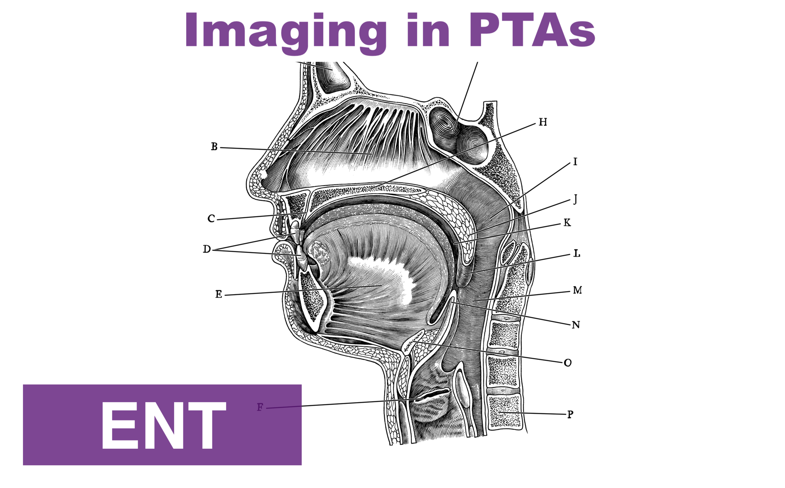
Imaging/Further Testing:

  • Chest radiograph

  • CT brain

  • CT C-spine

  • CTA head/neck

  • Can consider soft-tissue neck x-ray, if CT not immediately available

Further Management:

  • In patients with signs of hanging/strangulation, there should be a low threshold to obtain diagnostic imaging/testing as discussed above.

  • Expect pulmonary complications early.

    • They are a major cause of delayed mortality in near-hanging victims, as stated above.

  • Early intubation and airway management are important.

  • Non-intubated patients with pulmonary edema may benefit from positive end-expiratory pressure ventilation.

  • Patients with symptoms of laryngeal or tracheal injury (e.g. dyspnea, dysphonia, aphonia, or odynophagia), should undergo laryngobronchoscopy with ENT. [4,6]

  • Tracheal stenosis has been reported during the hospital course. Address cerebral edema from anoxic brain injury, using strategies to reduce intracranial pressure or seizure prophylaxis. [4]

  • Address vascular complications seen on CTA and coordinate intervention with the appropriate specialty at your institution.

  • Therapeutic Hypothermia

    • There is some evidence for therapeutic hypothermia in those with cardiac arrest from hanging injury [7,8] and those who are comatose from hanging injury. [9-11] While the evidence is weak, in the absence of better evidence, it is reasonable to consider hypothermia treatment in all comatose near-hanging victims. [1,12,13]

  • When suicide is suspected, evaluate patients for other methods of self-harm (e.g. wrist lacerations, self-stabbing, ingestions). It is also important to consider drug and alcohol intoxication. [4]

Disposition:

  • Admit critically ill patients to the appropriate ICU-level care.

  • Admit patients with abnormal radiologic or endoscopic imaging to the appropriate service and level of care.

  • Even if the initial presentation is clinically benign, all near-hanging victims should be observed for 24 hours, given the high risk of delayed neurologic, airway and pulmonary complications. [14]

  • Observe asymptomatic patients with normal imaging.

  • Psychiatry/Crisis Team consult on all suspected intentional cases.

  • Emphasize strict return precautions as well as education about possible delayed respiratory and neurologic dysfunction when discharging patients.

  • Some patients may require transfer to a trauma center if the required services are not available at the initial receiving facility. [1]

Prognostication:

  • GCS 3/GCS 3T is a predictor of very poor outcome, [15-19] but there is mixed evidence on the GCS as a predictor of outcomes in GCS scores greater than 3, especially with regard to neurologic intactness. [3,19]

  • Recovery of patients with neurology symptoms is unpredictable. [4]

  • Patients presenting with cardiac arrest have a very poor prognosis, and might be the strongest predictor of poor prognosis. [4,8,16,18,20]

  • Other predictors of poor clinical outcome include:

    • Anoxic brain injury or cerebral edema on head CT [3,19]

    • Prolonged hanging time [18]

    • Cardiopulmonary arrest [8,11,19]

    • Cervical spine injury

    • Hypotension on arrival


Expert Commentary

We’ve all certainly been involved with a patient with reported hanging injury at some point in our time in the ED. They are usually unimpressive if a person does it as more of a gesture rather than a true suicide attempt. When they are unfortunately done “correctly,” they usually result in a trip to the morgue instead of the ED. When the swiss cheese holes align and a true hanging attempt results in a serious but not fatal presentation, things can get quite hairy. I’ve been a part of one such case, and will never forget it. Here are my two cents.

Airway

This should undoubtedly be treated as a predicted difficult airway, not only due to likely cervical spine trauma, but also possibly due to airway edema. Get your ducks in a row for this unless this patient is crashing in front of you. Get your consultants/help (if available), preoxygenate, airway adjuncts open and ready, backup airway supplies if your first plan fails. Most importantly, have a plan and discuss this with your team beforehand. Don’t be afraid to take an awake look with a hyperangulated video laryngoscope, especially if this patient presents with stridor. Ketamine can be your friend here. This should be an airway that you do not undertake without a scalpel, finger, and bougie ready just in case. I like to draw a line on the patient’s skin overlying the cricothyroid membrane beforehand.

Trauma

Self explanatory, but don’t be stingy here. Light this patient up from head to pelvis, including the neck angiogram. Document a repeat neuro exam every time you move this patient.

Overdose/psych

Don’t forget your Tylenol and salicylate levels, EKG in this suicide attempt. If you feel the need to add the useless urine drug screen, I suppose this is fine as well.

Kevin Emmerich, MD, MS

Emergency Medicine Physician

Methodist Hospital

Gary, Indiana


How To Cite This Post:

[Peer-Reviewed, Web Publication] Karalius, V. Porras, N. (2021, Aug 9). Hanging Injuries. [NUEM Blog. Expert Commentary by Emmerich, K]. Retrieved from http://www.nuemblog.com/blog/hanging-emergencies


Other Posts You May Enjoy

References

1. Walls RM, Hockberger RS, Gausche-Hill M. Rosen's emergency medicine : concepts and clinical practice. Ninth edition. ed. Philadelphia, PA: Elsevier; 2018.

2. Aufderheide TP, Aprahamian C, Mateer JR, et al. Emergency airway management in hanging victims. Ann Emerg Med. 1994;24(5):879-884.

3. Salim A, Martin M, Sangthong B, Brown C, Rhee P, Demetriades D. Near-hanging injuries: a 10-year experience. Injury. 2006;37(5):435-439.

4. Tintinalli JE, Stapczynski JS, Ma OJ, Yealy DM, Meckler GD, Cline DM. Tintinalli's emergency medicine: a comprehensive study guide. 9th. ed. New York: McGraw-Hill Education; 2019.

5. Tugaleva E, Gorassini DR, Shkrum MJ. Retrospective Analysis of Hanging Deaths in Ontario. J Forensic Sci. 2016;61(6):1498-1507.

6. Hackett AM, Kitsko DJ. Evaluation and management of pediatric near-hanging injury. Int J Pediatr Otorhinolaryngol. 2013;77(11):1899-1901.

7. Hsu CH, Haac B, McQuillan KA, Tisherman SA, Scalea TM, Stein DM. Outcome of suicidal hanging patients and the role of targeted temperature management in hanging-induced cardiac arrest. J Trauma Acute Care Surg. 2017;82(2):387-391.

8. Kim MJ, Yoon YS, Park JM, et al. Neurologic outcome of comatose survivors after hanging: a retrospective multicenter study. Am J Emerg Med. 2016;34(8):1467-1472.

9. Jehle D, Meyer M, Gemme S. Beneficial response to mild therapeutic hypothermia for comatose survivors of near-hanging. Am J Emerg Med. 2010;28(3):390.e391-393.

10. Lee BK, Jeung KW, Lee HY, Lim JH. Outcomes of therapeutic hypothermia in unconscious patients after near-hanging. Emerg Med J. 2012;29(9):748-752.

11. Hsu CH, Haac BE, Drake M, et al. EAST Multicenter Trial on targeted temperature management for hanging-induced cardiac arrest. J Trauma Acute Care Surg. 2018;85(1):37-47.

12. Borgquist O, Friberg H. Therapeutic hypothermia for comatose survivors after near-hanging-a retrospective analysis. Resuscitation. 2009;80(2):210-212.

13. Sadaka F, Wood MP, Cox M. Therapeutic hypothermia for a comatose survivor of near-hanging. Am J Emerg Med. 2012;30(1):251.e251-252.

14. McHugh TP, Stout M. Near-hanging injury. Ann Emerg Med. 1983;12(12):774-776.

15. Kao CL, Hsu IL. Predictors of functional outcome after hanging injury. Chin J Traumatol. 2018;21(2):84-87.

16. La Count S, Lovett ME, Zhao S, et al. Factors Associated With Poor Outcome in Pediatric Near-Hanging Injuries. J Emerg Med. 2019;57(1):21-28.

17. Martin MJ, Weng J, Demetriades D, Salim A. Patterns of injury and functional outcome after hanging: analysis of the National Trauma Data Bank. Am J Surg. 2005;190(6):836-840.

18. Matsuyama T, Okuchi K, Seki T, Murao Y. Prognostic factors in hanging injuries. Am J Emerg Med. 2004;22(3):207-210.

19. Nichols SD, McCarthy MC, Ekeh AP, Woods RJ, Walusimbi MS, Saxe JM. Outcome of cervical near-hanging injuries. J Trauma. 2009;66(1):174-178.

20. Gantois G, Parmentier-Decrucq E, Duburcq T, Favory R, Mathieu D, Poissy J. Prognosis at 6 and 12months after self-attempted hanging. Am J Emerg Med. 2017;35(11):1672-1676.

Posted on August 9, 2021 and filed under Airway, Critical care, Trauma.

Scalpel Finger Bougie

Written by: Em Wessling, MD (NUEM ‘22) Edited by: Therese Whipple (NUEM ‘20) Expert Commentary by: Joseph Posluszny, MD

Written by: Em Wessling, MD (NUEM ‘22) Edited by: Therese Whipple (NUEM ‘20) Expert Commentary by: Joseph Posluszny, MD


Expert Commentary

Establishing an airway via a cricothyroidotomy is a stressful and tense experience.  In almost all of these cases, experienced airway staff have already attempted advanced airway maneuvers in patients typically at high risk for inability to intubate.  As the oxygen saturation drops and the patient becomes unstable, the most adept proceduralist present (whether emergency department physicians or surgeons) are asked to step in to secure a surgical airway.

The scalpel-finger-bougie technique is one proven and reliable method to secure a surgical airway via a cricothyroidotomy.  Some additions to the technique described above are:

  • Use a vertical incision through the skin and soft tissues.  If you are too superior or inferior with your initial incision, then this incision can be easily extended as needed.  A horizontal incision commits you to that cranial-caudal level.  It is often more of a struggle to identify the cranial-caudal orientation of the cricothyroid membrane rather than the medial-lateral orientation. 

  • In a patient with a stable and flexible neck, retract the neck via cranial pressure on the chin to bring the neck structures better into your working field. Insert a shoulder roll if available (unlikely) to augment this positioning.

  • After the tube is advanced, listen for bilateral breath sounds.  It is common, in this adrenaline fueled procedure, to advance the endotracheal tube too far, leading to a right main stem intubation.  This can limit your ventilation and oxygenation and can lead to confusion about the airway placement in the neck.  If there are no left lung field breath sounds, then pull the tube back until bilateral breath sounds are confirmed with auscultation.

  • Always verify tube placement with capnography.

  • Persistent, moderate volume bleeding is often from injury to the anterior jugular vein. Gentle, directed pressure on the area can control this bleeding while the patient is being transported to the operating room for a more definitive airway.

Joseph Posluszny, MD

Assistant Professor of Surgery (Trauma and Critical Care)

Northwestern Memorial Hospital


How To Cite This Post:

[Peer-Reviewed, Web Publication] Wessling, E. Whipple, T. (2021, Aug 2). Scalpel Finger Bougie. [NUEM Blog. Expert Commentary by Posluszny, J]. Retrieved from http://www.nuemblog.com/blog/scalpel-finger-bougie


Other Posts You May Enjoy

Posted on August 2, 2021 and filed under Trauma, Procedures.

Seasonal Influenza

Written by: Logan Wedel, MD (NUEM ‘22) Edited by: Laurie Aluce, MD (NUEM ‘21) Expert Commentary by: Gabrielle Ahlzadeh, MD (NUEM ‘19)

Written by: Logan Wedel, MD (NUEM ‘22) Edited by: Laurie Aluce, MD (NUEM ‘21) Expert Commentary by: Gabrielle Ahlzadeh, MD (NUEM ‘19)


Seasonal Influenza PNG.png

Expert Commentary

Thank you for the concise and informative resource guide. Don’t we all yearn for the days when patients with myalgias and fever were typically diagnosed with influenza and sent on their merry way? Believe it or not, there are some interns who may go their entire first winter as physicians without diagnosing influenza. 

 A large part of our jobs as emergency medicine physicians is reassurance. Patients want to know they are okay, but they always want something to make them feel better faster, which is true for influenza as well as coronavirus. And while most young, healthy patients do not need treatment for influenza, another consideration is time lost from work, even though symptom duration is really only decreased by one day. However, for some individuals, those one to two days of work may be essential, in which case, a prescription should be considered if the patient presents within 48 hours of symptom onset. For all other healthy adults, I discuss the side effects of the medication. Evidence from a Cochrane Review published in 2014 suggests that in healthy adults, the risks of side effects including nausea, vomiting, headaches and psychiatric symptoms likely outweighs any benefit, which again, does NOT include decreased hospitalizations rates or complications. I usually frame it as “yes, your flu symptoms may get better about 16 hours sooner, but you may also have nausea, vomiting and headaches.” Most people, in my experience, will then pass on a prescription for Tamiflu. 

Screening for chronic medical conditions, pregnancy, high risk household members is essential in knowing which patients require treatment. High level athletes is another patient population where treatment may be considered to allow them to get back to training faster but also to minimize spread to the rest of the team. 

Visits for influenza are also a great time to discuss vaccination with patients, especially during the current pandemic. Emphasis should be placed on how vaccines prevent life threatening complications from influenza and diminish symptom severity. This is a perfect time for vaccine education to hopefully prevent future pandemics.


References

  1. Jefferson T et al. Oseltamivir for Influenza in Adults and Children: Systematic Review of Clinical Study Reports and Summary of Regulatory Comments. BMJ 2014. PMID: 24811411.

Gabrielle Ahlzadeh, MD

Clinical Assistant Professor of Emergency Medicine

University of Southern California


How To Cite This Post:

[Peer-Reviewed, Web Publication] Wedel, L. (2021, May 17). Seasonal Influenza. [NUEM Blog. Expert Commentary by Ahlzadeh, G]. Retrieved from http://www.nuemblog.com/blog/seasonal-influenza


Other Posts You May Enjoy

Posted on May 17, 2021 and filed under Pulmonary.

Cocaine Chest Pain

Written by: Maren Leibowitz, MD (NUEM ‘23) Edited by: Zach Schmitz, MD (NUEM ‘21) Expert Commentary by: David Farman, MD

Written by: Maren Leibowitz, MD (NUEM ‘23) Edited by: Zach Schmitz, MD (NUEM ‘21) Expert Commentary by: David Farman, MD


CCP1.png

Expert Commentary

Cocaine chest pain was something that was frequently discussed but rarely encountered during my training and during my 11 years practicing in suburban and metropolitan Indiana (we're more of a heroin/meth state).  I suspect there is significant regional and local variation in the epidemiology of cocaine chest pain, likely influenced by the economics and local availability or popularity of intoxicants.

When cocaine is more expensive, we tend to see more methamphetamine use.  And vice-versa.  I was once told by a local sheriff that "people will get high on whatever they can get for less than $20" and I have found that to be true in practice.  I wouldn't expect that an emergency physician would be intimately familiar with the local micro-economics of the drug trade, but one should expect there to be a periodic waxing/waning of cocaine chest pain presentations.  Similarly, it may be a more frequent complaint dependent on cocaine's local popularity and availability.

When consulting with a Cardiologist about a cocaine chest pain case, it is important for the emergency physician to avoid letting premature closure or psychosocial biases unduly influence the patient's disposition.  It is not unheard of for physicians to minimize objective findings (ST segment abnormalities or biomarker elevation) and attribute them to the vasoactive properties of cocaine.  I have certainly been tempted to do so myself.  However, the article wisely points out the physiologic changes induced by cocaine use, both acutely and chronically.  Platelet aggregation and atherogenesis can absolutely promote a scenario that would require PCI in even the most frequent of 'frequent fliers' with cocaine chest pain. 

In short, I would have a low threshold to involve Cardiology in a patient who has objective findings regardless of their use of cocaine.  Similarly, a Cardiology request for a Urine Drug Screen shouldn't delay a patient's trip to the cath lab if they have a STEMI.  An exception to this may be a patient who has had recent coronary angiography that objectively demonstrates normal coronaries.  In that scenario I would consider serial markers, conservative management and strong consideration of non-cardiac causes of the pain (dissection, pneumothorax, etc.).

David Farman, MD FACEP

Emergency Medicine Physician

Franciscan Health Lafayette East


How To Cite This Post:

[Peer-Reviewed, Web Publication] Leibowitz, M. Schmitz, Z. (2021, May 10). Cocaine Chest Paine. [NUEM Blog. Expert Commentary by Farman, D]. Retrieved from http://www.nuemblog.com/blog/cocaine-chest-pain


Other Posts You May Enjoy

Posted on May 10, 2021 and filed under Cardiovascular.

Eliminating health disparities in LGBT individuals begins in the ED

Written by: David Feiger, MD (NUEM ‘22) Edited by: Vidya Eswaran, MD (NUEM ‘20) Expert Commentary by: Will Laplant, MD, MPH '20

Written by: David Feiger, MD (NUEM ‘22) Edited by: Vidya Eswaran, MD (NUEM ‘20) Expert Commentary by: Will Laplant, MD, MPH '20


Introduction

18% of lesbian, gay, bisexual, transgender, or questioning (LGBTQ) individuals avoided seeking medical attention for fear of discrimination according to a 2017 joint poll conducted by NPR, the Robert Wood Johnson Foundation, and Harvard T.H. Chan School of Public Health [1]. These fears are not unfounded—decades of anti-lesbian, gay, and bisexual (LGB) prejudice in medicine, despite greater social acceptance in the United States have tainted medicine’s perception in the LGB community.

Only in 1987 was “sexual orientation disturbance” removed from the DSM while conversion therapy, the scientifically-unfounded exercise of converting one’s sexual orientation to heterosexual, continues to be legal across much of the United States. In 2015, a pediatrician in Michigan declined to see a child of a lesbian couple which spurred a national debate of refusal of care on the basis of religious freedom [2]. Just this year the Conscience and Religious Freedom Division of the Department of Health and Human Services was established [3]. It is clear why LGB individuals may want to avoid routine medical attention.

As a disclaimer, LGBT individuals are often lumped into one category. We will focus this post on LGB individuals as we believe that the transgender experience, while occasionally overlapping with that of LGB individuals, deserves its own recognition in another post. Many studies and data grouped LGB and transgender individuals as one group and will be cited as such in this post.


Health Disparities

34% of LGBT individuals reported having been a victim of bullying, instituting a fear of discrimination and coming out that can lead to a variety of emotional and psychological consequences [4]. In the National Health Interview Study, 26% and 28% of gay men and women, respectively, and 41% and 47% of bisexual men and women, respectively, reported moderate or severe levels of mental distress compared to 17% of straight men and 22% of straight women [6]. These levels of psychological stress make LGB individuals more prone to emotional disorders leading to suicidal ideation, homelessness, depression, and substance abuse [4].

Furthermore, 18% of LGBT individuals reported having been coerced into sex and 23% sexually assaulted [4]. This victimization increases their risk of unsafe sexual behavior than their peers resulting in a twice greater likelihood of contracting sexually transmitted diseases when compared to straight-identifying men [5]. The fear of seeking medical attention only exacerbates these health disparities.

While much of the focus has been on the modern experience of LGB youth, it is important to be aware the circumstances of LGB seniors that may contribute to health disparities. Due to the social climate in which they grew up, they are less likely to be partnered, have children, or other social supports, increasing their barriers to health care. Furthermore, they often reside in senior communities where they often face continued discrimination [7].


Creating a More Welcoming Environment for All

The emergency department is often the first point of healthcare contact for the vulnerable, and is therefore a prime location to make healthcare more approachable for the LGB community.

The Waiting Room

Patients often spend hours in the waiting room, providing an opportune time to set an inviting tone for LGB patients. LGB individuals constantly seek subtle indications of acceptance in unknown environments [11]. At the most basic level, triage forms can include questions pertaining to sexual orientation and gender identity [7]. In fact, collection of this data has been recommended by the Institute of Medicine and the Joint Commission [8]. These forms can also include the hospital’s non-discrimination policy on the basis of sexual orientation and offer a contact for a patient advocate for those who have been unfairly treated. Staff can wear rainbow flag pins and waiting rooms can offer pamphlets that highlight LGB health among other health topics [7]. These small additions can make LGB individuals feel more welcome.


With the Physician
Despite 78% of emergency physicians believing that patients would refuse to reveal their sexual orientation in the emergency department, only 10% of patient respondents of all sexual orientations (n=1516) to the EQUALITY study reported that they would not answer the question [8]. As summarized by Dr. Adil Haider, the principal investigator of the study, “your patients want to be asked.”

When asking patients about their sexual orientation, it is important to use gender-neutral language. Ask “are you in a relationship?” or “do you have sex with men, women, or both?” to delve into a social history. These types of questions may comfort LGB patients to allow them to expand on details they find relevant [9]. Of course, patients who are not comfortable with their identity may continue to conceal their sexual orientation. Each individual’s coming out experience varies and it is crucial that the physician allows the patient to take his or her own time to reveal their sexual orientation, even if not on this visit. Simply asking the broad questions without judgment may begin to change the patient’s apprehensive attitude towards medicine.

Going Above and Beyond

While important to make the clinical encounter more inviting, more actions can be taken to make a hospital a leader in LGB care.

1.   Partner with local LGB organizations

Hospitals can partner with organizations that support the LGB community. Having a presence in health clinics targeting LGB individuals and other local LGB organizations will also allow the hospital to better understand and adapt to the needs specific to the community. Celebrating LGBT awareness months and having staff march in local LGB Pride events is a very public and visible way of showing support for the community [9].

2.   Actively recruiting and maintaining LGB staff

Health care providers should actively recruit LGB staff by ensuring equal employment benefits as their heterosexual colleagues by offering supplemental packages that include benefits for both married and unmarried same-sex partners. After hiring, ensure that LGB employees continue to receive support and mentoring by sponsoring LGB employee groups and functions [9].

3.   Striving for and achieving a perfect score on the Human Rights Campaign Healthcare Equality Index

The Human Rights Campaign is the largest organization supporting LGBT rights in the United States. Each year, the Human Rights Campaign scores and publishes a list of many hospitals in the United States and grades them on hospitals’ ability to provide inclusive care regardless of sexual orientation. The list evaluates a hospital’s patient and employment non-discrimination policies, visitation rights, LGBT-focused training offering, presence of patient services to LGBT individuals, employee benefits, and commitment to the community for a total score on a scale from 0 to 100.10 For healthcare institutions not achieving a perfect 100 score, this is a great tool to ensure progression to full equity of care for LGB patients.


Summing it Up

Despite rapid social acceptance of the LGB community in the United States within the past decade, remnants of fear and distrust in healthcare remain, exacerbating existing health disparities. While it may take several decades to fully eradicate the apprehension, taking the steps above will certainly make strides to achieve that goal.


Expert Commentary

Thank you for writing about such an important topic. I think we all understand the special position of the emergency department beyond the care of emergencies; it is a point of access for many marginalized communities that have been unable to receive care through other venues. It is our duty as emergency physicians to be able to provide competent and appropriate care for all who walk through our doors.

I think it makes sense to divide the topic of LGBTQ care into two: sexual minorities (those who identify as lesbian/gay/bisexual/queer, etc) and gender minorities (those who identify as trans, genderqueer or otherwise gender nonconforming). While they have much in common, the barriers and healthcare disparities they face are unique and different, and they each warrant a lengthy discussion. I look forward to your article on the care of gender minorities!


Normalizing Care in the Emergency Department

  • Identifying Those in the Room

Asking “how are the two of you related?” to figure out who else is in the room can prevent mishaps of assuming someone is with their friend, when really they are with their partner. As a major support system after discharge, it’s important to include a patient’s partner in the discussion and plan.

  • Taking a Sexual History

It’s important to not conflate sexual orientation with sexual behaviors. For example, there is a well defined subsect of men who have sex with men (MSM) but do not identify as gay/bisexual. By asking “do you have sex with men, women, or both?” you ensure that you capture the data you need to treat the patient appropriately. Some people get hung up on the follow up questions to further identify how to treat a patient:

               “Do you have anal intercourse?”

               “Do you have receptive, penetrative, or both?”

The same model can be used for oral and vaginal intercourse.

  • Addressing the Sexual Health of Sexual Minorities

If you are addressing sexual health needs in the emergency room, either because of chief complaint (eg. sore throat, rectal pain, vaginal discharge, abdominal pain) or exposure, try and be as comprehensive as possible. The majority of syphilis cases in the US are amongst MSM [1], making it an important consideration. I suppose this blog is also as good a place as any to highlight the recent CDC recommendations for treatment of STIs, including [2]:

  1. Monotherapy with ceftriaxone for confirmed gonorrhea given increasing azithromycin resistance

  2. Increased dosing of ceftriaxone (500mg from 250mg) for gonorrhea treatment, and 1g of ceftriaxone for those >150kg

  3. Doxycycline 100mg BID for 7 days for chlamydia infections

    *Notably, compliance with a 7 day course of treatment should be addressed and factored into the decision (with regards to the previous standard of azithromycin 1g as a single dose).

Identifying Bias

Bias comes in two forms: explicit biases, which we are cognizant of, and implicit, which we are not. Implicit bias stems from the confluence of your life experiences and the society you are a part of. If you have been raised in a society entrenched in systemic racism, sexism, ableism and heteronormativity, you have been exposed to stereotypes and prejudices which may subconsciously shape the way you make decisions. Healthcare professionals have been shown to have a similar level of implicit bias compared to the general population [3], and this implicit bias has been correlated with significant patient outcomes. [4] What shapes your decision in who receives narcotic pain medication or who stays in the hospital for observation? I highly recommend taking at least a few tests of implicit bias which are freely available and, in my opinion, highly informative: https://implicit.harvard.edu/implicit/takeatest.html

References

  1. CDC. Sexually transmitted disease surveillance 2013. Atlanta: US Department of Health and Human Services; 2014.

  2. St. Cyr S, Barbee L, Workowski KA, et al. Update to CDC’s Treatment Guidelines for Gonococcal Infection, 2020. MMWR Morb Mortal Wkly Rep 2020;69:1911–1916. DOI: http://dx.doi.org/10.15585/mmwr.mm6950a6external icon.

  3. FitzGerald, C., Hurst, S. Implicit bias in healthcare professionals: a systematic review. BMC Med Ethics 18, 19 (2017). https://doi.org/10.1186/s12910-017-0179-8

  4. William J. Hall et al. “Implicit Racial/Ethnic Bias Among Health Care Professionals and Its Influence on Health Care Outcomes: A Systematic Review”, American Journal of Public Health 105, no. 12 (December 1, 2015): pp. e60-e76.

Will Laplant MD, MPH

Emergency Medicine Physician

Good Samaritan Medical Center

Brockton, MA


How To Cite This Post:

[Peer-Reviewed, Web Publication] Feiger, D. Eswaran, V. (2021, May 2). Eliminating health disparities in LGBT individuals begins in the ED. [NUEM Blog. Expert Commentary by Laplant, W]. Retrieved from http://www.nuemblog.com/blog/lgbt-disparities


Other Posts You May Enjoy

References

  1. “Discrimination in America: Experiences and Views of LGBTQ Americans.” www.npr.org, National Public Radio, Nov. 2017, www.npr.org.

  2. Pear, Robert, and Jeremy W. Peters. “Trump Gives Health Workers New Religious Liberty Protections.” The New York Times, 18 Jan. 2018.

  3. Phillip, Abby. “Pediatrician Refuses to Treat Baby with Lesbian Parents and There’s Nothing Illegal about It.” The Washington Post, 19 Feb. 2015.

  4. Hafeez, Hudaisa, et al. “Health Care Disparities Among Lesbian, Gay, Bisexual, and Transgender Youth: A Literature Review.” Cureus, 2017, doi:10.7759/cureus.1184.

  5. Robinson, Joseph P., and Dorothy L. Espelage. “Peer Victimization and Sexual Risk Differences Between Lesbian, Gay, Bisexual, Transgender, or Questioning and Nontransgender Heterosexual Youths in Grades 7–12.” American Journal of Public Health, vol. 103, no. 10, 2013, pp. 1810–1819., doi:10.2105/ajph.2013.301387.

  6. Gonzales, Gilbert, et al. “Comparison of Health and Health Risk Factors Between Lesbian, Gay, and Bisexual Adults and Heterosexual Adults in the United States.” JAMA Internal Medicine, vol. 176, no. 9, 2016, p. 1344., doi:10.1001/jamainternmed.2016.3432.

  7. Understanding the Health Needs of LGBT People. Understanding the Health Needs of LGBT People, National LGBT Health Education Center, 2016.

  8. Haider, Adil H., et al. “Emergency Department Query for Patient-Centered Approaches to Sexual Orientation and Gender Identity.” JAMA Internal Medicine, vol. 177, no. 6, 2017, p. 819., doi:10.1001/jamainternmed.2017.0906.

  9. Ten Things: Creating Inclusive Health Care Environments for LGBT People. Ten Things: Creating Inclusive Health Care Environments for LGBT People, National LGBT Health Education Center, 2015.

  10. Human Rights Campaign. “Healthcare Equality Index 2018.” Human Rights Campaign, Human Rights Campaign, 2018, www.hrc.org/hei.

  11. Eliason, Michele J., and Robert Schope. “Does ‘Don't Ask Don't Tell’ Apply to Health Care? Lesbian, Gay, and Bisexual People's Disclosure to Health Care Providers.” Journal of the Gay and Lesbian Medical Association, vol. 5, no. 4, Dec. 2001.

Posted on May 3, 2021 and filed under Advocacy.

Bariatric Emergencies

Written by: Maurice Hajjar, MD, MPH (NUEM ‘22) Edited by: Philip Jackson(NUEM ‘20) Expert Commentary by: Gabby Ahlzadeh, MD

Written by: Maurice Hajjar, MD, MPH (NUEM ‘22) Edited by: Philip Jackson(NUEM ‘20) Expert Commentary by: Gabby Ahlzadeh, MD


bariatric1.png
bariatric2.png
bariatric3.png

Expert Commentary

Thanks for this great post. Being familiar with the anatomy of these various procedures is essential to understanding the complications and why you cannot be reassured by a benign abdominal examination. 

With laparoscopic band procedures, complications are more common early on and are more related to band erosion or migration. Migration of the lap-band is best evaluated with an upper GI series with Gastrografin. Another important question to ask patients is if their lap band has been inflated recently. This is typically done in a progressive manner, where normal saline is instilled within the subcutaneous port. This can also be a source of obstruction, and if emergent, the band can be deflated by aspirating fluid from the port. This should be done under the guidance of a surgeon, if possible. 

Asking about dietary indiscretions can also sometimes clue you in to why a person is having abdominal pain or nausea. Especially immediately post-op, these patients have specific dietary guidelines and are often limited to liquids or pureed foods. Dumping syndrome can also occur in this patient population, more common with gastric bypass surgery, due to rapid gastric emptying. Typically, these patients have bloating, sweating, facial flushing, diarrhea, nausea, early satiety about 30 to 60 minutes after a meal. Dumping syndrome is typically diagnosed clinically, though laboratory testing should be performed to rule out electrolyte derangements. Dumping syndrome is typically treated with dietary modifications and should be discussed with the patient’s surgical team. 

As these procedures become more common and advanced, some are also performed endoscopically, which comes with its own set of complications. Another newer procedure is the intragastric balloon, which is a saline filled silicone balloon that is inflated in the stomach. In the first few days after placement, patients may experience abdominal pain, nausea, and vomiting. Other complications include balloon rupture, bowel obstruction, gastric outlet obstruction, gastric ulcer, pancreatitis, nonalcoholic steatohepatitis and cholecystitis.  

Overall, management of these patients in the emergency department should be done in consultation with the surgeon.

Gabrielle Ahlzadeh, MD.PNG

Gabrielle Ahlzadeh, MD

Clinical Assistant Professor of Emergency Medicine

University of Southern California


How To Cite This Post:

[Peer-Reviewed, Web Publication] Hajjar, M. Jackson, P. (2021, Apr 26). Bariatric Emergencies. [NUEM Blog. Expert Commentary by Ahlzadeh, G]. Retrieved from http://www.nuemblog.com/blog/bariatric-emergencies


Other Posts You May Enjoy

Posted on April 26, 2021 and filed under Gastrointestinal.

Thyroid Storm ED Diagnosis and Management

Written by: Shawn Luo, MD (NUEM ‘22) Edited by: Steve Chukwuelebe, MD (NUEM '19) Expert Commentary by: Samia Farooqi, MD (NUEM '16)

Written by: Shawn Luo, MD (NUEM ‘22) Edited by: Steve Chukwuelebe, MD (NUEM '19) Expert Commentary by: Samia Farooqi, MD (NUEM '16)


Thyrotoxicosis Infographic.jpeg

Expert Commentary

For those of you who have not yet cared for a patient in Thyroid Storm, I guarantee that the experience will humble you.  As far as endocrine emergencies go, this one is (quite literally) a killer.  In 1993, Burch and Wartofsky cited mortality rates as high as 20 to 50% [1].  In more recent literature, the mortality rate of this condition is still at a whopping 10% [2].  As you can see from the beautifully presented infographic above, there are a number of complexities that factor into the diagnosis and treatment of Thyroid Storm.  This is in part due to the fact that thyroid hormone affects all organ systems, with clinical manifestations of disease involving everything from cardiac dysrhythmia to profound GI losses secondary to vomiting and diarrhea to acute psychosis. To ground this infographic in clinical reality, where we know that diagnoses are not always easy to make and the decision between one therapeutic option versus another is not always clear, I offer you just a few additional points:


1.  Making this diagnosis requires that you actually think about it in the first place. There is no single diagnostic test or image that will clinch this diagnosis for you.  The Burch and Wartofsky criteria are clinical findings that you as the provider must use in the correct context for them to be useful. This is a rare condition, and your availability bias will be working against you here.  Patient presentations can be vague and there is incredible overlap with other disease processes, with chief complaints ranging from anxiety to vomiting and abdominal pain to leg swelling.


2. Once this diagnosis has been considered or made, understand that these patients have incredibly complex hemodynamics, with multiple potential factors in play at any given time: hypovolemia from profound GI losses, concomitant sepsis, high output cardiac failure, and cardiac systolic dysfunction.  With regards to this last point, there is a high incidence of decompensated heart failure in patients with thyroid storm, and those with cardiogenic shock are at some of the greatest risk of mortality [2, 3, 4].  As such, it behooves you to re-assess these patients’ volume status frequently.  Examine them closely, and then re-examine them.  Use your bedside ultrasound to assess their cardiac output, IVC, and lung windows.  While they may be profoundly hypertensive when you first administer a beta blocker, concomitant sepsis and hypovolemia may surface very quickly as the patient’s blood pressure plummets.


3. This leads to my next point: when given the choice between propranolol and esmolol in managing tachydysrhythmia (whether profound sinus tachycardia or atrial fibrillation) in the context of Thyroid Storm, strongly consider esmolol.  The Japan Thyroid Association and Japan Endocrine Society Task Force specifically cites increased mortality rates in patients for whom propranolol was used versus esmolol or landiolol (super short-acting beta blocker used in Japan) [3].  Much of the benefit to using esmolol over propranolol is in its much shorter half-life. The alpha and beta half-life for propranolol are 10 minutes and 2.3 hours, respectively, whereas the alpha and beta half-life for esmolol are 2 minutes and 9 minutes, respectively.  Once the infusion is stopped, the effect of esmolol will have completely disappeared by 18 minutes [3]. 


4. When asked during oral boards how you would manage a patient with Thyroid Storm, a response involving antithyroid agents, inorganic iodide, corticosteroids, and beta blockers would score you full marks.  However, I would encourage you to also consider thoughtful administration of IV fluids (with frequent re-assessment of volume status), early empiric antibiotics, inotropic agents for patients in cardiogenic shock, and psychotropic medications as needed for restlessness/delirium/psychosis (with olanzapine preferred over haldol) [3].


Diagnosis and management of Thyroid storm in the Emergency Department requires us to draw upon so many of our expert skills in approaching undifferentiated and critically ill patients.  I hope that these points help to provide you with just a few more tools to include in your arsenal as you go forth. 


References

[1] Burch, HB, Wartofsky, L. (1993). Life-threatening thyrotoxicosis: Thyroid storm. Endocrinology and Metabolism Clinics of North America, 22(2): 263-77.

[2] Akamizu, T. (2018). Thyroid Storm: A Japanese Perspective. Thyroid, 28:1.

[3] Satoh, T, et al. (2016). 2016 Guidelines for the Management of Thyroid Storm from The Japan Thyroid Association and Japan Endocrine Society (First edition). Endocrine Journal, 63: 12 (1025-1064).

[4] Bourcier, S, et al. (2020). Thyroid Storm in the ICU: A Retrospective Multicenter Study. Critical Care Medicine, 48:1 (83-90).

dr-samia-siyar-farooqi-profile-8b03a076 (1).png

Samia Farooqi, MD

Assistant Professor

Department of Emergency Medicine

UT Southwestern


How To Cite This Post:

[Peer-Reviewed, Web Publication] Luo, S. Chukwuelebe, S. (2021, April 19). Thyroid Storm ED Diagnosis and Management. [NUEM Blog. Expert Commentary by Farooqi, S]. Retrieved from http://www.nuemblog.com/blog/thyroid-storm.


Other Posts You May Enjoy

Posted on April 19, 2021 and filed under Endocrine.

ED Clinical Decision Making Units

Written by: Mitchell Blenden, MD (NUEM ‘24) Edited by: Em Wessling (NUEM ‘22) Expert Commentary by: Tim Loftus, MD, MBA

Written by: Mitchell Blenden, MD (NUEM ‘24) Edited by: Em Wessling (NUEM ‘22) Expert Commentary by: Tim Loftus, MD, MBA


CDU1.png
CDU2.png
CDU3.png

Expert Commentary

Thank you to Dr’s Blenden and Wessling for the excellent overview of ED CDUs including some background and indications for their use. 

 Several points to highlight and elaborate upon include the following:

The background of the utility of CDUs mostly stems from their early function as rapid diagnostic and treatment centers (RDTCs) for chest pain.  The function and utility of CDUs have since grown to demonstrate clinical benefits well-established across a variety of conditions as Dr’s Blenden and Wessling have mentioned, including not only chest pain (rule out acute coronary syndrome) but also TIA, CHF, asthma, COPD, cellulitis, pyelonephritis, pneumonia, etc. 

Value and Benefits

The utility and value of ED CDUs will continue to expand.  The percentage of all hospital admissions that start in the ED continues to grow -- 67% in 2019, up from 58% in 2004 according to the ED Benchmarking Alliance.  Additionally, EDs cared for approximately 158M people as of 2018 (EMNet/NEDI-USA), up 32% over a 10 year period.  As the number of admissions continues to grow, and considering that some of these inpatient stays are short, it follows that many of these short inpatient admissions are subject to recovery audit contractors and payor denials.  Many clinical conditions which are often subject to short inpatient stays can be cared for in dedicated short stay observation units without adversely affecting, and for the most part improving, the quality of care delivery, safety, satisfaction, cost savings, and reducing subsequent inpatient LOS.  

All stakeholders in the health care system benefit from CDU use: patients are more accurately diagnosed before leaving the ED and are discharged home faster, payors avoid costly inpatient admission charges, hospitals keep scarce inpatient bed capacity open for more appropriate patients and avoid audits and denials, and providers deliver care in a setting that more appropriately matches patient needs to resources.

Dedicated Units with Protocolized Care

Observation patients can be managed in a variety of settings and contexts, but best practice that leads to best outcomes would be in dedicated observation units adherent to protocols tailored to the patients’ conditions, the best available evidence, and local institutional resources.

Shorter hospitalizations are more likely to occur in dedicated observation units under protocols than with unstructured hospitalization on inpatient teams and simply billing status changes to observation.

Financial Considerations

Much of the existing evidence has demonstrated that CDUs can provide care that efficiently utilizes resources and results in shorter hospital lengths of stay relative to other projects to expand capacity.   Further, hospitals may realize decreased operating expenses for those patients subsequently discharged home from the CDU who have diagnoses or clinical conditions that are not as profitable for the hospital to manage in the inpatient setting - for example, CHF, which can often create a loss for the hospital.  That being said, hospitals should be careful about shifting too much acute care into CDUs, because any CDU stay that subsequently results in inpatient admission (about 20% or so) are only paid by a single DRG, which includes that care provided for in the ED, CDU, and hospital unit. Thus, you can risk incurring additional costs without additional revenue. Finally, the duration of observation should exceed 8 hours only to justify the added expense of operating the CDU, because payors, including Medicare, generally do not pay clinical or facility fees for observation stays less than 8 hours.

Another consideration when estimating value created by a CDU is the increasing use by those who would have otherwise been discharged from the ED.  It is important to consider the value of a CDU not only by the cost savings to the hospital and patient but also the possible supply-induced demand of health care services and overutilization of those services to a detriment.

CMS, and other payors, do not necessarily exclude payment from observation status patients whose stay lasted longer than 24 or even 48 hours. However, the profit margin and efficiency are reduced when patients are staying in the CDU that long, highlighting an opportunity to evaluate your particular unit’s effectiveness, efficiency, and patient selection.

Final Considerations

CDUs are not appropriate for all EDs, as only about 5-10% of ED patients have been found to be appropriate for a CDU, and in order to optimize operational and financial efficiency, a certain minimum number of beds and fixed costs would need to be overcome. 

It is worth mentioning that protocol-driven CDUs in proximity to an ED with dedicated diagnostic and treatment algorithms, patient selection criteria, predetermined outcomes and end points have demonstrated the best outcomes with respect to cost savings, patient satisfaction, safety, and reduction in hospital LOS.  For administrative and clinical operations leaders, tracking process and outcome metrics such as LOS, occupancy rate, discharge rate, and bed turns in addition to other clinical and quality outcomes will enable ongoing continuous optimization of the CDU.

Depending on the resources and throughout considerations of each hospital and health system, at times CDUs provide great benefit in being able to flexibly accommodate inpatient holds, pre or postoperative patients, or additional acute ED treatment space as the need allows.  Design and construction with this in mind may enable the hospital to best accommodate ever changing dynamics - COVID being one example.

References

  1. Emergency Medicine Network (EMNet). National Emergency Department Inventory – USA. https://www.emnet-usa.org/research/studies/nedi/nedi2018/. Accessed 1 Jan 2021.

  2. Emergency Department Benchmarking Alliance (EDBA). Before there was COVID - 2019 Emergency Department Performance Measures Report. Accessed 1 Jan 2021.

  3. Baugh CW, Liang L-J, Probst MA, Sun BC. National Cost Savings From Observation Unit Management of Syncope. Academic Emergency Medicine. 2015;22(8):934-941. doi: 10.1111/acem.12720.

  4. Baugh, C. W., Venkatesh, A. K., & Bohan, J. S. (2011). Emergency department observation units: a clinical and financial benefit for hospitals. Health care management review, 36(1), 28-37.

  5. Baugh and Granovsky - ACEP Now - https://www.acepnow.com/article/new-cms-rules-introduce-bundled-payments-for-observation-care/?singlepage=1

  6. Making Greater Use Of Dedicated Hospital Observation Units For Many Short-Stay Patients Could Save $3.1 Billion A Year. Health Affairs. 2012;31(10):2314-2323. doi: 10.1377/hlthaff.2011.0926.

  7. Ross MA, Hockenberry JM, Mutter R, Barrett M, Wheatley M, Pitts SR. Protocol-driven emergency department observation units offer savings, shorter stays, and reduced admissions. Health Aff (Millwood). 2013;32(12):2149-2156. doi: 10.1377/hlthaff.2013.0662.

  8. Rydman RJ, Zalenski RJ, Roberts RR, et al. Patient satisfaction with an emergency department chest pain observation unit. Ann Emerg Med. 1997;29(1):109-115. doi: 10.1016/s0196-0644(97)70316-0.

Tim Loftus.jpg

Timothy Loftus, MD, MBA

Assistant Professor

Department of Emergency Medicine

Northwestern University


How To Cite This Post:

[Peer-Reviewed, Web Publication] Blenden, M. Wessling, E. (2021, Apr 12). ED Clinical Decision Making Units. [NUEM Blog. Expert Commentary by Loftus, T]. Retrieved from http://www.nuemblog.com/blog/ed-clinical-decision-making-units.


Other Posts You May Enjoy

Posted on April 12, 2021 and filed under Administration.

Pelvic Inflammatory Disease

Written by: Niki Patel, MD, MD (NUEM ‘22) Edited by: Luke Neill, MD  (NUEM ‘20) Expert Commentary by: Gabby Ahlzadeh, MD

Written by: Niki Patel, MD, MD (NUEM ‘22) Edited by: Luke Neill, MD (NUEM ‘20) Expert Commentary by: Gabby Ahlzadeh, MD


PID1.PNG
PID2.PNG
PID3.PNG
PID4.PNG
PID5.PNG
PID6.PNG
PID7.PNG
PID8.PNG
PID9.PNG
PID10.PNG
PID11.PNG
PID12.PNG
PID13.PNG
PID14.PNG
PID15.PNG

Expert Commentary

Thanks for this clear and succinct post. The differential diagnosis of lower abdominal and pelvic pain is extremely broad in both premenopausal and post-menopausal women. This is when the sexual history becomes important. A question we often overlook as part of the sexual history is asking about dyspareunia, which may help differentiate gynecological from intra-abdominal causes of abdominal pain, specifically in the case of PID. 

Patients with PID are frequently misdiagnosed with a urinary tract infection because they may have urinary symptoms, but the urinalysis often shows sterile pyuria, which should raise your suspicion for PID. 

And while the utility of the pelvic exam is constantly scrutinized and questioned in patients with vaginal bleeding, it is impossible to diagnose PID without it. Having said that, the clinical diagnosis is only 65-90% specific so even minimal symptoms with no other explanation warrant antibiotic therapy to reduce further complications. 

Underdiagnosis is even more significant in the adolescent patient population, who are at highest risk for developing PID. Over 70% of PID diagnoses among adolescents are made in the ED, with approximately 200,000 adolescents diagnosed annually. If the patient is accompanied by a family member or friend, having them step out to better elicit a sexual history is essential. HIV and syphilis testing should also be considered while these patients are in the ED. 

Ensuring follow-up for these patients within 48-72 hours is essential and must be emphasized. Patients should understand the complications of PID and the importance of antibiotic compliance prior to discharge, especially in younger patients. 

Gabrielle Ahlzadeh, MD.PNG

Gabrielle Ahlzadeh, MD

Clinical Assistant Professor of Emergency Medicine

University of Southern California


How To Cite This Post:

[Peer-Reviewed, Web Publication] Patel, N. Neill, L. (2021, Apr 5). Pelvic Inflammatory Disease. [NUEM Blog. Expert Commentary by Ahlzadeh, G]. Retrieved from http://www.nuemblog.com/blog/pelvic-inflammatory-disease


Other Posts You May Enjoy

Ketamine Pain Control

Written by: Maren Leibowitz, MD (NUEM ‘23) Edited by: Matt McCauley, MD (NUEM ‘21) Expert Commentary by: Seth Trueger, MD, MPH

Written by: Maren Leibowitz, MD (NUEM ‘23) Edited by: Matt McCauley, MD (NUEM ‘21) Expert Commentary by: Seth Trueger, MD, MPH


Ketamine Pain Control Final Copy-min-page-001.jpg

Expert Commentary

Thank you both for your excellent overview of ketamine analgesia. For me, the biggest thing is recognizing it is just not a big deal, and the biggest challenge is people's hesitancy to use it because it seems like a bigger deal than it is.

The main principles I highlight:

It's not hard, basically just give 0.1-0.3 mg/kg (20 mg is a good dose for most people).

The key is to give it in a small bag of saline, eg just mix it in 100ml and hang wide open (over about 20 min). This was demonstrated nicely in by Motov and colleagues (https://pubmed.ncbi.nlm.nih.gov/28283340/) and also resonates with my experience with patients.

This slow infusion minimizes (but does not eliminate) dysphoria/"feeling of unreality" some people get, and a slow hand push just does not do the trick. I also aim for 0.2mg/kg as rounding up can get people in the K-hole which is no fun (this is probably the only exception to my general rule of always use more, not less, anesthesia)

Ideally it's nice to do that dose as a bolus and then the same dose as a drip (eg 20mg over 20 min, followed by 20 mg per hour) but many EDs are unable to do that.

The easy way I think about it: a single does essentially replaces a dose of IV morphine- it does not require any more monitoring or have any increased risk of resp depression etc over morphine; ACEP, SEMPA & ENA have a statement on this. (https://www.acep.org/patient-care/policy-statements/sub-dissociative-dose-ketamine-for-analgesia/) It's just simply not sedation or dissociation (hence the term "sub-dissociative") and I have gotten in the habit of calling it "ketamine analgesia."

Perhaps the biggest downside is that for really severe pain, it works well but sometimes only as it's actively dripping in.

The situations I think about using ketamine analgesia are severe pain with contraindications to opioids (eg a patient with a fracture with a history of OUD who does not want opioids) or patients with severe pain where high doses of opioids are not sufficiently helping (eg malignant fractures, severe burns).

Theoretically we could use SDK all the time but there's a chicken/egg problem: we don't use it much, so people aren't very comfortable (plus some other various institutional/historical discomfort with ketamine in some sectors); also, realistically it doesn't last as long as IV opioids. Ideally I would probably use bolus + drip frequently if there weren't barriers.

Seth Trueger (1).PNG

Dr. Seth Trueger, MD

Assistant Professor of Emergency Medicine
Northwestern Memorial Hospital


How To Cite This Post:

[Peer-Reviewed, Web Publication] Leibowitz, M. McMauley, D. (2021, March 29.) Ketamine Pain Control. [NUEM Blog. Expert Commentary by Trueger, S]. Retrieved from http://www.nuemblog.com/blog/ketamine-pain-control.


Other Posts You May Enjoy

Posted on March 29, 2021 and filed under Pain Management.

SonoPro Tips and Tricks for Acute Cholecystitis

Written by: John Li, MD (NUEM ‘24) Edited by: Amanda Randolph (NUEM ‘21) Expert Commentary by: John Bailitz, MD & Mike Macias, MD

Written by: John Li, MD (NUEM ‘24) Edited by: Amanda Randolph (NUEM ‘21) Expert Commentary by: John Bailitz, MD & Mike Macias, MD


SonoPro Tips and Tricks

Welcome to the NUEM Sono Pro Tips and Tricks Series where Sono Experts team up to take you scanning from good to great for a problem or procedure!

For those new to the probe, we recommend first reviewing the basics in the incredible FOAMed Introduction to Bedside Ultrasound Book and 5 Minute Sono. Once you’ve got the basics beat, then read on to learn how to start scanning like a Pro!

Point of care right upper quadrant ultrasound has been shown to be a highly sensitive (82-91%), specific (66-95%), cost effective and efficient modality for emergency medicine physicians to quickly and effectively identify biliary pathology [1-5]. But despite its widespread utility, right upper quadrant ultrasound can often be a technically difficult study for the beginner sonographer, as there are multiple factors that can influence its ease of acquisition ranging from patient body habitus to bowel gas shadowing, and sonographer experience has been shown to influence its efficacy [1, 6-7].

Beyond the classic patient with right upper quadrant pain, what other scenarios do Sono-Pros use right upper quadrant ultrasound?

  1. Epigastric abdominal pain being “diagnosed” and even over treated as GERD. Pick up the probe in the symptomatic patient taking their PPI, EGD negative, or already treated for H. pylori

  2. Unexplained right shoulder or back pain. 

  3. Colicky pain in the right flank but no urinary findings of nephrolithiasis. 

  4. My gallstones are back! But my gallbladder is gone. Look for choledocholithiasis.

  5. Chronically ill elderly or immunosuppressed patients with unexplained fever or sepsis. 

SonoPro Tips - How to scan like a Pro

Always Start Smart: To Fail to Prepare is to Prepare to Fail whether in ED POCUS or ED Thoracotomy.

  1. Start with the patient in either the left lateral decubitus position or supine with the bed at approximately 30 degrees.

  2. Let the patient know “I’ll be asking you throughout this brief exam to take medium to deep breaths and hold for 5 sec, then automatically breathe out.” 

Still not not getting great views? 

  1. Scan between the ribs to use the liver as an acoustic window and avoid bowel gas. Switch to a small footprint phased array probe if needed. 

    • Not sure which intercostal space to use? Try about 7 centimeters to the right of the patient’s xiphoid process!

  2. Ask the patient to position their arms above their head to open the intercostal space. 

  3. Ask the patients to bend their knees to relax the abdominal muscles.

  4. In young, thin patients, the gallbladder may be more anterior and superior-- if you are scanning subcostally, try flattening out the probe even more!

Even a Small Pain in the Neck can be a Big Problem!

  1. Don’t forget the neck. There is a reason the gallbladder was so nicely distended and  easy to find. Be sure to scan carefully in two orthogonal planes to pick up subtle stones in the neck of the gallbladder!

    • If there is a lot of nearby bowel gas, tell your tech to look for these stones if your surgeons require a confirmatory comprehensive radiology ultrasound before operating. 

In this GIF, you can see a long-axis view of the gallbladder. When you are initially looking at the body and the fundus of the gallbladder, there are no clear shadowing stones. However, as the sonographer fans to the neck of the gallbladder, they can visualize multiple stones, which are casting shadows posteriorly. Image courtesy of the POCUS Atlas.

SonoPro Tips - Pro Pick Ups!

  1. Is that a stone or is that something else in the gallbladder? Roll the patient and see if the “stone” moves! 

    • If the stone in the fundus or body moves, then it’s more likely a mobile stone. 

    • If it doesn’t move, then consider a polyp or a malignancy. Polyps or malignancies generally are non-shadowing while stones are shadowing!

    • Impacted, “non-mobile” Neck Stone = Big Problem and likely to progress to acute cholecystitis. 

  2. What’s causing that shadow?

    • Stones shadow posteriorly. 

    • Edges shadow on the sides. Edge artifact results when ultrasound beams scatter passing by a smooth-walled structure, creating an anechoic stripe that could be confused with true shadowing!

  3. What if the entire gallbladder is casting a shadow?

    • Think about a gallbladder FULL of stones! This will cause only the most anterior stones to show up on ultrasound.

Here, on the right side of the screen you see a cross section of the gallbladder that has a large stone in it-- this is casting a shadow so you do not see the posterior wall of the gallbladder at all. This is called the wall echo sign-- where you will only see the most anterior surface of the stone. Image courtesy of the POCUS Atlas.

4. What are some of those pesky mimics of acute cholecystitis?

  • Think about hepatic pathologies! Acute hepatitis can cause a clinical Murphy’s sign. You can also have patients who present similarly when they have a congestive hepatopathy from their CHF. Even cirrhotic patients can present with a tender RUQ!

Here, you can see a dilated gallbladder with a thickened anterior wall and a small amount of pericholecystic fluid, all of which are consistent with acute cholecystitis. Image courtesy of the POCUS Atlas.

In this still image, you can see a thickened gallbladder wall (although be sure to measure the anterior wall, as the posterior wall can be thickened due to posterior acoustic enhancement!) and a small amount of pericholecystic fluid.  Image courtesy of the POCUS Atlas.

Here, you can see a dilated gallbladder with an obstructing stone in the neck of the gallbladder. Image courtesy of the POCUS Atlas.

SonoPro Tips - What the Pro’s Do Next!

Infographic courtesy of Justin Seltzer, MD

  1. If you see nonshadowing masses in the gallbladder:

    • Measure it! If the polyp is >1cm, then there’s a ~50% chance that this could be malignant, so be sure to refer these patients for additional imaging and close follow up. 

  2. What if you’re hoping to be really thorough and get a beautiful image of the CBD, but despite your best efforts, you cannot find it?

    • Draw some LFTs! A number of our emergency medicine colleagues, including Becker et. al and Lahham et. al, have done studies on this and it has been shown to be very unlikely that the CBD will be pathologically dilated in the setting of normal LFTs. On the flip side, if the LFTs appear cholestatic in nature, that’s another indication for a right upper quadrant ultrasound! [9-10]

SonoPro Tips - Where to Learn More

Do you want to see more pathologic images that you may see when you are doing a right upper quadrant ultrasound? Be sure to check out The Pocus Atlas by our expert editor Dr. Macias! It’s a great resource that also shows some of the rarer etiologies of gallbladder pathology, such as emphysematous cholecystitis or choledocholithiasis.

If you’re interested in looking at some of the evidence behind the right upper quadrant ultrasound, be sure to check out the evidence atlas here as well!


Expert Commentary

Thank you to NWEM1 John Li for bringing this great idea for a NUEM Blog Series to life. And another thanks to NUEM Blog Founder Mike Macias for his help on both content and graphics!

This new series is intended to push your Sono skills from just good, to really great. We will not rehash the basics. There are already abundant great resources available that we are truly thankful for and utilize everyday. But instead, we will share SonoPro Tips to help you more quickly master challenging POCUS applications and procedures. 

And there is no place better to start than Acute Cholecystitis. This is a great differentiator between the average and the expert clinician sonographer. As John outlines, start smart by expanding your indications and positioning your patient properly from the get go. Then breath, not you, the patient. Breath and hold again and again to bring the gallbladder and even difficult to discern pathology into clear view. Go beyond getting stones, and work to pick up, and explain other pathologies, as well as the bile ducts when needed.  

Thanks again John and Mike! Looking forward to the next post in this new series...

John-Bailitz.png

John Bailitz, MD

Vice Chair for Academics, Department of Emergency Medicine

Professor of Emergency Medicine, Feinberg School of Medicine

Northwestern Memorial Hospital

michael macias.PNG

Michael Macias, MD

Global Ultrasound Director, Emergent Medical Associates

Clinical Ultrasound Director, SoCal MEC Residency Programs


How To Cite This Post:

[Peer-Reviewed, Web Publication] Li, J. Randolph, A. (20201 Mar 22). SonoPro Tips and Tricks for Acute Cholecystitis. [NUEM Blog. Expert Commentary by Bailitz, J. Macias, M]. Retrieved from http://www.nuemblog.com/blog/sonopro-tips-and-tricks-for-acute-cholecystitis


Other Posts You May Enjoy

References

  1. Jain A, Mehta N, Secko M, Schechter J, Papanagnou D, Pandya S, Sinert R. History, Physical Examination, Laboratory Testing, and Emergency Department Ultrasonography for the Diagnosis of Acute Cholecystitis. Acad Emerg Med. 2017 Mar;24(3):281-297. doi: 10.1111/acem.13132. PMID: 27862628.

  2. Miller, Adam H., et al. “ED Ultrasound in Hepatobiliary Disease.” The Journal of Emergency Medicine, vol. 30, no. 1, 2006, pp. 69–74., doi:10.1016/j.jemermed.2005.03.017. 

  3. Shekarchi B, Hejripour Rafsanjani SZ, Shekar Riz Fomani N, Chahardoli M. Emergency Department Bedside Ultrasonography for Diagnosis of Acute Cholecystitis; a Diagnostic Accuracy Study. Emerg (Tehran). 2018;6(1):e11. Epub 2018 Jan 20. PMID: 29503836; PMCID: PMC5827043.

  4. American College of Emergency Physicians: Emergency Ultrasound Imaging Criteria Compendium. Oct. 2014, www.acep.org/globalassets/new-pdfs/policy-statements/emergency-ultrasound-imaging-criteria-compendium.pdf. 

  5. Hilsden R, Leeper R, Koichopolos J, et al. Point-of-care biliary ultrasound in the emergency department (BUSED): implications for surgical referral and emergency department wait times. Trauma Surg Acute Care Open. 2018;3(1):e000164. Published 2018 Jul 30. doi:10.1136/tsaco-2018-000164

  6. Ma, John, et al. Ma and Mateer's Emergency Ultrasound. McGraw-Hill Education, 2020. 

  7. Mallin, Mike, and Matthew Dawson. Introduction to Bedside Ultrasound: Volume 2. Emergency Ultrasound Solutions, 2013. 

  8. Macias, Michael. TPA, www.thepocusatlas.com/. 

  9. Becker BA, Chin E, Mervis E, Anderson CL, Oshita MH, Fox JC. Emergency biliary sonography: utility of common bile duct measurement in the diagnosis of cholecystitis and choledocholithiasis. J Emerg Med. 2014 Jan;46(1):54-60. doi: 10.1016/j.jemermed.2013.03.024. Epub 2013 Oct 11. PMID: 24126067.

  10. Lahham S, Becker BA, Gari A, Bunch S, Alvarado M, Anderson CL, Viquez E, Spann SC, Fox JC. Utility of common bile duct measurement in ED point of care ultrasound: A prospective study. Am J Emerg Med. 2018 Jun;36(6):962-966. doi: 10.1016/j.ajem.2017.10.064. Epub 2017 Nov 20. PMID: 29162442.

Posted on March 22, 2021 and filed under Ultrasound.

Health Risks Imposed by the Beach

Written by: Alex Herndon, MD (NUEM ‘21) Edited by: Ashley Amick, MD, MS (NUEM ‘18) Expert Commentary by: Patrick Lank, MD

Written by: Alex Herndon, MD (NUEM ‘21) Edited by: Ashley Amick, MD, MS (NUEM ‘18) Expert Commentary by: Patrick Lank, MD


With warm weather fast approaching, it’s time to break out the sunscreen and beach gear. Besides protecting oneself from UV rays and heat exhaustion, there are other dangerous pathogens lurking in the sandy shores that are worth being aware of as patients begin to flood the Emergency Department during summer vacation. Here are a few dangerous diseases to consider that masquerade as common chief complaints.  

1. More than just swimmer’s itch 

A 17 year old female presents to the Emergency Department complaining of a patchy skin rash that developed only a couple days after her first swim of the summer. Freshwater lakes house trematode parasites that upon contact leads to cercarial dermatitis, otherwise known as “swimmer’s itch”. Symptoms typically develop 2 days after exposure and last a week. Relief can be easily obtained with antihistamines and corticosteroid cream. [1]

Now consider that same patient is returning from a trip from Key West, Florida for a Bachelorette Party.  You notice that her legs are shaved and there are several small nicks around her ankles.  She is presenting with a worsening red rash on her lower leg that is red, warm, blistering, and in some locations has formed superficial ulcers. While it can be easy to chalk this us to severe sunburn and possible superimposed cellulitis, it is important not to miss this deadly necrotizing skin infection caused by Vibrio vulnificus, commonly known as one of many “flesh-eating bacteria.” Unlike the more benign trematode, V vulnificus can be found in brackish or saltwater, and in North America most commonly in the Gulf of Mexico. [2] V vulnificus infects wounds and leads to skin breakdown and ulceration and if not treated immediately infection has a mortality rate of anywhere from 25-50%. [3] Given the virulence of the disease, it is important to treat early and aggressively. The mainstay treatment for V vulnificus includes intravenous 3rd generation cephalosporins along with a tetracycline such as doxycycline. Source control becomes prudent and may require surgical debridement. [4]

2. “It’s just a cough”

With the warm weather finally here, a 60 year-old retiree began breaking in his paddle board along the shores of Lake Michigan. To cool off afterwards, he would hit the public beach showers. One week later he shows up at the Emergency Department complaining of body aches, low grade fevers, and a cough that won’t quit. While the bacteria Legionella pneumophila is typically associated with hot tubs, don’t forget other warm freshwater places this microbe loves to grow, including beach showers, air-conditioning units, and outdoor misters like those seen at amusement parks and sporting events. [4] People fall ill after inhaling aerosolized droplets from the contaminated sources.

Pontiac fever is a mild form of Legionella infection, presenting as vague flu-like symptoms that typically resolve in 2 to 4 days without treatment. However, the more severe form of infection, commonly known as Legionnaires disease, presents as pneumonia with cough, fever and myalgias. Unlike other bacterial pneumonias, Legionnaires is also more commonly associated with gastrointestinal symptoms like nausea, vomiting, and diarrhea, and can also cause hyponatremia. On average 15% of cases per year have been fatal, thus never forget to start atypical coverage for pneumonia, such as azithromycin, which provides adequate coverage for Legionella infection. [5]  And if Legionella is diagnosed or highly suspected, alerting local health authorities is important because early containment of possible sources, such as public showers, is imperative to preventing a deadly outbreak.

3. Beyond febrile seizures

A 10 year-old boy is sent to sailing camp in Wisconsin.  While he was well upon arrival, after only 3 days his parents get a call that their son has been hospitalized. His camp counselors brought him to the ED after he became febrile and had a seizure a day after capsizing in the lake. They reported throughout the day the boy had been complaining of a headache and was increasingly lethargic. Typically the constellation fever, headache, altered mental status, and seizure heralds bacterial meningitis. However given this child’s unique summer camp experience, one must consider other environmental exposures that pose a risk. 

While rare, warm freshwater lakes can house the deadly Naegleria fowleri, more commonly known as “the brain-eating amoeba.” [6] This amoeba enters via the swimmer’s olfactory nerve, reaching the brain where it causes primary amebic meningoencephalitis (PAM). Patients present within 1 day to 2 weeks after exposure, first with flu-like symptoms including fever, headache and vomiting, that eventually progress to involve hallucinations and seizures. Similar to any patient presenting with symptoms concerning for meningitis, performing a lumbar puncture is key in making the diagnosis. N fowleri can be identified within cerebral spinal fluid either via direct visualization, antigen detection or PCR. While the majority of cases have been fatal, with a fatality rate of nearly 98%, survival is possible if identified and treated early with miltefosine, an anti-leishmania drug. [7]

During these warm summer months it is vital to understand where your patients have been and what they have been doing because knowing those details can end up saving their lives.


Expert Commentary

Thank you Drs. Herndon and Amick for these wonderful reminders that there are more things to be afraid of at the beach than sharks (and/or Sharknados). While this blog post contains great tidbits on three diagnoses, I think these cases also highlight times when a careful focused clinical history changes the emergent work-up and treatment. These patients could have easily been diagnosed with another condition and had their definitive care delayed, so thank you for these reminders. 

As a native Floridian who grew up within walking distance of the Atlantic Ocean, I think there are a few additional entities for the emergency physician to consider when treating beachgoers. My medical toxicology training is begging me to direct this commentary towards my wheelhouse, but I will resist and will be sure to mention some other diagnoses. 

But to start, I have to bring up intoxication. For those readers who do not live in the Midwest of the United States, I want to make you aware that Chicago has a wonderful series of beaches. Having been working in an emergency department in Chicago for 15 years now, I also have to point out that the number one reason patients are brought to the ED from a beach is for alcohol intoxication. Higher temperatures, increased thirst, increased physical activity, prolonged drinking, and possible co-ingestion of other mind-altering substances all increase the chances that a day at the beach will end in the ED. So be careful, warn your teenage/twenty-something family members, and consider checking an ethanol concentration in altered beachgoers.

The geographic proximity our ED has to the beach and Lake Michigan also means we see a lot of drownings. Some are intentional, others accidental; some are associated with traumatic injuries, others with intoxication; some patients are pediatric, some are geriatric. Despite their variations, all drowning should be taken seriously and involve aspects of resuscitation that are worth reviewing when you get a chance. Although it is now a few years old, I recommend reading the review article “Drowning” by D Szpilman, et al. from NEJM in 2012 (DOI: 10.1056/NEJMra1013317). It’s a great review with some helpful references for people interested in reading more. 

Finally I would recommend anyone working in a clinical environment where the weather is about to turn warmer should review the clinical features and resuscitation of patients with heat-related injuries and superficial burns. When I was a PGY-1 in Chicago and had my first patient check in with a sunburn, I was in complete shock. Why did this person not know homecare for a sunburn? Easy, I thought: lots of aloe, move like a mummy for a day, and bathe in self-loathing and regret. But years of experience in a northern clime have taught me that changes in seasons are particularly dangerous for these injuries – people are out of practice, they forget, or they simply don’t care. No matter the reason, these early parts of the season are when we see big upticks in significant presentations. 

In summary, thank you again for bringing up these infectious complications of having fun at the beach. But if you want to scare some sense into your 15-year-old nephew, don’t only tell him about Naegleria fowleri – please also terrify him with stories of overdoses, drowning, and severe hyperthermia.

Patrick_Lank-04.jpg

Patrick Lank, MD, MS

Assistant Professor of Emergency Medicine

Medical Toxicologist

Department of Emergency Medicine


How To Cite This Post:

[Peer-Reviewed, Web Publication] Herndon, A. Amick, A. (2021, Mar 15). Health Risks Imposed by the Beach. [NUEM Blog. Expert Commentary by Lank, P]. Retrieved from http://www.nuemblog.com/blog/health-risks-imposed-by-the-beach.


Other Posts You May Enjoy

References

  1. Parasites: Cercarial Dermatitis. Centers for Disease Control and Prevention. 2012 January. <https://www.cdc.gov/parasites/swimmersitch/faqs.html>

  2. Thompson, H. Eight diseases to watch out for at the beach: “Flesh-Eating” bacteria. The Smithsonian. 2014 August. <https://www.smithsonianmag.com/science-nature/diseases-watch-out-beach-18095234 6/>

  3. Horseman, M. Surani, S. A comprehensive review of Vibrio vulnificus: an important cause of severe sepsis and skin and soft-tissue infection. Int J Infectious Diseases. 2011 March: 15(3): 157-166. 

  4. Thompson, H. Eight diseases to watch out for at the beach: Pontiac Fever and Legionnaires Disease. 2014 August. <https://www.smithsonianmag.com/science-nature/diseases-watch-out-beach-180952346 /#mftUupdDj 5cwE00L.99>

  5. Healthy Swimming: Respiratory Infections. Centers for Disease Control and Prevention. 2016 May. <https://www.cdc.gov/healthywater/swimming/swimmers/rwi/respiratory-infections.html>

  6. Thompson, H. Eight diseases to watch out for at the beach: “Brain-Eating” Amoeba. The Smithsonian. 2014 August. <https://www.smithsonianmag.com/science-nature/diseases-watch-out-beach-180952346 /#mftUupdDj5cwE00L.99>

  7. Parasites: Naegleria fowleri - Primary Amebic Meningoencephalitis - Amebic Encephalitis. Centers for Disease Control and Prevention. 2017 February. <https://www.cdc.gov/parasites/naegleria/pathogen.ht ml> 

Posted on March 15, 2021 and filed under Environmental.

Felon

Written by: Daniel Levine, MD (NUEM ‘23) Edited by: Will LaPlant, MD, MPH (NUEM '20) Expert Commentary by: Matt Levine, MD

Written by: Daniel Levine, MD (NUEM ‘23) Edited by: Will LaPlant, MD, MPH (NUEM '20) Expert Commentary by: Matt Levine, MD


felon blog final.png

Expert Commentary

Thank you for the handy concise reference for felon management. Some key points of emphasis and elaboration:

A TENSE distal finger pad is what distinguishes a felon from other finger infections.  A paronychia can be the portal of entry (as in this case https://www.ortho-teaching.feinberg.northwestern.edu/cases/hand/case14/index.html) . However, a paronychia alone should not cause a TENSE finger pad so check the finger pad before diagnosing a finger infection as a simply a paronychia.  Flexor tenosynovitis is not localized to the distal pad and should be readily distinguishable from a felon, which is localized. Whitlow may be confused for a purulent infection but is cutaneous and again, will not have a TENSE pad.

The felon itself is an intense throbbing pain and the procedure is extensive so analgesia is a major point of emphasis for successful management.

The key to understanding the nature and treatment of a felon is knowledge of the anatomy of the distal finger. There are 15-20 discrete, noncompliant, tough fibrous septae of the finger pad which run from the periosteum to the skin.  The abscess of a felon affects these unforgiving septae, which is why is it so painful.  The incision must enter the septae and the dissection must break up the remaining septae to completely drain the infection.  Other abscesses we drain may have loculations but not the tough septations of a felon.  So the dissection must be firm enough to break these septations. 

There are many different (and interestingly named) incisions that have been described to decompress a felon, like the “hockey stick” (https://lacerationrepair.com/felon-hockey-stick/) or the “fish mouth” (https://lacerationrepair.com/felon-fish-mouth/).  Many sources prefer lateral approaches over volar approaches.  The volar approaches incise directly through the highly innervated pad and residual paresthesia affecting finger function is a concern. 

Be aware that you are dissecting just volar to the phalanx to get into these septae, so this is deep.  If the etiology of the felon is direct extension from a paronychia, drain the paronychia first and see if you can access the felon by dissecting through the paronychia tract.  Afterwards, insert packing or a drain.  I have even seen consultants make bilateral incisions and feed a drain all the way through to facilitate drainage.

Refer all of these patients to hand clinic.  One patient I had who was lost to follow up returned to the ED weeks later with osteomyelitis of the distal phalanx (https://www.ortho-teaching.feinberg.northwestern.edu/cases/bone-lesions/case4/index.html).

Matt_Levine-33.png

Matthew R Levine, MD

Associate Professor of Emergency Medicine

Northwestern Memorial Hospital


How To Cite This Post:

[Peer-Reviewed, Web Publication] Levine, D. LaPlant, W. (2021, March 8). Felon. [NUEM Blog. Expert Commentary by Levine, M]. Retrieved from http://www.nuemblog.com/blog/felon.


Other Posts You May Enjoy

Posted on March 8, 2021 and filed under Procedures.

Paronychia

Written by: Richmond Castillo, MD, MS, MA (NUEM ‘23) Edited by: Andra Farcas, MD (NUEM ‘21) Expert Commentary by: Matthew Kippenhan, MD

Written by: Richmond Castillo, MD, MS, MA (NUEM ‘23) Edited by: Andra Farcas, MD (NUEM ‘21) Expert Commentary by: Matthew Kippenhan, MD


Paronychia.Final.png

Expert Commentary

While paronychia is commonly seen in Urgent Care and Fast Track settings, this is a condition that some EM residents may not get much exposure to during their training.  Knowledge of minor conditions such as this may not match the excitement of a critically ill patient, but it is an essential skill set for every EM physician. 

The biggest pitfall I have seen in treating paronychia is failure to drain a patient who has a clear fluid collection.  Providers not trained to perform the procedure will often give oral antibiotics as treatment, but once purulence has developed these will be ineffective without drainage.  We tend to see a fair number of patients who have already been on antibiotics but have failed to improve.

If you see a patient without signs of abscess, provide proper instruction above on soaking and topical antibiotic ointment, but be sure to provide anticipatory guidance that it may still worsen and require drainage - just not yet.  Patients appreciate knowing your thought process on why you are providing specific treatment based on their presentation.  

When draining a paronychia, I prefer to use an 11 blade laid flat on the nail to make a small incision, then lifting the skin slightly away from the nail.  While some tolerate the procedure well, I do offer a digital block to alleviate any apprehension.  For those with more extensive involvement, you may place a wick with a short piece of 1/4" iodoform and have the patient remove it in 1-2 days.  Be sure to offer a wound check in 48 hours  - either in the ED or an urgent care if available.  Counsel the patient that the finger will not look completely normal for 1-2 weeks but we want to see a steady improvement over that time.

Although infrequent, patients with diabetes or vascular disease can have further complications, such as an ascending cellulitis, evolution into a deep space infection of the digit, or even osteomyelitis.  While the amount of pain typically drives patients to seek care sooner, patients with certain conditions (ie substance abuse, poor access to care, AMS) may go unrecognized for a longer period leading to more serious infection.  Complicated cases like this should have imaging (xray) and consultation with a hand surgeon.

Matthew Kippenhan.PNG

Matthew Kippenhan, MD

Medical Director, Emergency Department

Northwestern Memorial Hospital

Assistant Professor, Feinberg School of Medicine

Northwestern University


How To Cite This Post:

[Peer-Reviewed, Web Publication] Castillo, R. Farcas, A. (2021, Mar 1). Paronychia. [NUEM Blog. Expert Commentary by Kippenhan, M]. Retrieved from http://www.nuemblog.com/blog/paronychia.


Other Posts You May Enjoy

Posted on March 1, 2021 .

Knee Dislocation

Written by: Andrew Rogers, MD, MBA (NUEM ‘22) Edited by: Amanda Randolph, MD (NUEM ‘21) Expert Commentary by: Matt Levine, MD

Written by: Andrew Rogers, MD, MBA (NUEM ‘22) Edited by: Amanda Randolph, MD (NUEM ‘21) Expert Commentary by: Matt Levine, MD


Introduction

Tibiofemoral dislocations are a relatively uncommon injury with high risk of morbidity to patients, and therefore represent an injury that the Emergency Physician should familiar with diagnosing and treating.  The incidence of knee dislocation is quite low, representing approximately 0.02% of all orthopedic injuries. [1] Morbidity is high, with rates of vascular injury of 7-40%, neurologic injury of 5-40%, and amputation rates of around 12%. [2,3]  Delays in identifying vascular injury >8 hours can result in higher rate of amputation approaching 86%. [3,4] Timely identification, treatment, and disposition directly impacts patient’s lives and limbs. 

Anatomic Review

The knee is stabilized by ligaments, tendons, muscles, menisci, and cartilage.  Figure 1 highlights key anatomic structures that stabilize the knee joint, including the major ligaments of the ACL, PCL, MCL, and LCL.  These ligaments, in various combinations, are disrupted in knee dislocations.

The neurovascular anatomy is also important to review and understand (Figure 2).  The popliteal artery courses posterior to the joint and is tethered both proximally (at the tendinous hiatus of the adductor magnus) and distally (at the tendinous arch of the soleus muscle), making it susceptible to injury. [2]  The sciatic nerve divides into the tibial and common peroneal nerves proximal to the popliteal fossa, with the common peroneal nerve tethered about the fibular neck. This attachment similarly increases the risk of common peroneal nerve injury. [2]

Figure 1: Structural Anatomy of the Knee [5]

Figure 1: Structural Anatomy of the Knee [5]

Figure 2: Neurovascular Anatomy of the Knee [6]

Figure 2: Neurovascular Anatomy of the Knee [6]

Mechanism of injury

The mechanism of a knee dislocation can be high-energy, low-energy, or ultra-low energy. [2] 

  • High energy: MVCs, falls from heights, crush injuries –  ~50% of knee dislocations

  • Low energy: Sports injuries – ~33% of knee dislocations

  • Low energy: Falls from standing - ~10% of knee dislocations

  • Ultra-low energy: ADLs in morbidly obese (BMI >40) population [7, 8] 

Classifications of Knee Dislocations

Two main classifications are used to categorize knee dislocations. [2] The Kennedy Classification defines the injury based on the direction of displacement of the tibia relative to the femur (Figure 3)  The Schenck Classification is based on the pattern of ligamentous injury, with the Wascher modifications specifying lateral ligaments ruptured (Figure 4).  

Figure 3: Kennedy Classification of knee dislocations with example illustrations [9]

Figure 3: Kennedy Classification of knee dislocations with example illustrations [9]

Figure 4: Schenck Classification System with Wascher Modification [2]

Figure 4: Schenck Classification System with Wascher Modification [2]

ED Evaluation

Approximately 50% of knee dislocations spontaneously reduce so the Emergency Physician should maintain spontaneously reduced knee dislocation on their differential in all patients presenting with knee pain, especially for the obese patient with an ultra-low energy mechanism. [2, 3] Figure 5 is a summary diagram from Gottlieb, et al summarizing the algorithm for evaluation and management of knee dislocations.

Figure 5: Algorithm for the evaluation and management of knee dislocations in the Emergency Department [10]

Figure 5: Algorithm for the evaluation and management of knee dislocations in the Emergency Department [10]

History

Some historical details to inquire about include:

  • Mechanism of injury

  • Sensation of instability

  • Deformity at any point in time

  • History of injury or surgery to the joint

  • Timing of injury

Initial Physical Exam

As 50% of dislocations spontaneously reduce or due to an obese patient population, there may not be an obvious physical deformity.  Key points to include (and document) on the physical exam include: [3, 10]

  • Gross deformity

  • Vascular exam

    • Assess for presence of dorsalis pedis and posterior tibialis pulses

    • Look for hard signs of vascular injury: pallor, coolness, pulsatile hematoma, pulsatile hemorrhage, palpable thrill, audible bruit, absent or diminished pulses

  • Neurologic exam

    • Sensory and motor deficits

    • Signs of common peroneal nerve injury:

      • Sensory deficit to lateral leg and dorsal foot

      • Inability or weakness in eversion and dorsiflexion of foot

  • Skin exam

    • Look for pinched, discolored, tented, or threatened skin

    • Assess for open dislocation or fracture

    • Bruising without effusion suggests capsule disruption – may hint toward dislocation [11]

  • Ligamentous laxity

    • May be limited by pain, effusion, or deformity

  • Compartments exam

 In high-energy mechanisms, consider other injuries as a knee dislocation may be distracting, or consider careful examination of the knee if other injuries limit history (for example a head injury sustained in an MVC).


Initial imaging

  • AP and lateral radiographs of the knee

  • Recommended to also obtain radiographs of femur, tibia and fibula, as well as ankle and hip joints, although additional radiographs should not delay closed reduction if indicated, and may be obtained after reduction of the knee [11]

  • If patient cannot tolerate plain films, can consider urgent CT

  • Assess for dislocation and fractures (Figure 6)

  • Some subtle signs of spontaneously reduced knee dislocation include: [11]

    • Widening of medial joint space on AP film

    • Segond fracture – avulsion fracture of lateral tibial plateau which is frequently associated with ACL disruption (Figure 7)

    • Fibular head avulsion fracture (AKA arcuate fracture) – avulsion of LCL or arcuate ligament complex (Figure 7)

Figure 6: Lateral knee dislocation [12]

Figure 6: Lateral knee dislocation [12]

Figure 6: Posterior knee dislocation [13]

Figure 6: Posterior knee dislocation [13]

Figure 7: Segond fracture with red circle showing lateral tibial plateau avulsion fracture [14]

Figure 7: Segond fracture with red circle showing lateral tibial plateau avulsion fracture [14]

FIgure 7: Fibular head avulsion fracture with white arrow showing avulsed fragment [15]

FIgure 7: Fibular head avulsion fracture with white arrow showing avulsed fragment [15]

Reduction

Once the diagnosis of knee dislocation is made, reduction should be attempted in the Emergency Department under adequate analgesia and conscious sedation. Early orthopedics consultation is recommended. Reduction technique requires at least two team members to perform, and involves reversing the mechanism of injury (Figure 8). [3, 10] One team member stabilizes the distal femur while the other team member provides in-line traction on the lower leg. If this is unsuccessful, then apply anterior or posterior pressure to the proximal tibia and/or distal femur with in-line traction still applied to facilitate relocation. Avoid applying pressure to the popliteal fossa, which may cause or worsen neurovascular injury. Some knee dislocations may not be reducible in the Emergency Department and may require open reduction by orthopedics in the operating room. 

Figure 8: Technique for reduction of knee dislocation [20]

Figure 8: Technique for reduction of knee dislocation [20]

Post reduction

After successful reduction, splint in 20 degrees of flexion and/or use a knee immobilizer to stabilize the joint. [3, 10] Cut out windows in the splint to perform regular neurovascular checks. Prior to splint placement, consider full ligamentous examination while the patient is under conscious sedation prior to splinting to assess ligamentous injury. 

Repeat evaluation of neurovascular status is imperative. Evaluation of peripheral pulses is key, but normal pulses may not rule out vascular injury due to collateral flow about the knee. [16, 17] An Ankle-Brachial Index (ABI) should be obtained in all patients (Figure 9). An ABI is <0.9 has been shown to be 100% sensitive for vascular injury requiring operative repair. [3] Patients with an ABI >0.9 still require close monitoring and repeat exams.

Figure 9: Ankle brachial Index [18]

Figure 9: Ankle brachial Index [18]

Some authors advocate for angiography in all knee dislocations while others advocate for angiography only in those with an abnormal ABI. [3, 17, 19] Given the time-sensitivity of a vascular injury threatening the limb, early consultation with vascular surgery in patients with concern for vascular injury is recommended.  Vessel imaging should never delay operative intervention if indicated. [3]

Options for vessel imaging include:

  • CTA – quick and readily available in the ED with high sensitivity (95-100%) and specificity (99.7-100%) [10]

  • Direct or selective angiography – considered standard of care but is invasive and introduces risks of vessel cannulation.

  • Duplex ultrasonography – has good sensitivity (95-100%) and specificity (97-100%) but is operator dependent, may miss small intimal injuries, and availability may be limited. [10]


Disposition

Emergency surgery is indicated for:

  • Open dislocation

  • Irreducible dislocation

  • Ischemic limb

  • Vascular injury

  • Compartment syndrome

All patients not taken to the operating room for emergent exploration should be admitted for at least 24 hours of neurovascular and compartment checks. [3,10] The knee should be immobilized as described above, with the leg made non-weight bearing. If evidence of common peroneal nerve injury, will need ankle-foot orthotics and physical therapy. Nearly all knee dislocations will require eventual reconstruction 2-3 weeks post-injury with nerve exploration and repair as indicated at that time. [19]


Summary and Key Points

  • Knee dislocations are rare but significant injuries that require time-sensitive diagnosis and management.

  • There is high morbidity, with up to 40% vascular and/or nerve injury, and up to 10% leading to amputation.

  • Consider early consultation with orthopedic surgery and vascular surgery, as appropriate

  • Early closed reduction in the Emergency Department is key, with appropriate post-reduction immobilization

  • The neurovascular exam is critical, both pre- and post-reduction

  • All patients should have an ABI check post-reduction, regardless of the presence of a pulse, with vessel imaging pursued in those with abnormal ABI

  • All patients require admission for neurovascular and compartment checks


Expert Commentary

Thank you, Dr. Rogers, for providing an excellent comprehensive review of knee dislocations. Over the course of my career in the last 20+ years, imaging workup for knee dislocations has evolved substantially.  It used to be dogma that all knee dislocations get traditional IR angiography.  This was based on reports of undiagnosed popliteal artery thrombosis, where patients presented with a clinically and radiographically reduced knee and a palpable dorsalis pedis pulse that progressed to ischemic compartment syndrome and amputation. This paradigm has shifted because:

  • More recent studies indicate that the true incidence of popliteal artery injury is 7.5-14%. This is lower than initial older data suggested. [1, 2, 3]

  • The vast majority of intimal tears do not progress

  • CT angiography has emerged as a less invasive and highly sensitive option.

  • Emerging data have shown that observation periods for serial pulse checks and ABIs are highly sensitive for detecting clinically significant vascular lesions.

Remember that the first priorities in knee dislocations are rapid diagnosis and reduction. Reduction can restore absent pulses.  Remember the algorithm provided by Dr. Rogers depends on what the vascular exam is AFTER reduction, not before.

Time sensitivity for those cases with vascular deficits cannot be emphasized enough. Delay dramatically affects outcome. Residual amputation rates post-surgery are 10%. [1] In cases of delay exceeding 8 hours, amputation rates have been reported to reach as high as 86%. [4] If required, on-table angiography in the operating theatre, rather than in radiology, has been reported to save 3 hours. [5, 6] So patients with ischemia/pulselessness after reduction need to be taken emergently to the OR, not to the CT suite.

Magnetic resonance (MR) angiography has been proposed as an alternative to define the vascular anatomy and diagnose asymptomatic vascular lesions, particularly as all these patients are likely to have MRI at some point to define the extent of ligamentous injury. In a small series of knee dislocations, findings were comparable to traditional angiography. Consider advocating for this when your consultant suggests CTA for patients with normal vascular exams and ABIs.


References:

  1. Boisrenoult P, Lustig S, Bonneviale P, et al. Vascular lesions associated with bicruciate and knee dislocation ligamentous injury  Rev Chir Orthop Traumatol, 9 (2009), 621-626.

  2. Harner CD, Waltrip RL, Bennett CH, et al. Surgical management of knee dislocations  J Bone Joint Surg Am, 86 (2004), 262-273.

  3. Rios A, Villa A, Fahandezh H, et al. Results after treatment of traumatic knee dislocations: a report of 26 cases  J Trauma, 5 (2003), 489-494.

  4. Green NE, Allen BL. Vascular injuries associated with dislocation of the knee  J Bone Joint Surg Am, 5 (1977), 236-239.

  5. Lim LT, Michuda MS, Flanigan DP, Pankovich A.  Popliteal artery trauma 31 consecutive cases without amputation  Arch Surg, 11 (1980), 1307-1313.

  6. Ottolenghi CE.  Vascular complications in injuries about the knee joint  Clin Orthop Relat Res (1982), 148-156.

Matt_Levine-33.png

Matthew R Levine, MD

Associate Professor of Emergency Medicine

Northwestern Memorial Hospital


How To Cite This Post:

[Peer-Reviewed, Web Publication] Rogers, A. Randolph, A. (2021, Feb 22). Knee Dislocation. [NUEM Blog. Expert Commentary by Levine, M]. Retrieved from http://www.nuemblog.com/blog/knee-dislocation.


Other Posts You May Enjoy

References

  1. Rihn, et al. “The acutely dislocated knee: Evaluation and management.” Journal of the American Academy of Orthopaedic Surgeons. 2004;12(5):334-346

  2. Medina, et al. “Vascular and Nerve Injury After Knee Dislocation: A Systematic Review.” Clinical Orthopaedics and Related Research. September 2014. 472 (9): 2621-2629.

  3. Boyce, et al. “Acute Management of Traumatic Knee Dislocations for the Generalist.” Journal of the American Academy of Orthopaedic Surgeons.” December 2015. 23(12):761-768

  4. Patterson et al. “LEAP Study Group: Knee dislocations with vascular injury: Outcomes in the Lower Extremity Assessment Project (LEAP) Study.” Journal of Trauma 2007;63(4):855-858

  5. “Knee Diagram.” Wikimedia Commons. https://commons.wikimedia.org/wiki/File:Knee_diagram.svg

  6. Morcos, et al. “Popliteal lymph node dissection for metastatic squamous cell carcinoma: A case report of an uncommon procedure for an uncommon presentation.” World Journal of surgical Oncology. October 2011. 9(1):130

  7. Azar, et al. “Ultra-Low Velocity Knee Dislocations.” American Journal of Sports Medicine. October 2011. 29(10):2170-4.

  8. Vaidya, et al. “Low-Velocity Knee Dislocations in Obese and Morbidly Obese Patients” Orthopaedic Journal of Sports Medicine. April 2015. 3(4).

  9. Lasanianos, et al. “Knee Dislocations”. Trauma and Orthopaedic Classifications. Springer-Verlag London 2015. pp339-341.

  10. Gottleib et al. “Evaluation and Management of Knee Dislocation in the Emergency Department.” The Journal of Emergency Medicine. November 2019. 58(1) 34-42.

  11. Lachman, et al. “Traumatic Knee Dislocations: Evaluation, Management, and Surgical Treatment.” Orthopedic Clinics of North America. October 2015. 46{(4):479-93

  12. Murphy, Andrew. “Lateral Knee Dislocation”. Wikimedia Commons. 20 May 2017. https://commons.wikimedia.org/wiki/File:Lateral-knee-dislocation-1.jpg

  13. Duprey K and Lin M. “Posterior Knee Dislocation”. Wikimedia Commons. 2 February 2010. https://commons.wikimedia.org/wiki/File:PosteriorKneeDIsclocation.jpg

  14. Ellisbjohns “Segond Fracture”. Wikimedia Commons. 2 November 2009. https://commons.wikimedia.org/wiki/File:SegondFracture.JPG

  15. Thrush, et al. “Fractures Associated with Knee Ligamentous Injury.” Complex Knee Ligament Injuries, pp149-159. January 2019.

  16. McDonough, E. and Wojtys E. “Multiligamentous injuries of the knee and associated vascular injuries”. American Journal of Sports Medicine. January 2009, 37 (1):156-9

  17. Barnes et al. “Does the pulse examination in patients with traumatic knee dislocation predict a surgical arterial injury? A meta-analysis.” Journal of Trauma; Injury, Infection, and Critical Care. December 2002. 53(6):1109-14.

  18. Jmarchn. “Ankle-Brachial Index” Wikimedia Commons. 14 February 2014.https://commons.wikimedia.org/wiki/File:Pad_abi_ENG.svg

  19. Fanelli, Gregory C. “”Knee Dislocation and Multiple Ligament Injuries of the Knee.” Sports Medicine and Arthroscopy Review. December 2018. Issue: Volume 26(4), p150-152.

  20. Henretig, et al. “Textbook of Pediatric Emergency Procedures. Philadelphia, PA: Williams & Wilkins. 1997. P1098. https://aneskey.com/knee-dislocation-and-reduction/

Posted on February 22, 2021 and filed under Orthopedics.

What to expect when you're expecting a concussion

Written by: Kelsey Green, MD (NUEM ‘23) Edited by: Jordan Maivelett, MD (NUEM ‘20) Expert Commentary by: Jake Stelter, MD

Written by: Kelsey Green, MD (NUEM ‘23) Edited by: Jordan Maivelett, MD (NUEM ‘20) Expert Commentary by: Jake Stelter, MD


Mild TBI Final Draft-1.png

Expert Commentary

This is a great review of anticipatory guidance when counseling patients who have been diagnosed with a concussion.  As noted, “mild traumatic brain injury (mTBI)” is often used synonymously with “concussion.” A better way to conceptualize this is to view concussion as a form of mTBI, realizing that mTBI can represent a spectrum of conditions.  One of the most important treatments of concussion from the Emergency Department (ED) perspective is to counsel patients on what to expect and how to best control their symptoms.  Concussions can present with a wide range of symptoms as detailed and can be quite distressing and disruptive to patients. As correctly pointed out, the presence of vestibular symptoms (i.e. dizziness or gait instability) as well as pre-existing mental health diagnoses, such as depression or anxiety, are associated with a protracted symptom course. Setting expectations of the symptoms they may develop and the possible timeline of symptom duration is important for patients as they manage their condition.  Early conservative treatment with adequate sleep and relative cognitive and physical rest will help manage and reduce the intensity of symptoms.  In our current society, it is nearly impossible to completely avoid screens and reading.  Hence, “everything in moderation” is appropriate when counseling these patients.  If the patient has to work at a computer, advise them to take frequent breaks for at least 10 minutes for every 30 minutes of screen time.  In addition, it is recommended that patients with a concussion avoid alcohol.  It is also advisable to avoid excessive caffeine.  However, if a patient already uses caffeine on a daily basis, they should not stop completely, as that can lead to withdrawal headaches.  Over-the-counter pain relievers, such as naproxen, ibuprofen or acetaminophen are appropriate for headache treatment, provided there are no contraindications to use.

 There are multiple return-to-learn, -work and -play protocols that have been published.  This is particularly applicable to athletes who have sustained a sport-related concussion (SRC).  Most schools and athletic programs have protocols that have been developed in conjunction with athletic trainers and team physicians.  It is important to remember that as an ED provider, you should not clear a patient to return to play.  That process needs to be conducted by the school athletic trainer in collaboration with the team physician after they have had the opportunity to evaluate the patient. You should consider referring your concussion patients to a Primary Care Sports Medicine or Neurology provider for follow-up if they do not have a team physician to visit.

There are multiple free resources available to providers who are interested in learning more about concussion and educating patients.  The Sport Concussion Assessment Tool – 5th Edition (SCAT5) is an in-depth evaluation tool that is often used by Sports Medicine clinicians when evaluating the extent and severity of a patient’s concussion syndrome.  These resources are listed here:

References

American Medical Society for Sports Medicine position statement on concussion in sport:

https://bjsm.bmj.com/content/53/4/213

SCAT5:

https://bjsm.bmj.com/content/bjsports/early/2017/04/26/bjsports-2017-097506SCAT5.full.pdf

27447 (1).jpg

Jacob Stelter, MD

Emergency Medicine, Primary Care Sports Medicine

Division of Emergency Medicine

NorthShore University HealthSystem


How To Cite This Post:

[Peer-Reviewed, Web Publication] Green, K. Maivelett, J. (2021, Feb 14). What to expect when you're expecting a concussion. [NUEM Blog. Expert Commentary by Stelter, J]. Retrieved from http://www.nuemblog.com/blog/concussion.


Posted on February 15, 2021 and filed under Neurology.

Pericardiocentesis

Written by: David Feiger, MD (NUEM ‘22) Edited by: Abiye Ibiebele, MD (NUEM ‘21) Expert Commentary by: Daniel Schimmel, MD, MS

Written by: David Feiger, MD (NUEM ‘22) Edited by: Abiye Ibiebele, MD (NUEM ‘21) Expert Commentary by: Daniel Schimmel, MD, MS


Introduction

An emergent pericardiocentesis may be a life-saving procedure and is indicated in patients with pericardial effusion and associated hemodynamic instability. The degree of pericardial effusion severity lies on a continuum and when associated with hemodynamic instability, is known as cardiac tamponade. The volume and rate at which the effusion develops most affects a patient’s hemodynamics. The clinical exam findings – hypotension, distant heart sounds, and jugular venous distention (known as Beck’s Triad) – often do not occur simultaneously if cardiac tamponade is suspected and confirmed early.

Given the urgency of the situation and severity of the patient’s clinical status, blind insertion of a needle into the pericardial space using anatomic landmarks has historically been the method of choice to restore hemodynamic stability. The availability of bedside ultrasound in many emergency departments more recently has led many physicians to pursue an image-guided pericardiocentesis. Various studies have demonstrated decreased mortality and morbidity with an ultrasound-guided approach when compared to a blind approach. However, depending on the clinical context and tools available, a blind approach may be the optimal choice. 

Indications and Contraindications

Hemodynamic instability secondary to pericardial effusion, is the number one reason to perform an emergent pericardiocentesis in the emergency room. Pericardiocentesis for patients with symptomatic pericardial effusion but without hemodynamic instability may be deferred to inpatient management. 

An emergent pericardiocentesis has few contraindications. Aortic dissection or cardiac free wall rupture is sometimes considered an absolute contraindication to pericardiocentesis, but in the absence of immediately available life-saving procedures, pericardiocentesis should be strongly considered. Relative contraindications include use of anticoagulation, platelets < 50K, and uncorrected coagulopathy. Furthermore, trauma patients with hemopericardium should preferentially undergo surgical pericardial drainage or emergency thoracotomy. 

Blind Emergent Pericardiocentesis

Relevant Anatomy to Keep in Mind

Internal thoracic artery (internal mammary artery) – artery running cephalad to caudal on the anterior chest wall parallel to the sternum bilaterally

Neurovascular bundle – a collection of an intercostal vein, artery, and nerve running caudal to each rib

Materials

  • Sterile gloves, gown

  • Chlorhexidine swab

  • At least a 7 cm 18-gauge spinal needle or introducer needle if planning for continuous access to pericardial space

  • Syringes (10mL and 60-80mL)

  • Three-way stopcock

  • Plastic drainage tubing

  • Surgical clamp (optional)

1) Position the patient appropriately.

Provide respiratory support with nasal cannula or mechanical ventilation as indicated. Placing patients upright at 30 degrees to enhances comfort and allows gravity-dependent pooling of pericardial fluid.

2) Select an entry site.

Left parasternal and apical approaches are the most commonly pursued and have been shown to be superior to the classic subxiphoid approach in observational studies. When selecting an entry point, recall the location of the internal thoracic artery and subcostal neurovascular bundle. Cleanse a large area of the chest and upper abdomen with the chlorhexidine swab.

3) Insert and advance the spinal needle.

Insert the spinal needle into skin. Once the bevel is in the skin, remove the stylet, attach a three-way stopcock with a large volume syringe attached and maintain negative pressure as you advance the needle. Avoid sliding the needle laterally to prevent lacerating tissues. If a different trajectory is required, retract the needle, keeping the bevel in the skin and drive the needle at the desired angle maintaining negative pressure on the syringe. Momentary resistance may be met as the needle approaches the pericardium, but with continued advancement, a “pop” may be felt followed by aspirate in the syringe and improving patient hemodynamics. See below for more detailed instructions for each approach.

subxyphoid.PNG

Subxiphoid

Insertion: 1 cm inferior to the left xiphocostal angle, 30 degrees with the patient’s chest

Direction: Towards left mid-clavicle. If unsuccessful, retract the spinal needle and redirect 10 degrees towards the patient’s right

parasternal.PNG

Parasternal

Insertion: Fifth intercostal space at the left parasternal border no more than 1 cm lateral, cephalad to the inferior rib and perpendicular to the patient’s chest

Direction: Posterior

apical.PNG

Apical

Insertion: Fifth, sixth, or seventh left intercostal space approximately 6 cm from the parasternal border, cephalad to the inferior rib

Direction: Patient’s right shoulder

4) Draining the pericardial effusion.

Steady the needle with a surgical clamp at the needle shaft closest to the surface of the skin effectively preventing further needle advancement. Attach the plastic tubing to the stopcock allowing emptying of the syringe contents into a collecting vestibule without exchanging syringes. If planning to place a more permanent line (see “Establishing Continuous Access to the Pericardial Space”), consider aspirating just enough fluid to stabilize the patient’s hemodynamics and leave the remaining pericardial fluid to provide space for placing a line.

Tips for Pericardiocentesis with Ultrasound Guidance

Additional materials:

  • Bedside ultrasound

  • Sterile ultrasound probe cover

  • Skin marker

The safety and success of the steps above can be enhanced with bedside ultrasound. Bedside ultrasound can help prior to the procedure by finding the largest effusion nearest the skin and during the procedure by visualizing the needle trajectory in order to avoid important organs and other structures.  There are several methods of ultrasound use in pericardiocentesis.

In static guidance, ultrasound is only used for procedure planning. The subxiphoid, parasternal, and apical views can be explored to find the largest effusion and determine the optimal entry. Often, the needle entry point is marked with a skin marker and another mark is made for the planned trajectory. Distance and angle from the skin to the effusion is also noted.

In dynamic guidance, the needle is passed through the skin parallel to the 2D plane created by the ultrasound probe after finding an effusion pocket. The needle can be visualized as it is advanced towards the pericardial effusion and enters the pericardial space.

No ultrasound? Hook up an EKG!

Additional materials:

  • Continuous EKG monitor

  • Wire with alligator clips

A continuous EKG can be used to prevent inadvertent traversing of the myocardium with the needle without an ultrasound. Attach one alligator clip to the needle and the other to an anterior lead on a continuous EKG. ST-elevations will be apparent on the EKG if the myocardium is touched. If ST-elevations are noted, simply retract the needle.

Confirmation of Pericardial Access with Ultrasound Guidance

Additional materials:

  • Bedside ultrasound

  • Two 10 mL syringes (one with 4 mL of saline, one with 0.5 mL of air)

Confirming success in accessing the pericardial space can also be made injecting agitated saline and visualizing bubble artifact on ultrasound. To do so, attach the two saline syringes to the three-way stopcock. Turn off access towards the patient and rapidly push the contents from one syringe to the other until the fluid appears opacified. When all the saline is in one syringe, close off the access to the empty syringe and push the fluid towards the patient, visualizing it on the ultrasound. Confirmation is especially important when blood is aspirated and helps distinguish between pericardial versus ventricular placement.

Establishing Continuous Access to the Pericardial Space

Additional materials:

  • Flexible or curved-tip (J) guidewire

  • 6-8 Fr drainage catheter (pigtail, sheath, or central venous catheter)

  • 7 cm or longer 18-gauge introducer needle (as opposed to spinal needle)

  • Dilator

  • 11 blade scalpel

  • Suture

  • Needle driver

Many of the materials above may be found in a central venous kit. Using the Seldinger technique, a line can be placed for continuous access to the pericardial space. Ensure that an introducer needle is used when initially accessing the pericardial space. Keeping the surgical clamp and needle in place, remove the stopcock and syringe, and gently advance the guidewire just beyond the bevel of the needle. Remove the introducer needle, ensuring the guidewire does not move, and use the scalpel to make a short incision at the guidewire’s entry into the skin. Advance the dilator over the guidewire to loosen the tissue. Remove the dilator leaving the guidewire in place and advance the drainage catheter just 1 cm beyond the guidewire into the pericardial space. Retract the guidewire while maintaining the position of the catheter, aspirate fluid to confirm placement, and secure the drain’s position with sutures and placement of a sterile dressing. Further confirmation of proper placement can be made using the agitated bubble study as described in “Confirmation of Pericardial Access with Ultrasound” above.

Conclusion

There are very few contraindications for an emergent pericardiocentesis in a patient with pericardial effusion and hemodynamic instability. While ultrasound-guided pericardiocentesis have lower morbidity and mortality rates, clinical context and emergent patient decompensation may make an image-guided procedure infeasible. Apical and parasternal access with a blind procedure have fewer complications than a subxiphoid approach. A pericardiocentesis may be a life-saving intervention as even a small amount of fluid aspirated may dramatically improve a patient’s hemodynamics.


Expert Commentary

Thank you Dr. Feiger for this excellent summary of pericardiocentesis.  From center to center there can be variability in the expertise and mechanism through which pericardiocentesis is performed.  Some institutions may have an echo focused pericardiocentesis service, while some institutions may perform the bulk of their pericardiocentesis in an interventional suite with the assistance of fluoroscopic imaging.  However, there are times when pericardiocentesis must be performed as an emergency procedure with landmark guidance.  Luckily, point of care ultrasound has been very commonplace in the emergency department and intensive care units facilitating visualization of fluid pockets that can be identified for safer access and to demonstrate successful drainage at the end of the procedure.  

The relevant anatomy and associated complications from tissue injury during needle advancement for pericardiocentesis changes depending on the planned access route.

Subcostal

  • Liver laceration or puncture

  • Pneumothorax

  • Right atrial or ventricular laceration

Apical

  • Pneumothorax

  • Left or right ventricular laceration

Parasternal

  • Pneumothorax

  • Right ventricular laceration

In each location, careful use of ultrasound can avoid potential life-threatening complications.  How the ultrasound is used also varies depending on location.  Generally the ultrasound can be used to identify a path and I will have trainees hold the ultrasound the exact same way as they would hold their needle to mimic the path they will use when advancing into the pericardial space.  

Common errors that I have seen are listed below.

  1. Moving the needle side to side while it is in the body to try and find the appropriate space.  I am impressed at the body’s ability to tolerate a straight in and out movement of a needle.  But moving a needle tip back and forth creates lacerations that are difficult to heal and may result in tissue damage and uncontrollable bleeding requiring surgical intervention.

  2. Moving the needle to find it under ultrasound, rather than moving the probe to find the needle can be dangerous.  If the needle is in the wrong location, it should be moved.  Otherwise, do not bounce the needle within the body to try and identify it on ultrasound.

  3. A common subcostal error is needle path facing towards the spine through the torso.  The subcostal position can be more successful with the patient upright at 30 degrees so that the fluid layers to the bottom portion of the heart, increasing the pocket size for needle entry.  However, this position then requires the physician to aim slightly up, almost moving parallel to the ribs, to avoid needle entry being too low and passing underneath the effusion.

  4. Also from the subcostal position, the initial position angle for subcostal pericardiocentesis should be to the left middle cervical bone as Dr. Feiger mentioned.  However, many performing subcostal pericardiocentesis overcompensate initially and head too laterally to capture fluid. 

  5. From the apical position, the fluid is likely best obtained with the patient in the left lateral decubitus position.  A drop door that is often present on an sonographer’s bed for performing echocardiograms is nice but not necessary.

As mentioned, the cardiac probe is not generally used to watch needle entry but to identify the most optimal path.  However, if also equipped with a vascular probe, in the parasternal location, the vascular probe can easily watch the needle enter into the pericardial space while avoiding delicate structures like the internal thoracic artery.

Using fluoroscopic guidance, needle location can be identified in relation to the movement of the cardiac border and wire advancement can be used to identify a course consistent with the pericardial space and not limited by other cardiac structures.  A small amount of contrast can be injected into the space and seen to highlight the cardiac borders allowing confirmation of the pericardial space.   

But in the absence of fluoroscopy, and a more specific confirmation, an echo with agitated bubbles injected can verify presence of the needle, or a microcatheter, in the pericardial space.  If after injection, bubbles are seen within the cardiac chambers, the needle should be withdrawn.  If possible to obtain a pressure measurement during the procedure, this is one other guide to inform the operator of the needles location.  If a right ventricular waveform Is present, the needle has entered the ventricular space.  It may be possible to withdraw the needle until the high pressures of the ventricle reduce and if the needle aspirates, another agitated bubble injection can be performed.

An urgent pericardiocentesis with a large effusion can be easily performed at the bedside, particularly with the aid of an ultrasound and knowing the anatomy with immediate improvement in hemodynamics.  Send the fluid for analysis and exchange the needle for a drain to ensure patient stability until the evaluation is complete.

Daniel Schimmel.PNG

Daniel Schimmel, MD, MS

Interventional Cardiologist

Northwestern Memorial Hospital

Associate Professor

Feinberg School of Medicine


How To Cite This Post:

[Peer-Reviewed, Web Publication] Feiger, D. Ibiebele, A. (2021, Feb 8). Pericardiocentesis. [NUEM Blog. Expert Commentary by Schimmel, D]. Retrieved from http://www.nuemblog.com/blog/pericardiocentesis.


Other Posts You May Enjoy

References

  1. Gueria, Rajesh. “Ultrasound Guided Procedures in Emergency Medicine Practice - Pericardiocentesis.” Sonoguide, 2008, www.acep.org/sonoguide/pericardiocentesis.html.

  2. Heffner, Alan C. “Emergency Pericardiocentesis.” Edited by Allan B Wolfson et al., UpToDate, 29 May 2019, www.uptodate.com/contents/emergency-pericardiocentesis?search=pericardiocentesis&source=search_result&selectedTitle=1~65&usage_type=default&display_rank=1.

  3. Konnoff-Phillips, Kelly and Janis Provinse, directors. Agitated Saline Bubble Study. Highland Hospital Emergency Department, 2015.

  4. Maisch, Bernhard, and Sabine Pankuweit. Interventional Pericardiology: Pericardiocentesis, Pericardioscopy, Pericardial Biopsy, Balloon Pericardiotomy, and Intrapericardial Therapy. Springer, 2011.

  5. Nicks, Bret A, et al. Emergency Pericardiocentesis. New England Journal of Medicine, 22 Mar. 2012, www.nejm.org/doi/full/10.1056/NEJMvcm0907841.

Posted on February 8, 2021 and filed under Procedures.

Kawasaki Disease

Written by: Richmond Castillo, MD (NUEM ‘23) Edited by: Andra Farcas (NUEM ‘21) Expert Commentary by: Wee-Jhong Chua, MD

Written by: Richmond Castillo, MD (NUEM ‘23) Edited by: Andra Farcas (NUEM ‘21) Expert Commentary by: Wee-Jhong Chua, MD


kawasaki disease.png

Expert Commentary

A great summary of a rare, but important illness to consider in children with prolonged fevers. Kawasaki disease is a vasculitis to small and medium vessels, often occurring in childhood. While most children recover, there are important cardiac complications that need to be considered. Treatment with IVIG within the first 10 days of illness has been shown to reduce the prevalence of coronary artery aneurysms. In fact, untreated disease has been associated with an incidence of coronary artery aneurysm as high as 25%. [1]

The diagnosis of this disease process is challenging as there are no definitive testing methodologies and even the etiology of the systemic inflammatory process remains unknown. While most guidelines include 5 days of fever, the 2017 American Heart Association Scientific Statement indicates that 4 days of fever with the corresponding clinical features can be diagnostic. Moreover, in rare instances experienced clinicians have been able to make the diagnosis as early as 3 days. [1] In clinical practice, the clinical pathways at leading children’s hospitals use 4 days as a threshold for evaluation with the presence of 4 or 5 principal clinical features are present. [2,3] However, the standard practice continues to be 5 days for the classic diagnosis.

With high clinical suspicion for Kawasaki Disease, transfer to a pediatric hospital should be considered with consultations with Infectious Disease, Cardiology, and Rheumatology. It is important to have a high clinical suspicion for Kawasaki disease in children with prolonged fevers.

References

  1. McCrindle BW, Rowley AH, Newburger JW, et al. Diagnosis, treatment, and long-term management of Kawasaki disease: A scientific statement for health professionals from the American Heart Association. Circulation. 2017;135(17):e927-e999. doi:10.1161/CIR.0000000000000484

  2. D. Whitney, MD; K. Dorland, BSN; J. Beus, MD; J. Brothers, MD; L. Buckley, MD; S. Burnham MD; D. Campeggio, MSN; K. DiPasquale, MD; H. Ghanem MD; J. Hart MD; J. Lavelle, MD; C. Law PharmD; S. Natarajan, MD; J. Ronan, MD; V. Scheid, MD; S. Swami, MD; H. Ba C. Kawasaki Disease or Incomplete Kawasaki Disease Clinical Pathway — Emergency Department and Inpatient | Children’s Hospital of Philadelphia. Accessed October 20, 2020. https://www.chop.edu/clinical-pathway/kawasaki-disease-incomplete-kawasaki-disease-clinical-pathway

  3. Seattle Children’s Hospital, M Portman, M Basiaga, E Beardsley, R Engberg, K Hayward, K Kazmier, M Leu, R Migita, J Rasiah, R Sadeghian, S Sundermann, S Vora, S Zaman, 2018 February. Kawasaki Disease Pathway. Available from: http://www.seattlechildrens.org/pdf/Kawasaki-Disease-Pathway.pdf

Wee Chua.PNG

Wee-Jhong Chua, MD

Attending Physician, Pediatric Emergency Medicine

Ann & Robert H. Lurie Children's Hospital of Chicago


How To Cite This Post:

[Peer-Reviewed, Web Publication] Castillo, R. Farcas, A. (2021, Feb 1). Kawasaki Disease. [NUEM Blog. Expert Commentary by Chua, W]. Retrieved from http://www.nuemblog.com/blog/kawasaki-disease.


Other Posts You May Enjoy

Posted on February 1, 2021 and filed under Pediatrics.

Apple Heart Study

Written by: Em Wessling, MD (NUEM ‘22) Edited by: Dana Loke, MD (NUEM ‘19) Expert Commentary by: Rod Passman, MD

Written by: Em Wessling, MD (NUEM ‘22) Edited by: Dana Loke, MD (NUEM ‘19) Expert Commentary by: Rod Passman, MD


Chief Complaint: My watch thinks I have Atrial Fibrillation!

 As technology advances, medicine must continue to advance in pace. Wearable technology has been evolving for decades. The information gathered from a wide range of these devices may someday help to provide healthcare workers with valuable information about a patient’s condition. However, for now, there is limited research on their utility within the healthcare field.

 Thus far, both Apple Watch and Fitbit have been shown to correctly identify tachycardia during atrial tachyarrhythmias, but their accuracy to the heart rate varied with the type of arrhythmia (1,2). Apple Watch has been shown to be more accurate than Fitbit (1,2). The WATCH AF trial demonstrated it was possible with reasonable sensitivity (93.7%) and specificity (98.2) to use smart watches to diagnose Atrial Fibrillation (3). 

How Apple Watch is tracking atrial fibrillation:

-        Photoplethysmography: the use of light to determine volume within a structure at a given time

-        Pulse is estimated by time between peak volume seen by photoplethysmography.

-        When the pulse is highly variable between consecutive beats, irregular heart beat is suspected.


Apple Heart Study: The plan and the preliminary data (4, 5)

Study Design: Prospective, single arm pragmatic study

-        Enrolled 419,093 participant  

-        Inclusion Criteria: appropriate Apple technology, Age≥22 years, US resident, proficient in English, valid phone number and email.

-        Exclusion criteria: self-reported atrial fibrillation , atrial flutter, or anticoagulation

-        Methods:  “Irregular Pulse Notification” (indication of possible atrial fibrillation) sent to participants  if 5/6 irregular pulses within  48 hour period, at which point participant was instructed to wear EKG patch for up to 7 days.

-        Primary Outcome: Proportion of patients alerted with “Irregular Pulse Notification” who were found to have atrial fibrillation or atrial flutter on EKG patch, in the 65+ population as well as in all-comers.

-        Secondary Outcomes: Positive predictive value (PPV) of irregular heart rhythm notification; percentage of those with irregular notification who contacted a health care professional within 3 months.

Preliminary Data presented at ACC:

-        Participants who received “Irregular Pulse Notification”:  2,161 (0.52% all comers)

-        Participants age >65 who received “Irregular Pulse Notification”: >3%

-        EKG Patches sent to 658 participants; 450 returned.

  • 34% of those returned showed atrial fibrillation

  • PPV for Tachogram: 71%

  • PPV for “Irregular Pulse Notification”: 84%

-        Notification to doctor - approx. 50%

Limitations:

  • Small sample size for EKG patches, despite high enrollment

  • Self-reported data

  • Self-selecting group, i.e.may not be able to extrapolate prevalence data to those who do not wear smart watches


Potential Impact on Emergency Departments:

As more and more studies validate the accuracy of wearable technology to measure and recognize health conditions, the implications must be analyzed as well.

Prior to 2017, researchers began to predict that there would be an expansive increase in the rates of atrial fibrillation due to “worldwide aging” (6). While this review acknowledged there were “potential applications” for smart phone technology in the diagnosis, their predictions of the expanse of the epidemic of atrial fibrillation preceded definitive research showing increased diagnosis rates with wearable technology, which will likely only further expedite this growing patient population.  The mSToPS Trial showed that immediate in-home monitoring with an EKG patch had 3% greater rates of atrial fibrillation diagnosis compared to delayed EKG monitoring at 4 months. This led to increased use of anticoagulants and increased health care utilization (7). If this increase was seen with EKG patches, consider the influx of patients to primary care and cardiology clinics in addition to emergency departments that can be projected based on the rise of smart watch detection of atrial fibrillation. Researchers in Australia had begun studying this prior to the commencement of the Apple Heart Study (8). When cardiac patients were asked if they trusted smart watches to predict arrhythmia and measure their heart rate only 53% agreed; however, that did not stop 91% from reporting they would seek care if their watch alerted them about an abnormality (8). While the preliminary data from the Apple Heart study shows that a much smaller percentage of those who were not previously cardiac patients sought medical care when alerted by their Apple Watch, further study is needed to see the extent to which advances in smart watch health technology will lead to an influx in patients to the Emergency Department due to concerns of arrhythmia found by a smartwatch (5).

 While the accuracy of these methods of arrhythmia detection are still being studied, the potential for ED presentation with this chief complaint will continue to rise. In the fourth quarter of 2017 financial year, Apple alone sold greater than 8 million smart watches worldwide, making it the largest watch vender in the world (9).  With these increased sales, comes the potential for increased recognition of arrythmia by smartwatch. Healthcare organizations throughout the country must strive to develop effective and efficient clinical pathways in order to evaluate, potentially diagnose, and treat this patient population. Upon presentation to the Emergency Department, each patient should receive an EKG, telemetry monitoring while in the Emergency Department and screaming lab work: often including CBC, BMP + Mg, and troponin.  From there, the pathway may vary. Many would agree, if the patient is and has always been asymptomatic, work up is unremarkable, with normal sinus rhythm on their EKG, discharge home with an EKG patch and follow up with cardiology is reasonable. Conversely, an EKG showing atrial fibrillation would constitute a new diagnosis and further work up would proceed as with any other new diagnosis of Atrial Fibrillation. However, for those who fall in-between, the disposition is not as clear. What would you do?


Expert Commentary

More than 800 years ago, Maimonides described an irregular pulse that likely represented atrial fibrillation (AF). The development of the electrocardiogram by Einthoven 700 years later allowed surface recordings of human AF for the first time.1 With the recognition that AF is often asymptomatic and paroxysmal, the development of inexpensive, non-invasive, passive monitors for irregular rhythm identification has long been recognized as a potentially important tool for arrhythmia detection and management.

At its core (pun intended), the purpose of the Apple Heart Study was to assess the feasibility of AF screening in large populations by monitoring participants with a wrist-worn photoplethysmography (PPG) monitor.2 The PPG algorithm in the Apple Watch samples the pulse several times daily during periods of physical inactivity and increases the sampling rate if an irregular tachogram is detected. If 5 out of 6 tachograms are consistent with AF (requiring > 60 minutes of AF), the wearer receives an irregular rhythm notification. Since the version of the Apple Watch used in the study did not have the 30-second ECG feature (available in Series 4 watches and later), the Apple Heart Study protocol asked those who received the irregular rhythm notification to wear an ECG patch at a later date. 

Several important facts can be gleaned from the Apple Heart Study.  First, the study virtually enrolled 419,297 individuals in less than a year, a testament to the interest in the subject matter, the ease of remote enrollment when appropriate, and the enormous potential of digital health studies. Second, the fear that the healthcare system would be inundated with false positive AF notifications appears unfounded as 99.8% of participants under age 40 did not receive an irregular rhythm notification.  Third, the positive predictive value for the irregular rhythm notification was surprisingly high (84%) despite that fact that the patch was applied a mean of 13 days following the notification and was worn for less than 7 days on average. This last point is worth emphasizing since with paroxysmal AF, a negative monitor placed two weeks after an irregular rhythm notification may simply mean that AF was not present during both time periods.

The study also has some important caveats.  The Apple Heart Study did not report the sensitivity and specificity of the PPG algorithm for AF detection, a critical piece of missing data needed for clinical care and future research. Furthermore, only a minority of patients who received an irregular rhythm notification actually wore and returned the ECG monitor, showing that virtual enrollment doesn’t always translate into virtual protocol compliance. From a research perspective, wearable AF monitors have allowed for large-scale screening studies such as the Huawei  Heart and Heartline Studies aimed at understanding the true prevalence of AF and the risks and benefits of early detection and treatment.3,4 From a clinical perspective, a patient who says “my watch says I have AF” still requires ECG confirmation, but that too has been made easier with the new generation of wearables.

References

1. Prystowsky EN. The history of atrial fibrillation: the last 100 years. J Cardiovasc Electrophysiol. 2008;19(6):575-582. doi:10.1111/j.1540-8167.2008.01184.

2. Perez MV, Mahaffey KW, Hedlin H, et al. Large-Scale Assessment of a Smartwatch to Identify Atrial Fibrillation. N Engl J Med. 2019;381(20):1909-1917. doi:10.1056/NEJMoa1901183

3. Guo Y, Wang H, Zhang H, et al. Mobile Photoplethysmographic Technology to Detect Atrial Fibrillation. J Am Coll Cardiol. 2019;74(19):2365-2375. doi:10.1016/j.jacc.2019.08.019

4. www.heartline.com

WbGJlUXI_400x400.jpg

Rod Passman, MD

Professor, Feinberg School of Medicine

Cardiac Electrophysiology

Northwestern Memorial Hospital


How To Cite This Post:

[Peer-Reviewed, Web Publication] Wessling, E. Loke, D. (2021, Jan 25). Apple Heart Study. [NUEM Blog. Expert Commentary by Passman, R]. Retrieved from http://www.nuemblog.com/apple-heart.


Other Posts You May Enjoy

References

1. Koshy, Anoop N., et al. "Smart watches for heart rate assessment in atrial arrhythmias." International journal of cardiology 266 (2018): 124-127.

2. Koshy, A., et al. "Heart Rate Assessment by Smart Watch: Utility or Futility?." Heart, Lung and Circulation 26 (2017): S280-S281.

3. Dörr, Marcus, et al. "The WATCH AF trial: SmartWATCHes for detection of atrial fibrillation." JACC: Clinical Electrophysiology5.2 (2019): 199-208.

4Turakhia, Mintu P., et al. "Rationale and design of a large-scale, app-based study to identify cardiac arrhythmias using a smartwatch: The Apple Heart Study." American heart journal207 (2019): 66-75.

5. ACC News Story. “Apple Heart Study Identifies AFib in Small Group of Apple Watch Wearers.” American College of Cardiology: Latest in Cardiology, American College of Cardiology, 16 Mar. 2019, www.acc.org/latest-in-cardiology/articles/2019/03/08/15/32/sat-9am-apple-heart-study-acc-2019.

6. Morillo CA, Banerjee A, Perel P, Wood D, Jouven X. Atrial fibrillation: the current epidemic. J Geriatr Cardiol. 2017;14(3):195–203. doi:10.11909/j.issn.1671-5411.2017.03.011

7. Steinhubl SR, Waalen J, Edwards AM, et al. Effect of a Home-Based Wearable Continuous ECG Monitoring Patch on Detection of Undiagnosed Atrial Fibrillation: The mSToPS Randomized Clinical Trial. JAMA.2018;320(2):146–155.

8. Koshy, A., et al. "Cardiac Patients Likely to Seek Medical Assistance Based on Abnormal Heart Rate Readings on Smart Watches or Smartphone ECG Monitors." Heart, Lung and Circulation 26 (2017): S280.

9. Canalys Press Team. “18 Million Apple Watches Ship in 2017, up 54% on 2016.” Canalys Newsroom, Canalys, 6 Feb. 2018, www.canalys.com/newsroom/18-million-apple-watches-ship-2017-54-2016.

Posted on January 25, 2021 and filed under Cardiovascular.

Intubating the Pregnant Patient in the ED

Written by: Priyanka Sista, MD (NUEM ‘20) Edited by: Steve Chukwulebe, MD (NUEM ‘19) Expert Commentary by: Samir Patel, MD

Written by: Priyanka Sista, MD (NUEM ‘20) Edited by: Steve Chukwulebe, MD (NUEM ‘19) Expert Commentary by: Samir Patel, MD


Expert Commentary

Tip for #1 - While 3-5 minutes of 100% oxygen is ideal to achieve denitrogenation, in an emergency 8 vital capacity breaths (maximal inhalation and exhalation) with a high FiO2 source is sufficient in a cooperative patient.

Tip for #2 - Airway edema is even worse in preeclamptic patients, and Mallampati scores acutely worsen DURING labor. Don’t bother with direct laryngoscopy and go straight to the video laryngoscope if it’s available.

Tip for #3 - In this scenario, the ideal LMA or supraglottic airway is one that includes a port for passage of an OG tube. Your pregnant patient in the ER with increased aspiration risk is not likely to be NPO for 8 hours like they are for anesthesiologists before surgery.

Tip for #4 - The rapid sequence dose of rocuronium is 1.2 mg/kg. You can immediately reverse rocuronium with sugammadex 16 mg/kg if necessary. For cost purposes, succinylcholine is still the best choice unless medically contraindicated.

Tip for #5 - According to ACOG, if cardiac arrest occurs in a woman greater than 23 weeks gestation, and there is no return of spontaneous circulation after 4 minutes of correctly performed CPR, a perimortem c-section should be performed with the goal of delivering the fetus by the fifth minute.

Samir Patel.PNG

Samir K. Patel, MD

Assistant Professor

Northwestern University Feinberg School of Medicine

Department of Anesthesiology


How To Cite This Post:

[Peer-Reviewed, Web Publication] Sista, P. Chukwulebe, S. (2021, Jan 18). Intubating the pregnant patient in the ED. [NUEM Blog. Expert Commentary by Patel, S]. Retrieved from http://www.nuemblog.com/blog/intubating-the-pregnant-patient.


Other Posts You May Enjoy

Posted on January 18, 2021 and filed under ENT, Airway.