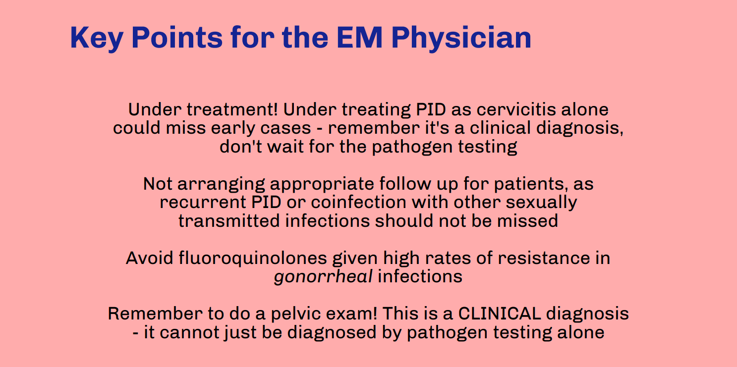
Written by: Diana Halloran, MD (NUEM ‘24) Edited by: Sean Watts, MD (NUEM ‘22) Expert Commentary by: Quentin Reuter, MD (NUEM ‘18)
The United States has been facing a debilitating opioid epidemic, which has been partially fueled by the over-prescription of these medications in the emergency department setting. In addition, the opioid epidemic has grown exponentially during the COVID-19 pandemic. More than 40 states have reported increases in opioid-related mortality, resulting in an increased burden on an already overstrained healthcare system. (1) Prescribing the medication Buprenorphine in the emergency department offers an opportunity to ameliorate these past faults and rising statistics.
The basics:
Buprenorphine, which goes by the trade name Subutex, works by acting as both a partial mu agonist and weak kappa antagonist on opiate receptors in the brain. (2) This mechanism of action enables buprenorphine to exert analgesic effects, as well as antagonistic effects when additional opiates are consumed. In addition, buprenorphine does not carry significant sedative effects, making respiratory depression extremely rare. (3) Buprenorphine is also safe in pregnancy – a 2016 meta-analysis found no difference in pregnant patients given methadone versus buprenorphine when assessing for congenital malformations. (4) The American College of Obstetrics & Gynecology has released a committee position statement, encouraging the use of buprenorphine in pregnant patients with opioid use disorder. (5)
How to prescribe:
While the DEA X-waiver is required to write a prescription for buprenorphine for addiction treatment, withdrawal, or detox, it is not required to order or administer a dose in the hospital or emergency department. (6) This exception, called the “three-day rule”, allows a patient to come to the emergency department for three consecutive days to obtain a dose of buprenorphine if found to be in opioid withdrawal. (7)
In order to dose buprenorphine in the emergency department, the patient must be in mild acute opioid withdrawal, with a Clinical Opiate Withdrawal Score (COWS) of at least 8. (8,9) Administration of buprenorphine should not occur if the patient does not appear to be clinically withdrawing, as administration in this setting could actually precipitate withdrawal.
Dosing: (10)
4mg of sublingual buprenorphine can be given initially, allowing 20-40 minutes for resolution of withdrawal symptoms with repeat dosing every 1-2 hours as needed. (10)
On Day 2, the patient’s response to Day 1 should be assessed. If the patient’s opioid withdrawal symptoms were controlled, the same dose can be continued. If not, the dose should be increased by 2-4mg. (10)
On Day 3, the patient’s response to Day 2 should be assessed. Again, if the patient’s withdrawal symptoms are controlled then the same dose can be continued. If not, the dose can be increased by 2-4mg for Day 3. (10)
After 3 days this dose should be continued for 3-7 days until steady-state levels are achieved (10)
Doses should be decreased by 2mg if the patient experiences opioid intoxication (10)
Use in the emergency department:
While buprenorphine and long-term treatment of opioid use disorder may seem confined to primary care physicians and psychiatrists, emergency medicine physicians have been shown to be successful providers for initiating buprenorphine treatment versus brief intervention and referral with a result of decreased self-reported illicit opioid use. (11) In addition, Dr. Gail D’Onofrio, chair of the Department of Emergency Medicine at Yale, found that emergency department initiated buprenorphine treatment was associated with the increased self-reported engagement of addiction treatment and reduced illicit opioid use within a two-month interval. (12) Increasing evidence demonstrates that the emergency department provides an opportunity to intervene on opioid use disorder, with more and more emergency medicine physicians becoming X-waiver certified.
References
Issue brief: Reports of increases in opioid and other drug-related overdose and other concerns during COVID pandemic. American Medical Association. https://www.ama-assn.org/system/files/2020-12/issue-brief-increases-in-opioid-related-overdose.pdf. Published December 9, 2020.
Wakhlu S. Buprenorphine: a review. J Opioid Manag. 2009 Jan-Feb;5(1):59-64. doi: 10.5055/jom.2009.0007.
Walsh SL, Preston KL, Stitzer ML, Cone EJ, Bigelow GE. Clinical pharmacology of buprenorphine: ceiling effects at high doses. Clin Pharmacol Ther. 1994 May;55(5):569-80. doi: 10.1038/clpt.1994.71.
Zedler BK, Mann AL, Kim MM, Amick HR, Joyce AR, Murrelle EL, Jones HE. Buprenorphine compared with methadone to treat pregnant women with opioid use disorder: a systematic review and meta-analysis of safety in the mother, fetus and child. Addiction. 2016 Dec;111(12):2115-2128. doi: 10.1111/add.13462.
Committee Opinion No. 711 Summary: Opioid Use and Opioid Use Disorder in Pregnancy. Obstetrics & Gynecology. 2017;130(2):488-489. doi:10.1097/aog.0000000000002229
Special Circumstances for Providing Buprenorphine. SAMHSA. https://www.samhsa.gov/medication-assisted-treatment/statutes-regulations-guidelines/special-circumstances. Published August 19, 2020.
Nagel L. Emergency Narcotic Addiction Treatment. https://www.deadiversion.usdoj.gov/pubs/advisories/emerg_treat.htm.
Wesson DR, Ling W. Clinical Opiate Withdrawal Scale. PsycTESTS Dataset. June 2003. doi:10.1037/t48752-000
D'Onofrio G, O'Connor PG, Pantalon MV, et al. Emergency department-initiated buprenorphine/naloxone treatment for opioid dependence: a randomized clinical trial. JAMA. 2015;313(16):1636-1644. doi:10.1001/jama.2015.3474
Dosing Guide For Optimal Management of Opioid Dependence. The National Alliance of Advocates for Buprenorphine Treatment.
D’Onofrio G, O’Connor PG, Pantalon MV, et al. Emergency Department–Initiated Buprenorphine/Naloxone Treatment for Opioid Dependence: A Randomized Clinical Trial. JAMA. 2015;313(16):1636–1644. doi:10.1001/jama.2015.3474
D'Onofrio G, Chawarski MC, O'Connor PG, Pantalon MV, Busch SH, Owens PH, Hawk K, Bernstein SL, Fiellin DA. Emergency Department-Initiated Buprenorphine for Opioid Dependence with Continuation in Primary Care: Outcomes During and After Intervention. J Gen Intern Med. 2017 Jun;32(6):660-666. doi: 10.1007/s11606-017-3993-2.
Expert Commentary
Thanks to Dr. Halloran and Watts for providing an informative discussion on buprenorphine prescribing from the ED. Buprenorphine continues to emerge as the state of the art treatment strategy for opioid use disorder (OUD) and thus, developing a working knowledge for when and how to use it is essential.
While there is little doubt that the medical field fueled the opioid epidemic through the prescribing of pain medications, EM is often given a disproportionate amount of blame for the current situation. In 2012, EM prescriptions made up only 4.3% of all opioids in circulation (1). Furthermore, I anticipate our specialty will continue to lead the fight against the opioid epidemic as practices such as naloxone prescribing, education around safe injecting practices, reduction and optimization of opioid prescribing efforts, and buprenorphine initiation gain further traction in the ED.
Obtaining a DEA X is the first step to prescribing buprenorphine. In April of this year guidelines for the administration of buprenorphine were updated to allow practitioners to treat up to 30 patients at a time with no extra training (2). While these changes will likely expand buprenorphine prescribing from the ED, it is vital that we do not operate in a silo.
To effectively manage this complex patient cohort, a coherent system of addiction medicine services is vital. EDs must partner with local community resources to make rapid addiction medicine appointments available. Our department utilizes specially trained addiction care coordinators, nurses with extensive training in addiction medicine to help evaluate OUD patients and navigate the fractured array of outpatient services.
Prior to the implementation of our Medication for Opioid Use Disorder (MOUD) program, our clinicians had relatively little to offer patients that directly addressed their underlying addiction. While anecdotal, we believe that by utilizing MOUD, we have begun to rebuild trust between OUD patients and the medical system. A once generally negative relationship between OUD patients and our ED staff has been replaced with a hopeful rapport, confident that recovery for these patients is a distinct possibility. This therapeutic relationship continues to grow and we believe will lead to long-term sustained recovery for many of our OUD patients in the surrounding community.
References
Levy B, Paulozzi L, Mack KA, Jones CM. Trends in Opioid Analgesic-Prescribing Rates by Specialty, U.S., 2007-2012. Am J Prev Med 2015;49:409-13.
Reuter Q, Smith G, McKinnon J, Varley J, Jouriles N, Seaberg D. Successful Medication for Opioid Use Disorder (MOUD) Program at a Community Hospital Emergency Department. Acad Emerg Med 2020.

Quentin Reuter, MD
Emergency Medicine Physician
Core Faculty at Summa Health
How To Cite This Post:
[Peer-Reviewed, Web Publication] Halloran D., Watts S. (2021, Sept 13). Buprenorphine Use in the ED. [NUEM Blog. Expert Commentary by Reuter Q.]. Retrieved from http://www.nuemblog.com/blog/buprenorphine


![Figure 1: Structural Anatomy of the Knee [5]](https://images.squarespace-cdn.com/content/v1/549b0d5fe4b031a76584e558/1613997183287-ADV18WC2TENS07IAKW85/Picture1.png)
![Figure 2: Neurovascular Anatomy of the Knee [6]](https://images.squarespace-cdn.com/content/v1/549b0d5fe4b031a76584e558/1613997188684-EJ3N1PMXE529KLK8HF5X/Picture2.png)
![Figure 3: Kennedy Classification of knee dislocations with example illustrations [9]](https://images.squarespace-cdn.com/content/v1/549b0d5fe4b031a76584e558/1613997416760-QW0N1ML3ECI1MOFL10CA/Picture3.gif)
![Figure 4: Schenck Classification System with Wascher Modification [2]](https://images.squarespace-cdn.com/content/v1/549b0d5fe4b031a76584e558/1613997441216-A4W0T4WFTEFRW9QKNGF3/Picture4.png)
![Figure 5: Algorithm for the evaluation and management of knee dislocations in the Emergency Department [10]](https://images.squarespace-cdn.com/content/v1/549b0d5fe4b031a76584e558/1613997537110-D0IQVUWEETPIQY53O7BZ/Picture5.png)
![Figure 6: Lateral knee dislocation [12]](https://images.squarespace-cdn.com/content/v1/549b0d5fe4b031a76584e558/1613998385205-AVMREK6NR6X9OK4U3NE3/Picture6.jpg)
![Figure 6: Posterior knee dislocation [13]](https://images.squarespace-cdn.com/content/v1/549b0d5fe4b031a76584e558/1613998412027-J92I2QTYDUU7MMPFLGWZ/Picture7.jpg)
![Figure 7: Segond fracture with red circle showing lateral tibial plateau avulsion fracture [14]](https://images.squarespace-cdn.com/content/v1/549b0d5fe4b031a76584e558/1613998585798-OB51SEXPTLTH98ZZHJDT/Picture8.jpg)
![FIgure 7: Fibular head avulsion fracture with white arrow showing avulsed fragment [15]](https://images.squarespace-cdn.com/content/v1/549b0d5fe4b031a76584e558/1613998622903-ZZWVB1KHKD3VLOPTOSDD/Picture9.jpg)
![Figure 8: Technique for reduction of knee dislocation [20]](https://images.squarespace-cdn.com/content/v1/549b0d5fe4b031a76584e558/1613998715615-OCFRYK6BGVNV5R1IX9FK/Picture10.jpg)
![Figure 9: Ankle brachial Index [18]](https://images.squarespace-cdn.com/content/v1/549b0d5fe4b031a76584e558/1613998836971-HGC56BHGLWIPTADPOLTU/Picture11.png)